Femoral inguinal hernia
Key Images
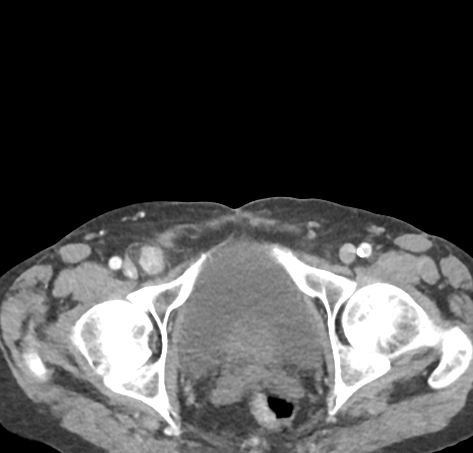
Look For First
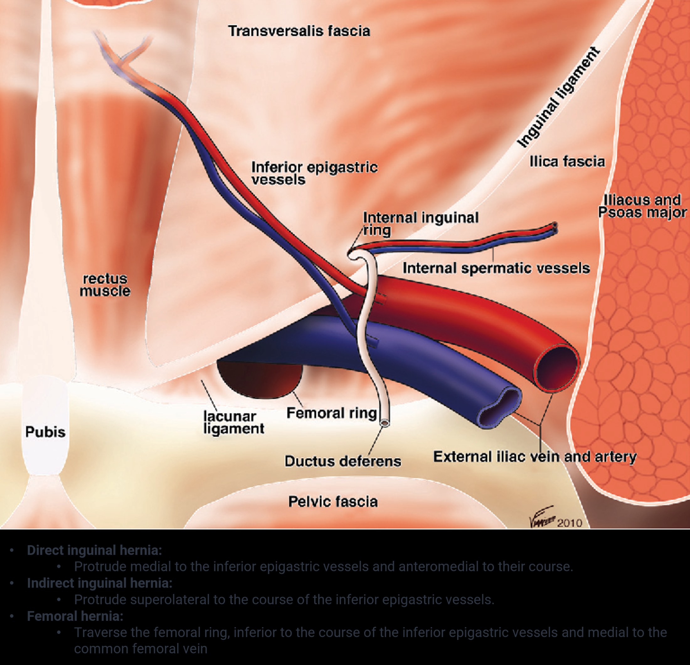
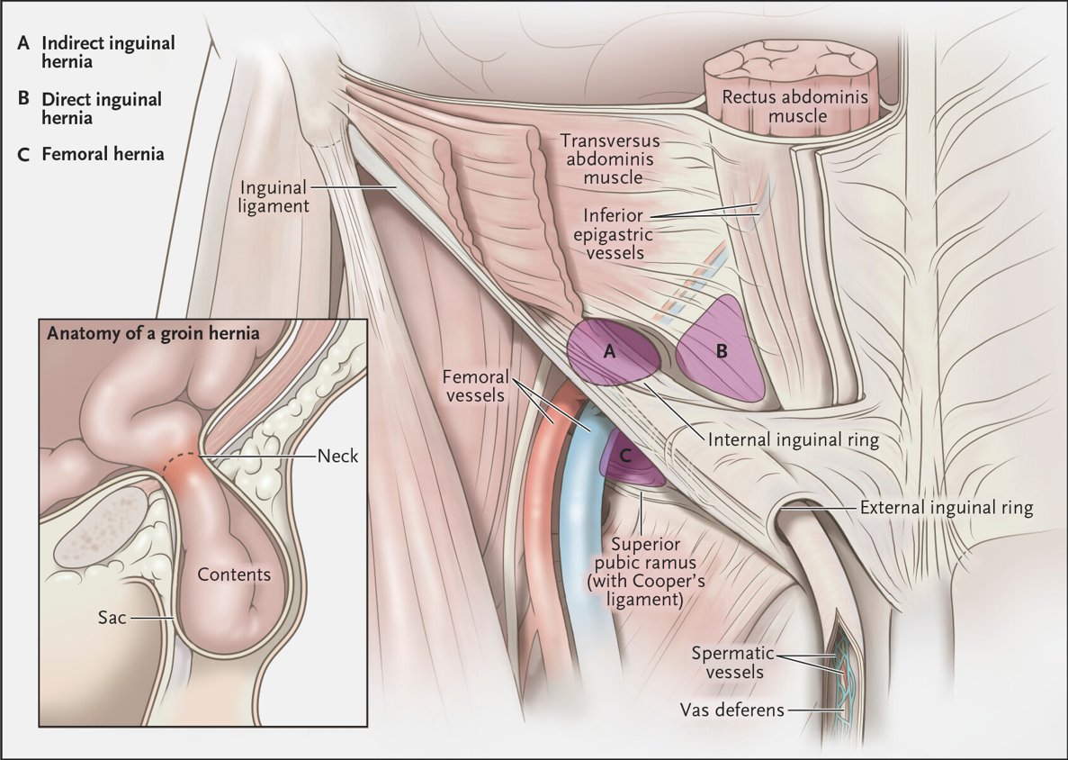
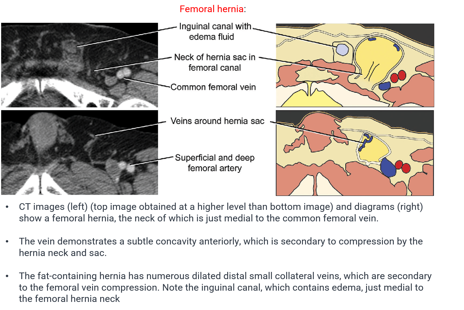
- Narrow funnel-shaped neck protruding through the femoral ring below the inguinal ligament
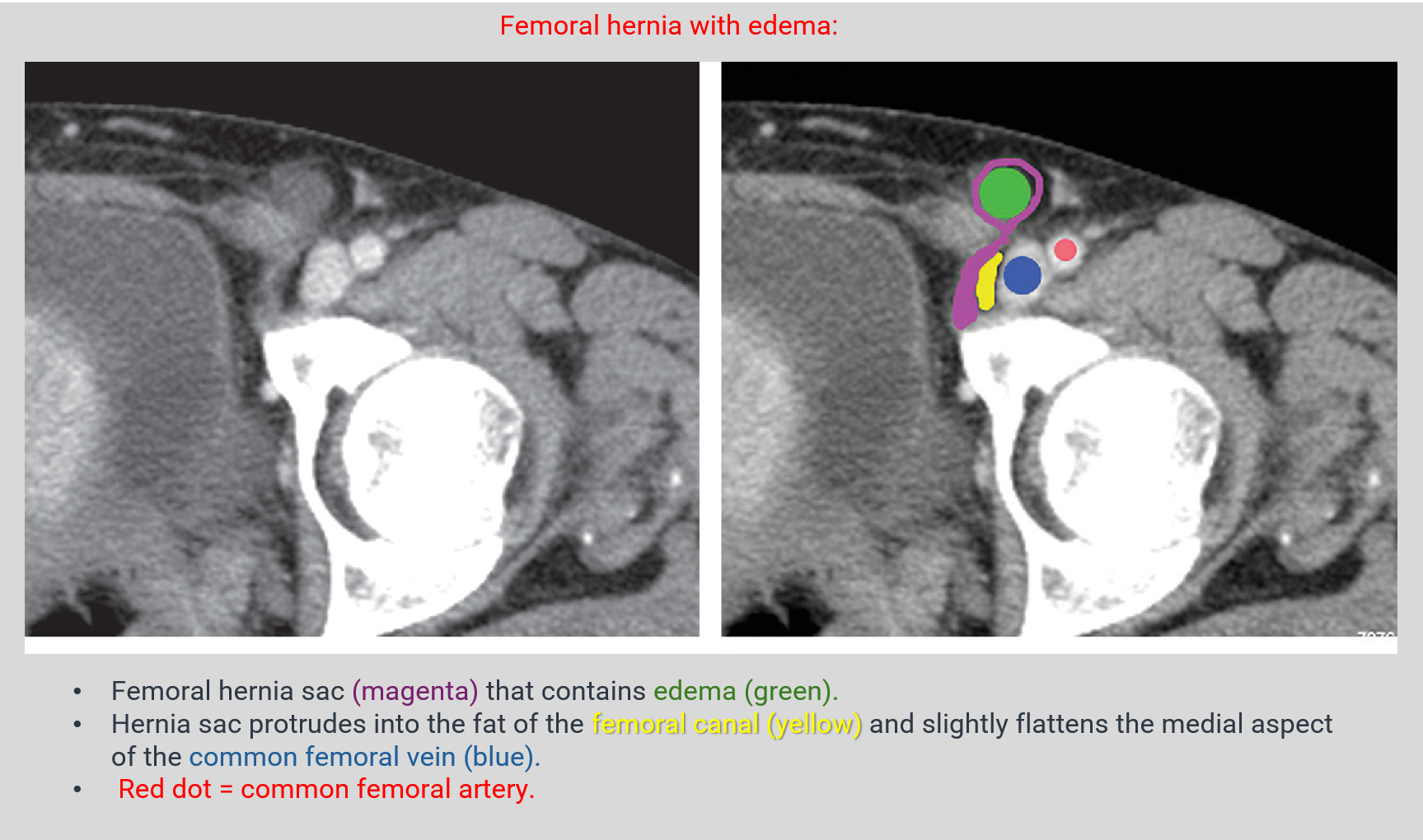
- Medial indentation or compression of the common femoral vein on axial imaging
- Hernia location lateral to the pubic tubercle, in the space medial to the femoral vein
Key Image Findings
- On ultrasound, the femoral vein is typically indented or compressed by the hernia sac when scanning below the inguinal ligament in the space medial to the femoral vein, and may become dilated caudal to the hernia sac.
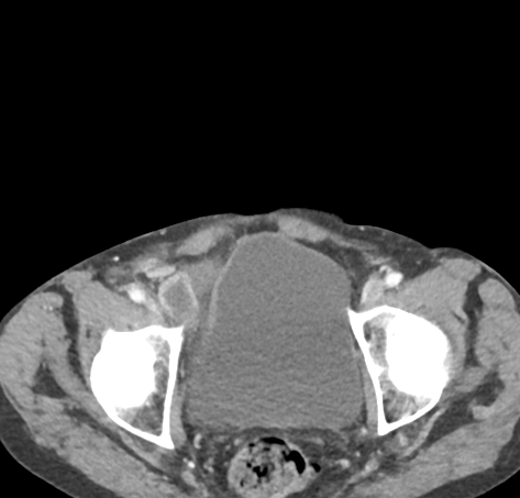
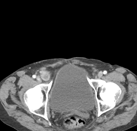
- Characteristic funnel-shaped neck is seen on CT, appearing as a narrow protrusion through the femoral ring on axial images.
- On axial CT, the neck originates below the origin of the inferior epigastric artery and is located medial to the common femoral vein.
- The femoral vein demonstrates medial wall concavity from compression by the hernia sac, with distal collateral vein engorgement as a secondary sign.
- On coronal CT, the hernia is visualized in the space containing fat between the inguinal ligament, common femoral vein, and adductor longus muscle.
- The hernia may contain preperitoneal fat, omentum, small bowel, or other peritoneal contents.
- Valsalva maneuver on ultrasound can dynamically provoke or accentuate the herniation, confirming the diagnosis.
- Right-sided predilection is noted, and femoral hernias show increased prevalence in females compared to inguinal hernias.
Differential Diagnosis
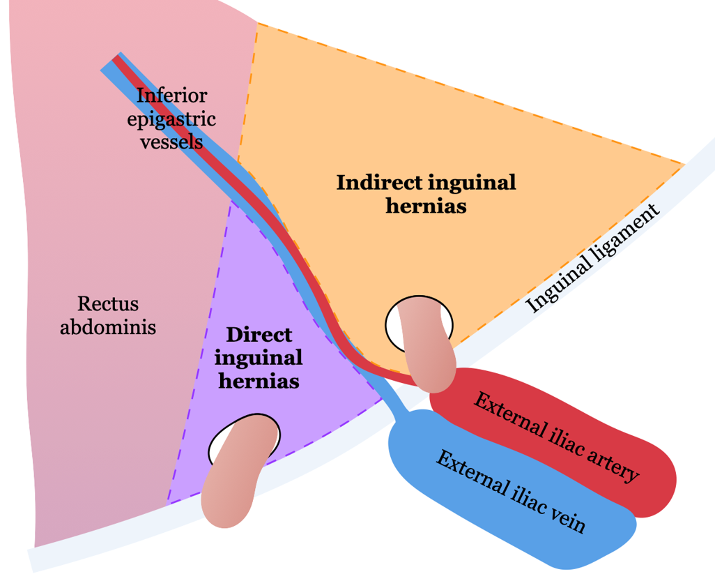
- Inguinal hernia: remains lateral to the pubic tubercle rather than medial, rarely compresses the femoral vein, and is more common in males.
- Lymphadenopathy: lacks the funnel-shaped neck and characteristic compression of the femoral vein seen with femoral hernias.
- Fluid collection or seroma: lacks the narrow funnel-shaped neck and does not compress the femoral vein.
- Laugier hernia (variant): protrudes through the lacunar ligament rather than the femoral ring proper.
- Cloquet hernia (variant): protrudes through the pectineal fascia rather than the femoral ring proper.
Discussion
Femoral hernias have a narrow funnel-shaped neck that predisposes them to compression and incarceration risk, particularly affecting the femoral vein.
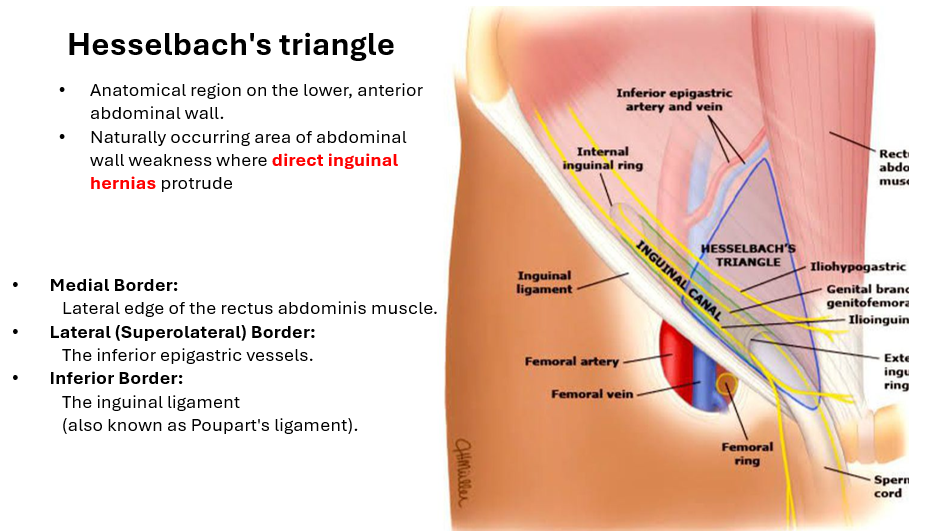
The diagnosis of femoral hernia hinges on recognizing the characteristic location below the inguinal ligament and medial to the femoral vein, which distinguishes it from inguinal hernias.
Femoral vein compression with distal collateral vein engorgement is an invariable finding in femoral hernias and serves as a key diagnostic clue.
Valsalva maneuver on ultrasound is a dynamic technique that provokes herniation and improves diagnostic confidence.
Coronal imaging is particularly useful for demonstrating the relationship of the hernia to the inguinal ligament, femoral vein, and adductor longus muscle.
Atypical variants (Laugier, Cloquet, Hesselbach, Velpeau, Serafini hernias) may have different anatomic relationships to the femoral vessels and should be recognized.
Reporting Pearls
Describe the hernia as a "narrow funnel-shaped sac protruding through the femoral ring, located below the inguinal ligament and medial to the common femoral vein, with compression causing medial wall concavity" to clearly convey both the morphology and hemodynamic significance.
Pitfalls
- Confusing femoral hernia with inguinal hernia on imaging; remember femoral hernias are medial to the femoral vein and compress it, while inguinal hernias are lateral to the pubic tubercle and rarely compress the vein.
- Overlooking the funnel-shaped neck on axial images, which is the pathognomonic morphologic feature; scan systematically below the inguinal ligament.
- Failing to assess for femoral vein compression and distal collateral engorgement, which are invariable findings and critical for risk stratification.
- Not performing Valsalva maneuver on ultrasound or dynamic imaging, which may be needed to provoke a borderline or intermittent herniation and increase diagnostic confidence.