Indirect inguinal hernia
Key Images
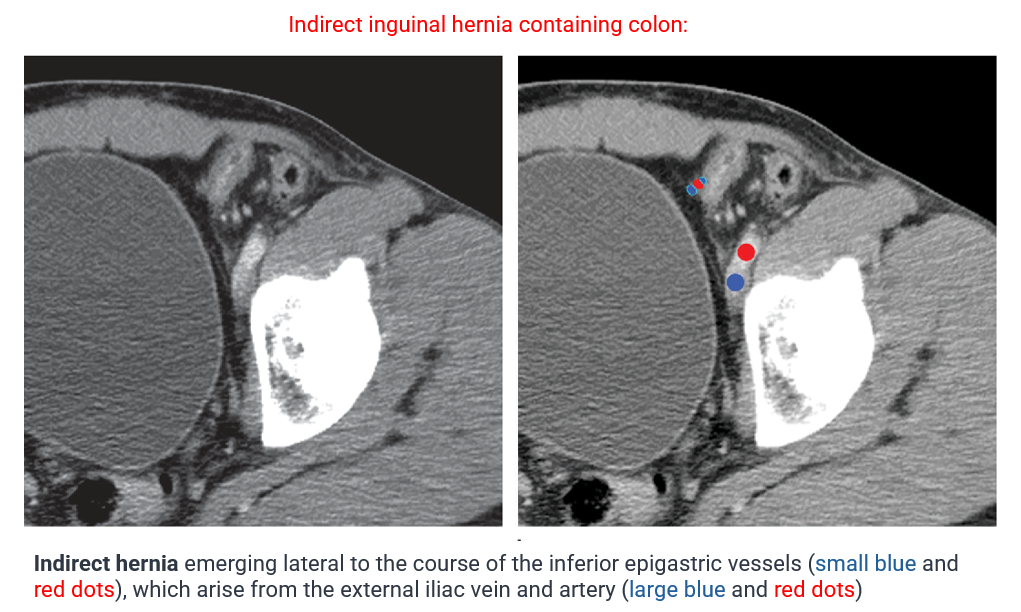
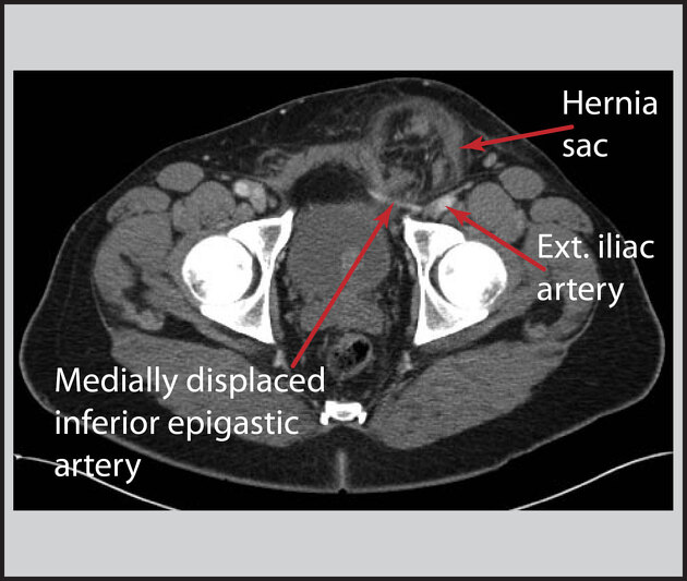
Look For First
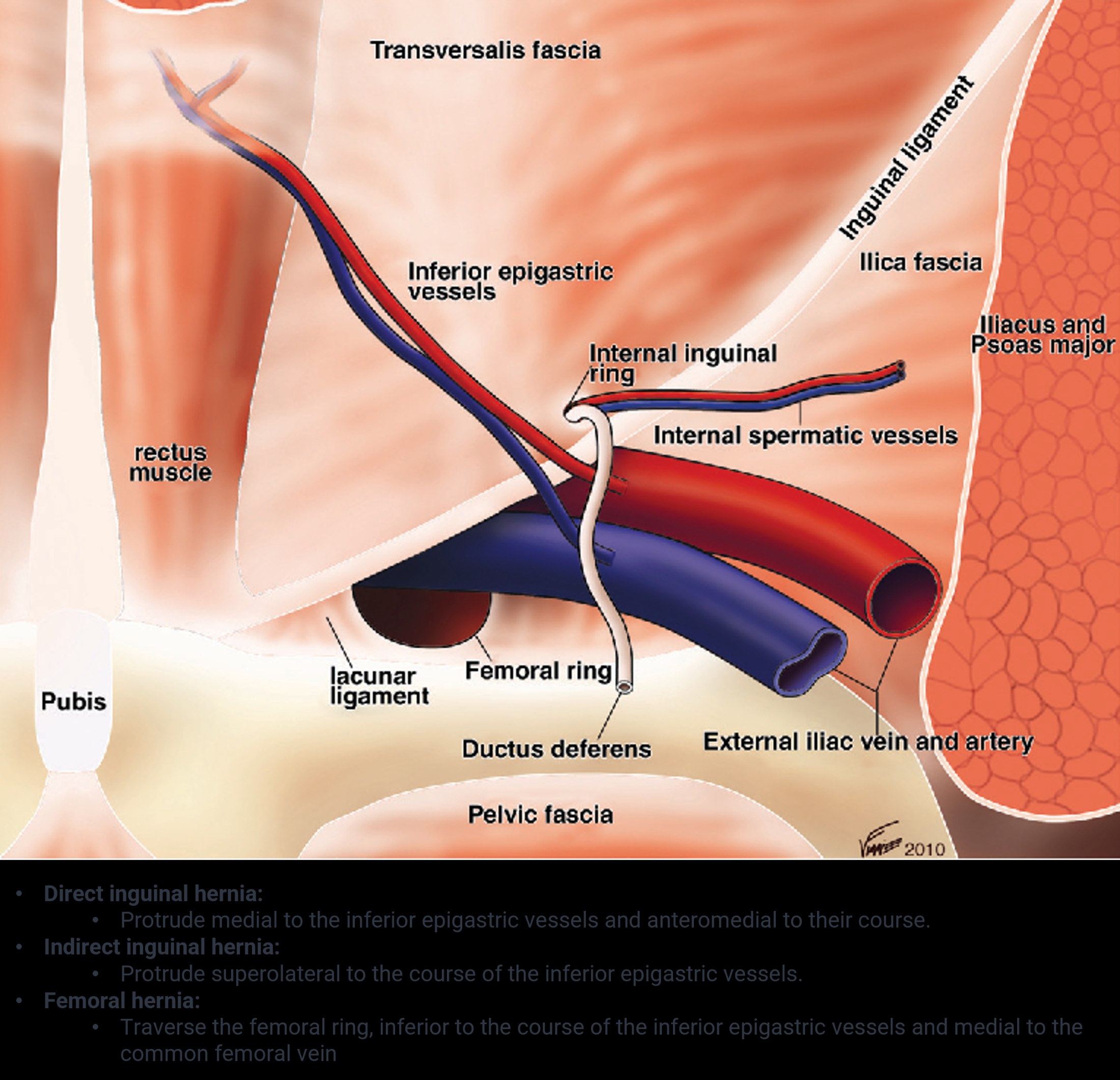
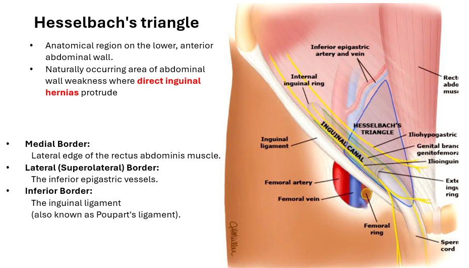
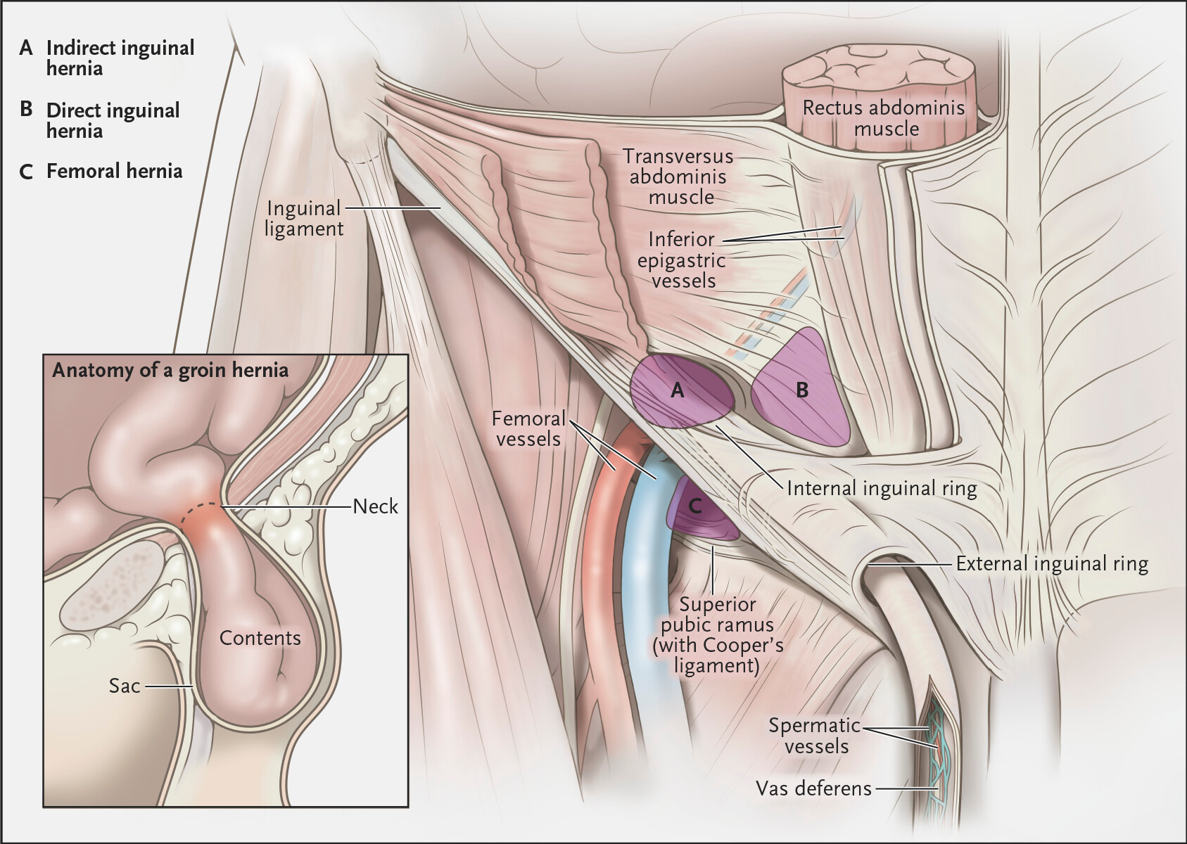
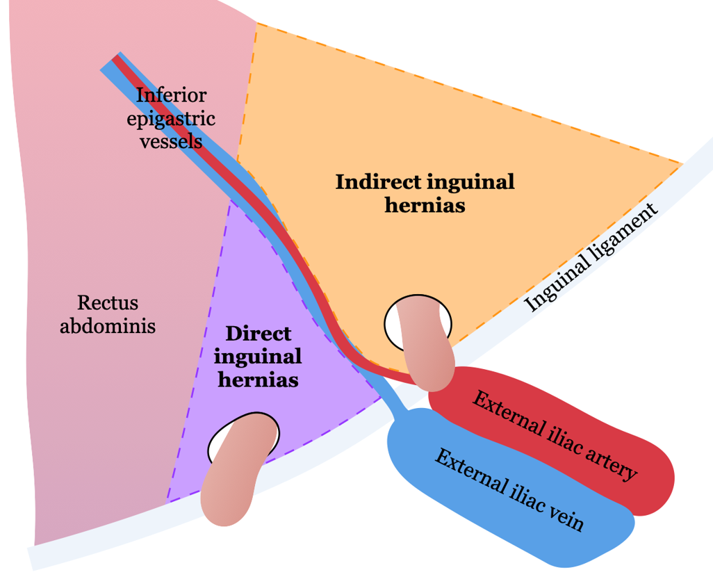
- Hernial sac lateral and superior to the inferior epigastric vessels (lateral to Hesselbach's triangle)
- Protrusion through the deep (internal) inguinal ring into the inguinal canal
- In males: content entering anterior to the spermatic cord and potentially extending into the scrotum; in females: following the round ligament into the labia majora
- Movement of fat or fluid within the hernia on dynamic imaging during Valsalva maneuver
Key Image Findings
- Hernial sac arises lateral and superior to the inferior epigastric vessels, distinguishing it from direct hernias which arise medial to these vessels.
- The sac protrudes through the deep (internal) inguinal ring and extends inferomedially within the inguinal canal to emerge via the superficial inguinal ring.
- In males, the hernial content enters the canal anterior to the spermatic cord and may extend through the superficial ring into the scrotum, creating a scrotal component.
- In females, the hernia tends to follow the round ligament trajectory into the labia majora.
- Common contents include mesenteric fat (most common), peritoneal fluid, small bowel loops, and occasionally mobile colon segments (sigmoid, cecum, appendix), bladder, or ureter.
- On ultrasound, dynamic imaging with Valsalva maneuver shows movement of hernial contents, confirming the hernia rather than a fixed mass such as a spermatic cord lipoma.
- Many indirect inguinal hernias are asymptomatic and longstanding, though large hernias may cause patient symptoms from mass effect.
- Complications on imaging may include signs of incarceration (fixed contents), strangulation (bowel wall thickening, mesenteric edema), or obstruction (bowel dilatation).
Differential Diagnosis
- Direct inguinal hernia: arises medially to the inferior epigastric vessels and above the inguinal ligament, with the inguinal canal typically compressed or displaced (lateral crescent sign), whereas indirect hernias arise lateral to these vessels.
- Femoral hernia: exits below the inguinal ligament and caudal to the inferior epigastric vessels, located in a different anatomical compartment than indirect inguinal hernias.
- Spermatic cord lipoma (on testicular ultrasound): may mimic an inguinal hernia containing omental fat, but lacks movement with Valsalva maneuver, whereas a true indirect hernia shows dynamic motion of contents.
Discussion
Indirect inguinal hernias result from persistence of the processus vaginalis during testicular descent, explaining the seven-fold higher incidence in males and the high prevalence in children.
Incarceration is the most common complication associated with inguinal hernias, with incidence as high as 30% in infants younger than 2 months, making early detection clinically important.
Strangulation with bowel ischemia, perforation, and intestinal obstruction are severe complications that may be evident on imaging as bowel wall changes, mesenteric fat stranding, or luminal dilatation.
The anatomy of hernia passage differs by gender: males have risk of scrotal extension (anterior to spermatic cord), while females follow the round ligament into the labia majora.
Dynamic imaging during Valsalva maneuver is essential for diagnosis, as movement of contents confirms a hernia and helps distinguish it from fixed masses.
Indirect inguinal hernias are five times more common than direct inguinal hernias and represent the vast majority of inguinal hernias in the pediatric population.
Reporting Pearls
Describe the hernia location precisely: "Indirect inguinal hernia arising lateral to the inferior epigastric vessels, protruding through the deep inguinal ring and extending inferomedially through the inguinal canal." Specify the contents (fat, bowel, fluid), note any gender-specific extension (scrotal in males, labia in females), and document whether contents are reducible or show signs of incarceration/strangulation on dynamic imaging.
Pitfalls
- Confusing indirect with direct inguinal hernia: indirect hernias arise lateral to the inferior epigastric vessels and deep ring, while direct hernias arise medial to these structures and bulge directly through the posterior inguinal wall.
- Mistaking a spermatic cord lipoma for an indirect inguinal hernia on ultrasound: perform Valsalva maneuver to demonstrate movement of true hernia contents and absence of movement in a lipoma.
- Failing to perform dynamic imaging (Valsalva) to confirm hernia: static images alone may underestimate or miss small hernias, particularly in the pediatric population.
- Missing scrotal or labial extension of the hernia: in males, always examine for content extending into the scrotum anterior to the spermatic cord; in females, assess for labia majora involvement.