Adenomyosis vs Endometriosis
Key Images
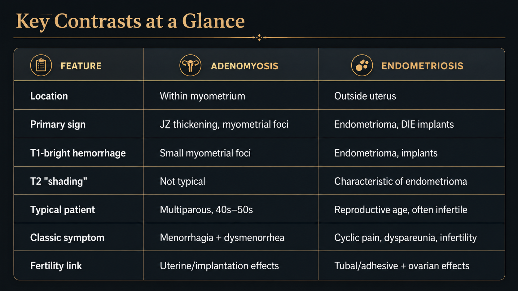
Look For First
- Junctional zone (JZ) thickening ≥12 mm on T2-weighted images for adenomyosis
- T1-hyperintense endometrioma with T2 'shading' on fat-saturated sequences for endometriosis
- Ill-defined low-T2 myometrial signal with small T1-hyperintense hemorrhagic foci within the uterine wall (adenomyosis) versus implants at extrauterine sites such as uterosacral ligaments, pouch of Douglas, or rectovaginal septum (endometriosis)
Key Image Findings
- Adenomyosis is characterized by junctional zone thickening ≥12 mm, representing hypertrophied smooth muscle responding to ectopic endometrial glands within the myometrium.
- Adenomyotic myometrium shows ill-defined low T2 signal (reflecting fibrosis and smooth-muscle hypertrophy) interspersed with small T2-hyperintense and T1-hyperintense (hemorrhagic) foci representing ectopic glandular and blood-containing spaces.
- Adenomyosis causes globular uterine enlargement with radiating striations extending from the endometrium into the myometrium, best seen on T2-weighted imaging.
- Endometrioma appears as a complex ovarian cyst with T1-hyperintense content that does not suppress on fat-saturated sequences (distinguishing it from fat or gadolinium), combined with characteristic T2 'shading' (peripheral bright T2 signal with central dark T2 signal) and possible hemosiderin deposits.
- Deep infiltrating endometriosis (DIE) manifests as low-T2 spiculated or stellate soft-tissue nodules and fibrosis at the uterosacral ligaments, rectovaginal septum, bowel wall, and bladder, best appreciated on multiplanar imaging with dedicated pelvic protocol.
- Endometriosis causes secondary findings including pelvic adhesions, tethering, angulated or kinked bowel loops, and 'kissing ovaries' (opposed adnexa due to adhesive traction), findings not typical of isolated adenomyosis.
- Adenomyosis is best assessed on T2-weighted and T1-weighted (with fat saturation) imaging focused on the uterine body and junctional zone, whereas endometriosis requires a comprehensive pelvic survey to detect peritoneal, ligamentous, and ovarian implants.
- Adenomyosis typically presents in multiparous women aged 40–50 years with menorrhagia and dysmenorrhea, while endometriosis typically presents in reproductive-age women with cyclic pain, dyspareunia, and infertility, though both are estrogen-dependent and often coexist.
Differential Diagnosis
- Uterine leiomyoma (fibroid) versus adenomyosis: fibroids are well-circumscribed, often homogeneously low T2, cause mass effect distorting the junctional zone, and may show degeneration or cystic change; adenomyosis shows ill-defined junctional zone thickening without a discrete nodule.
- Endometrial cancer versus adenomyosis: endometrial cancer presents as an echogenic or heterogeneous endometrial mass with abnormal junctional zone disruption and often elevated T2 signal; adenomyosis is diffuse myometrial involvement with preserved, though thickened, junctional zone.
- Ovarian cyst with hemorrhage versus endometrioma: hemorrhagic ovarian cysts may show T1 hyperintensity but typically resolve over time and lack the characteristic T2 shading pattern and persistent T1-bright non-fat-suppressing content of endometrioma.
- Bowel pathology (Crohn disease, malignancy) versus deep infiltrating endometriosis: bowel inflammation or neoplasm shows enhancement, wall thickening, and clinical/endoscopic correlation; DIE typically causes wall thickening without mucosal involvement or enhancement and is associated with peritoneal findings.
- Adenomyosis versus diffuse myometrial edema from infection or inflammation: myometritis shows acute T2 hyperintensity, clinical fever, and elevated inflammatory markers; adenomyosis is chronic with low T2 foci and no acute systemic symptoms.
- Normal thick junctional zone (reproductive age) versus pathologic adenomyosis: the junctional zone normally measures up to 10–12 mm in reproductive-age women and may transiently thicken with hormonal changes; adenomyosis is diagnosed when JZ ≥12 mm persists with associated T2 hyperintense and T1 hyperintense foci.
Discussion
Adenomyosis and endometriosis share a common origin (ectopic endometrial tissue) but differ fundamentally in location: adenomyosis is intrauterine (within the myometrium), while endometriosis is extrauterine, and this anatomic distinction determines clinical presentation, imaging findings, and treatment approach.
Coexistence of adenomyosis and endometriosis is common; identifying one entity should prompt careful search for the other, as presence of both adversely affects fertility and may alter therapeutic strategy.
Adenomyosis causes secondary smooth-muscle hypertrophy and fibrosis around ectopic endometrial glands, leading to the imaging hallmark of junctional zone thickening and globular uterine enlargement; this pathology impairs endometrial receptivity and implantation.
Endometriosis often triggers adhesive disease and fibrotic implants outside the uterus, particularly at the pouch of Douglas, uterosacral ligaments, and ovaries, explaining the classic symptoms of dyspareunia, cyclic pain, and tubal/ovarian-factor infertility.
Both adenomyosis and endometriosis are estrogen-dependent and may be amenable to hormonal therapy; however, adenomyosis is primarily a disease of multiparous women (40s–50s) whereas endometriosis typically affects younger reproductive-age women, though the latter may persist or develop later.
MRI is the gold standard for detecting and characterizing both adenomyosis (T2 junctional zone assessment) and endometriosis (T1-fat-sat and T2 imaging for endometriomas and DIE); a comprehensive pelvic protocol with multiplanar imaging and fat saturation is essential to avoid missing either diagnosis.
Reporting Pearls
Clearly separate adenomyosis from endometriosis in your report by stating the precise location of findings: "Junctional zone thickening (≥12 mm) with myometrial foci consistent with adenomyosis" for intrauterine disease, versus "T1-hyperintense ovarian cyst with T2 shading and non-fat-suppressing content consistent with endometrioma" or "Low-T2 nodular fibrosis at the uterosacral ligament/rectovaginal septum consistent with deep infiltrating endometriosis" for extrauterine disease, and explicitly note whether both entities are present, as this impacts counseling and treatment.
Pitfalls
- Mistaking a normal junctional zone (which can measure up to 10–12 mm in reproductive-age women) for pathologic adenomyosis; adenomyosis requires both JZ thickening ≥12 mm and associated T2-hyperintense and T1-hyperintense myometrial foci rather than isolated JZ measurement.
- Failing to perform dedicated fat-saturated sequences; without fat saturation, T1-hyperintense endometriomas cannot be confidently distinguished from fat, and endometriotic implants at peritoneal or ligamentous sites may be missed.
- Overlooking the characteristic T2 'shading' pattern (peripheral bright, central dark signal) of endometrioma; absence of shading should prompt consideration of other hemorrhagic cystic lesions and correlation with imaging over time.
- Finding adenomyosis and assuming this excludes endometriosis (or vice versa); substantial proportion of patients have both, so identification of one should trigger systematic evaluation of extrauterine sites and careful assessment of the myometrium.