Cerebral toxoplasmosis (neurotoxoplasmosis)
Key Images
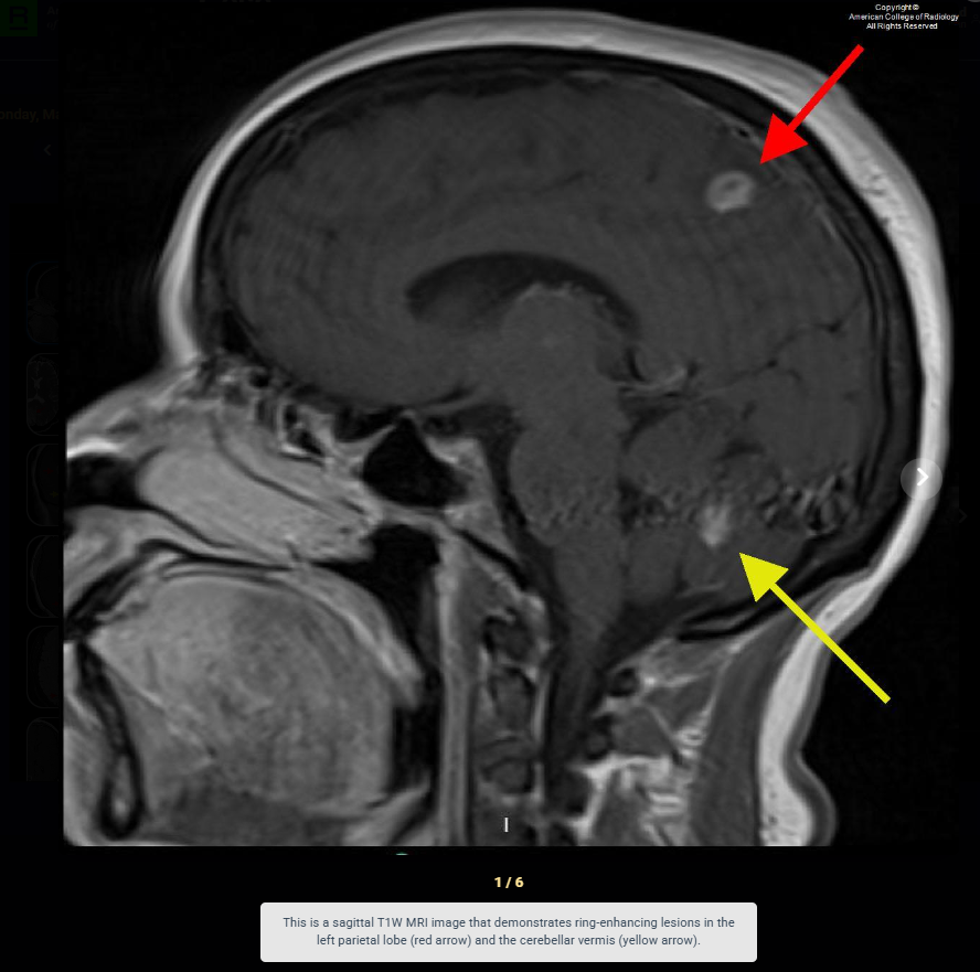
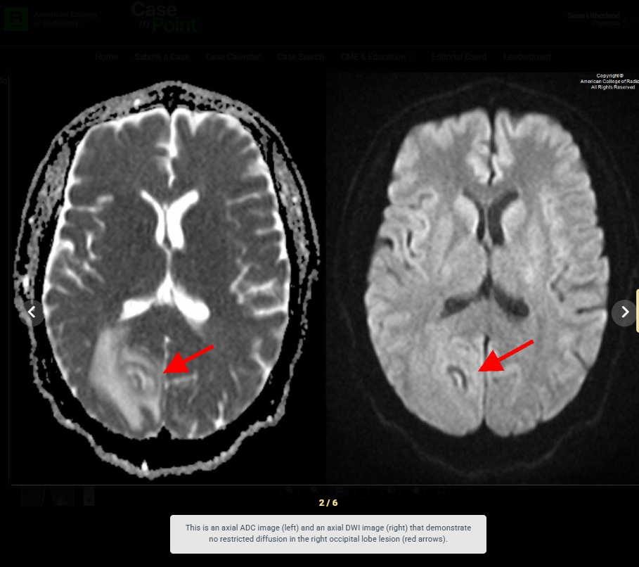
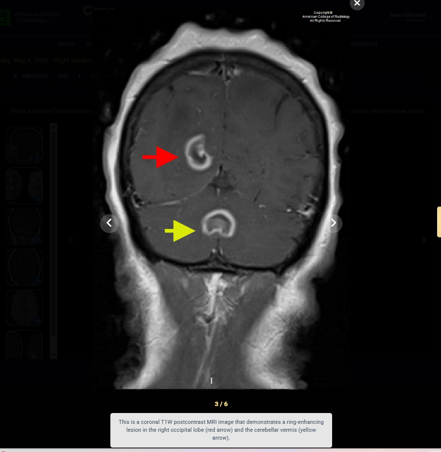
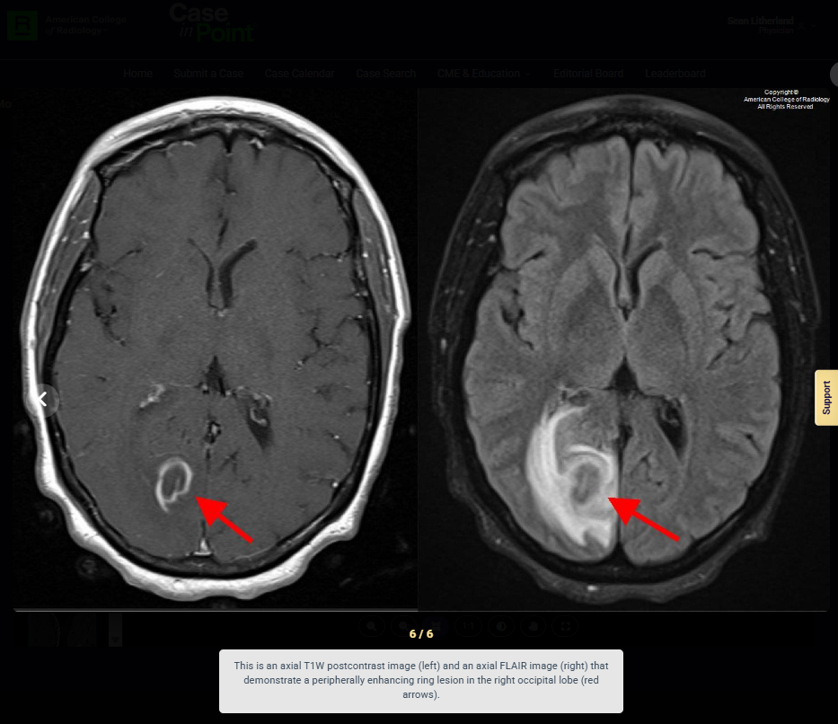
Look For First
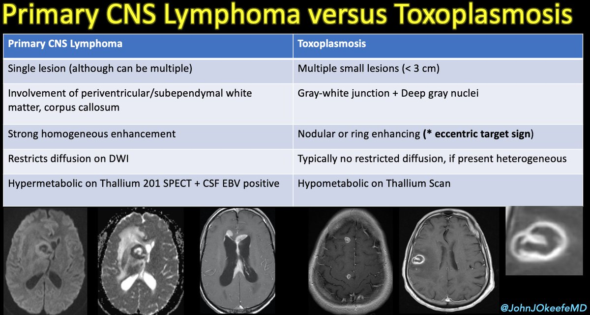
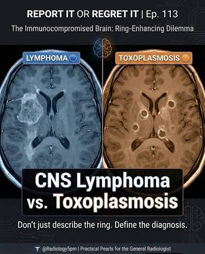
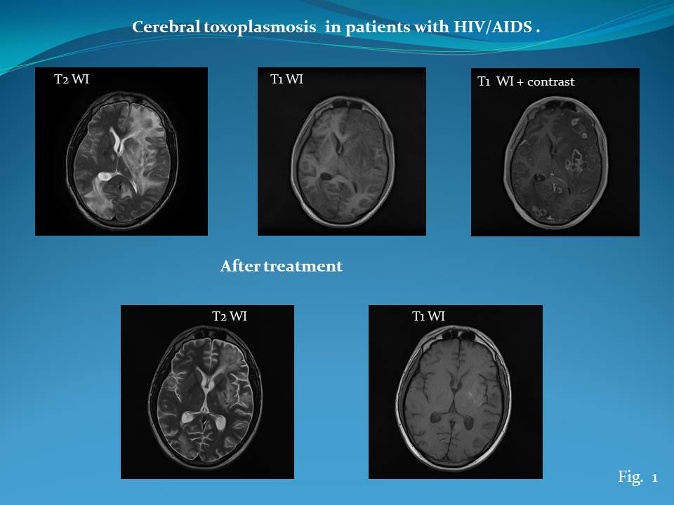
- Ring-enhancing lesions with concentric and eccentric 'target sign' enhancement on MRI
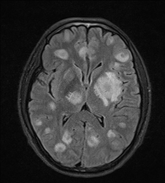
- Multiple lesions in basal ganglia and corticomedullary junction distribution
- Perilesional edema surrounding contrast-enhancing focal brain lesions
- Absence of restricted water diffusion on DWI/ADC (unlike CNS abscesses)
Key Image Findings
- On MRI, cerebral toxoplasmosis presents with characteristic ring-enhanced lesions with concentric and eccentric 'target sign' enhancement pattern accompanied by perilesional edema.
- Lesions typically locate in the basal ganglia and corticomedullary junction, with multiple lesions scattered throughout these regions rather than subependymal distribution.
- On CT, toxoplasmosis shows hypodense and contrast-enhancing focal brain lesions with mass effect, primarily in basal ganglia and corticomedullary junction.
- DWI/ADC imaging reveals higher ADC values (less restriction of water diffusion) in toxoplasma lesions, helping distinguish from pyogenic CNS abscesses which show marked diffusion restriction.
- MR spectroscopy shows lipid/lactate peaks without the prominent choline increase seen in lymphoma, supporting the diagnosis of toxoplasmosis.
- FDG-PET demonstrates relatively low FDG uptake in most toxoplasmosis cases, in contrast to the high hypermetabolic uptake seen in primary CNS lymphoma.
- Multiparametric MRI combining DWI/ADC, perfusion imaging, and MR spectroscopy provides the best noninvasive imaging differentiation from mimics like CNS lymphoma.
- Biopsy with immunohistochemistry is reserved for inconclusive imaging cases to definitively rule out malignant B-cells and confirm diagnosis when noninvasive testing is non-diagnostic.
Differential Diagnosis
- Primary CNS lymphoma: typically shows subependymal distribution near brain ventricles, marked diffusion restriction on DWI/ADC (high DWI, low ADC), elevated choline peak on MRS, and high FDG uptake on PET, with poor prognosis versus toxoplasmosis responding well to antibiotics.
- Cerebral metastasis: typically solitary or fewer lesions with different distribution pattern and clinical context of known primary malignancy; toxoplasmosis more commonly multifocal.
- Cryptococcus neoformans: typically presents as meningitis or gelatinous-appearing lesions in basal ganglia; toxoplasmosis shows ring enhancement rather than minimal enhancement.
- CNS tuberculosis: typically affects meninges and produces hypodense lesions with ring enhancement but with different clinical presentation and CSF findings.
- Pyogenic CNS abscess: shows marked restricted water diffusion on DWI/ADC unlike toxoplasmosis; additionally, clinical presentation and organism-specific findings differ.
- HIV-related dementia or other opportunistic infections: distinguished by imaging characteristics and clinical context.
Discussion
Toxoplasmosis infection is very common, but symptomatic toxoplasmic encephalitis occurs in less than 10% of infected patients and primarily affects immunocompromised populations including HIV/AIDS patients, transplant recipients, and other immunosuppressed individuals.
The pathophysiology involves reactivation of dormant brain cysts from Toxoplasma gondii in immunocompromised hosts, presenting with characteristic CNS symptoms including headaches, confusion, weakness, lack of coordination, and seizures.
Diagnosis is supported by the Sabin-Feldman dye test (gold standard) and ELISA (most common) detecting anti-toxoplasma IgG and IgM antibodies, with immunohistochemistry used on biopsy tissue for definitive confirmation.
The distinction from primary CNS lymphoma is clinically critical because toxoplasmosis responds well to antibiotics while lymphoma carries poor prognosis and requires high-toxicity chemotherapy.
Multiparametric MRI sequences including DWI/ADC, perfusion imaging, and MR spectroscopy combined with functional imaging like FDG-PET provide high accuracy in noninvasive differentiation from similar-appearing lesions.
Biopsy remains the definitive diagnostic tool when noninvasive imaging and serologic tests are inconclusive, particularly important before committing to toxic lymphoma therapy.
Reporting Pearls
Report the characteristic ring-enhancing lesions with concentric and eccentric 'target sign' enhancement in the basal ganglia and corticomedullary junction distribution, emphasize the absence of diffusion restriction on DWI/ADC, and note that while imaging is highly suggestive of toxoplasmosis, definitive diagnosis requires correlation with serology (Sabin-Feldman dye test or ELISA) or histopathology, particularly to exclude primary CNS lymphoma.
Pitfalls
- Primary CNS lymphoma is the most important mimic and can appear nearly identical on conventional MRI; failure to use multiparametric sequences (DWI/ADC, MRS) or FDG-PET may lead to misdiagnosis with serious clinical consequences given the different treatment and prognosis.
- Assuming the diagnosis based on imaging alone without serologic confirmation (Sabin-Feldman, ELISA) or biopsy in inconclusive cases can delay appropriate treatment or lead to incorrect diagnosis.
- Overlooking the distribution pattern difference: toxoplasmosis lesions scatter throughout basal ganglia and corticomedullary junction, while lymphoma typically shows subependymal distribution adjacent to ventricles; location alone can be a helpful clue.
- Confusing the DWI/ADC findings: remembering that toxoplasma lesions show higher ADC values (less restriction) while CNS lymphoma and abscesses show marked diffusion restriction is essential for accurate differentiation.