CNS Cell Types and Tumors

A practical cell-of-origin framework for CNS tumors, integrated with modern molecular classification.

Reference Images

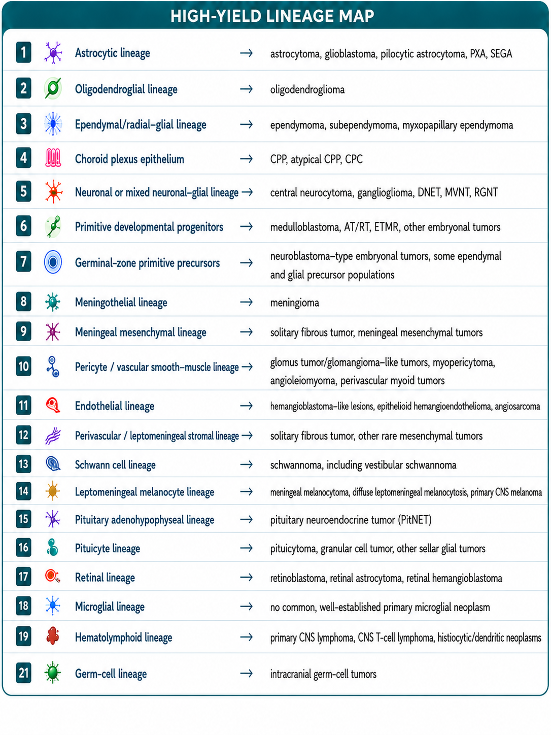
High-yield CNS tumor lineage map
High-Yield Lineage MapClick for a scrollable, zoomable viewOpen original image
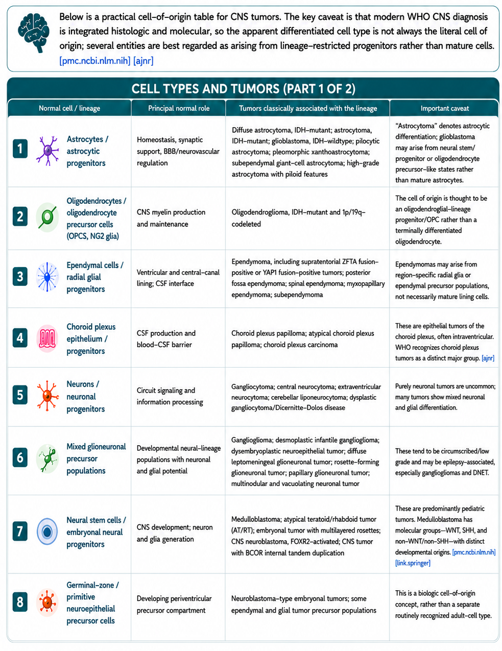
CNS cell types and tumors, part 1
Cell Types and Tumors — Part 1Click for a scrollable, zoomable viewOpen original image
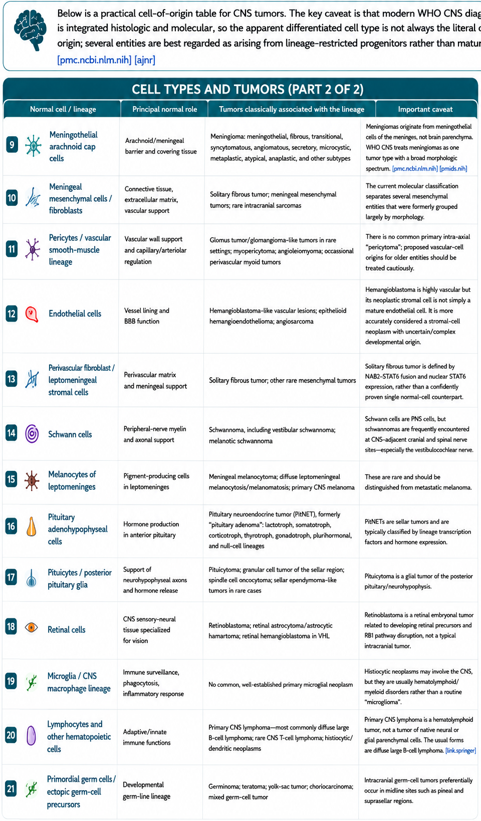
CNS cell types and tumors, part 2
Cell Types and Tumors — Part 2Click for a scrollable, zoomable viewOpen original image

Interpretation Caution

The traditional phrase “tumor arising from cell X” is clinically useful, but it is an oversimplification. Modern WHO CNS diagnosis integrates histology and defining molecular alterations; the apparent differentiated cell type is not always the literal cell of origin. Several entities are best understood as arising from lineage-restricted progenitors rather than mature cells.

For example, an adult infiltrating glioma with oligodendroglial morphology is classified as oligodendroglioma only when it is IDH-mutant and 1p/19q-codeleted.

Neural and Glial

Neurons

  • Excitatory, inhibitory, neuromodulatory, sensory, projection, and local interneuron populations.
  • Associated tumors include gangliocytoma, central neurocytoma, DNET, ganglioglioma, MVNT, and cerebellar liponeurocytoma.

Astrocytic lineage

  • Astrocytes regulate homeostasis, synapses, metabolism, and neurovascular coupling.
  • Classic associations include diffuse astrocytoma, glioblastoma, pilocytic astrocytoma, pleomorphic xanthoastrocytoma, and SEGA.

Oligodendrocyte lineage

  • OPCs, newly formed oligodendrocytes, and mature myelinating oligodendrocytes.
  • Oligodendroglioma requires IDH mutation and 1p/19q codeletion.

Ventricular and Developmental

Ependymal and barrier cells

  • Ependymal and radial-glial lineage: ependymoma, subependymoma, and myxopapillary ependymoma.
  • Choroid plexus epithelium: choroid plexus papilloma, atypical papilloma, and carcinoma.

Progenitor populations

  • Mixed glioneuronal precursor populations: ganglioglioma, desmoplastic infantile ganglioglioma, DNET, RGNT, and diffuse leptomeningeal glioneuronal tumor.
  • Primitive developmental progenitors: medulloblastoma, AT/RT, ETMR, and other embryonal tumors.
  • Germinal-zone precursors are a developmental concept and not a separate routinely recognized adult cell type.

Meningeal and Vascular

Meningeal lineages

  • Meningothelial/arachnoid-cap cells: meningioma spectrum.
  • Meningeal mesenchymal and perivascular fibroblast lineages: solitary fibrous tumor and rare mesenchymal tumors.
  • Leptomeningeal melanocytes: meningeal melanocytoma, melanocytosis, and primary CNS melanoma.

Neurovascular unit

  • Endothelium, pericytes, vascular smooth muscle, perivascular fibroblast-like cells, macrophages, and astrocyte endfeet maintain the BBB.
  • Associated lesions include hemangioblastoma-like lesions, epithelioid hemangioendothelioma, angiosarcoma, and rare perivascular myoid tumors.

Boundary and Specialized Cells

Schwann cells

Schwann cells are PNS cells found at CNS-adjacent cranial and spinal nerve transition zones. They are associated with schwannoma, including vestibular schwannoma.

Sellar and retinal lineages

  • Adenohypophyseal cells: pituitary neuroendocrine tumor (PitNET).
  • Pituicytes/posterior pituitary glia: pituicytoma, granular cell tumor, and spindle cell oncocytoma.
  • Retinal lineage: retinoblastoma, retinal astrocytoma, and retinal hemangioblastoma.

Immune and Germ-Cell Lineages

Microglia and macrophages

Microglia are resident CNS macrophage-lineage cells involved in surveillance, phagocytosis, synaptic pruning, and inflammation. There is no common, well-established primary microglial neoplasm.

Hematolymphoid cells

Primary CNS lymphoma, CNS T-cell lymphoma, and histiocytic/dendritic neoplasms reflect hematolymphoid lineages rather than routine glial differentiation.

Primordial germ cells

Ectopic germ-cell precursors can produce intracranial germ-cell tumors, classically in midline sites such as pineal and suprasellar regions.
Clinical Takeaway

  • Use lineage as an organizing framework, not as proof of literal cell of origin.
  • Pair morphology and imaging phenotype with molecular criteria.
  • Recognize that many tumors likely arise from progenitor populations.
  • Do not group epithelial choroid plexus tumors with ependymal tumors solely because both are ventricular.
  • Distinguish Schwann-cell tumors from intrinsic CNS glial tumors.

Further reading

Secondary Links