Dysembryoplastic neuroepithelial tumor (DNET)
Key Images
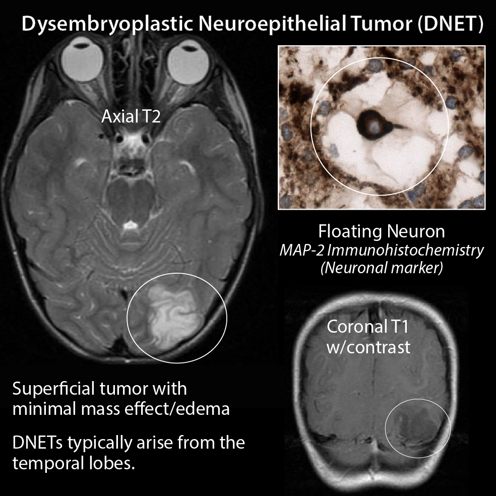
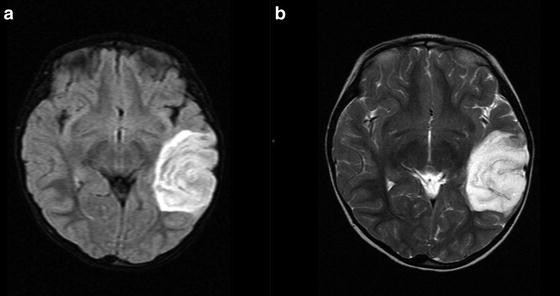
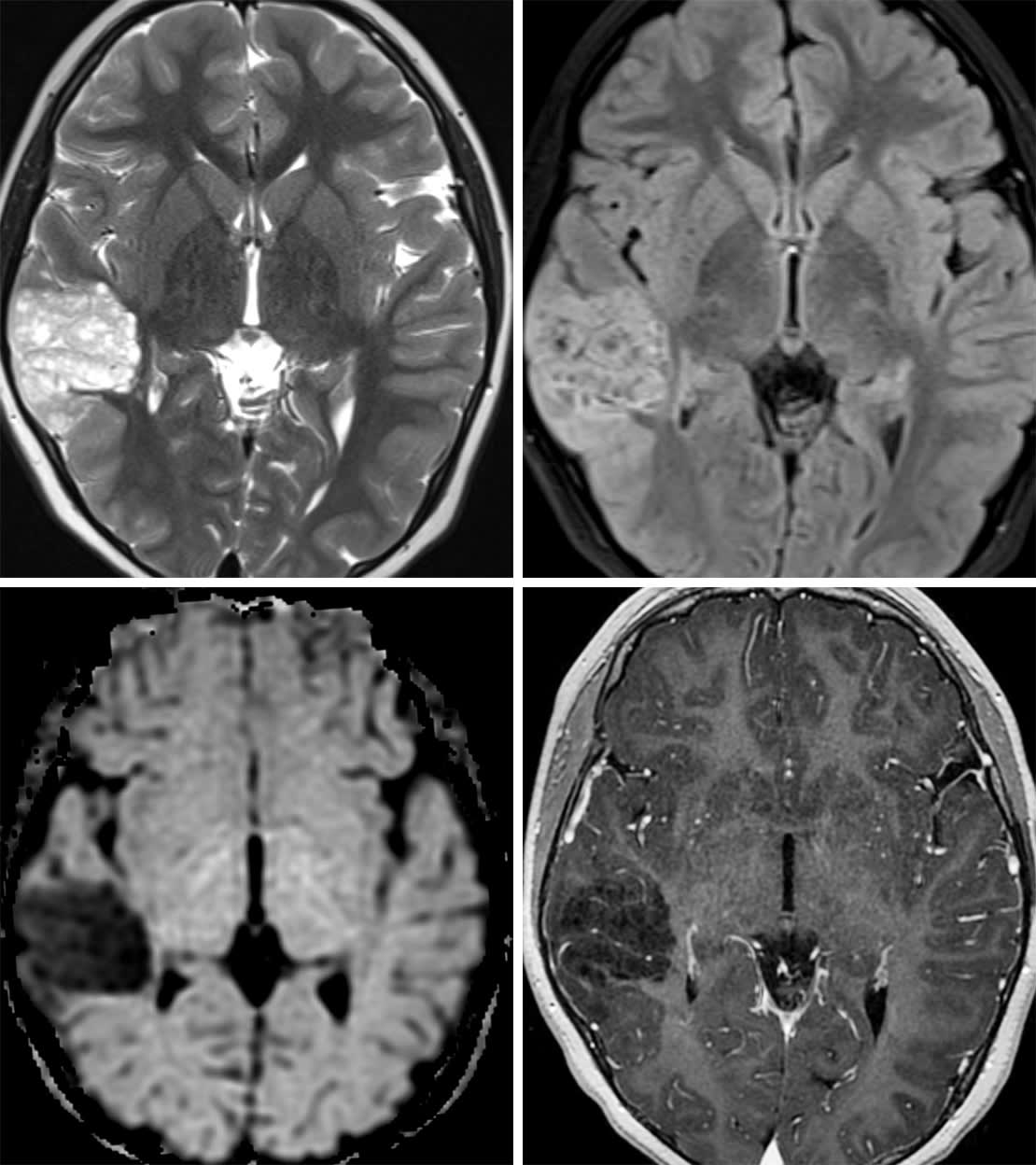
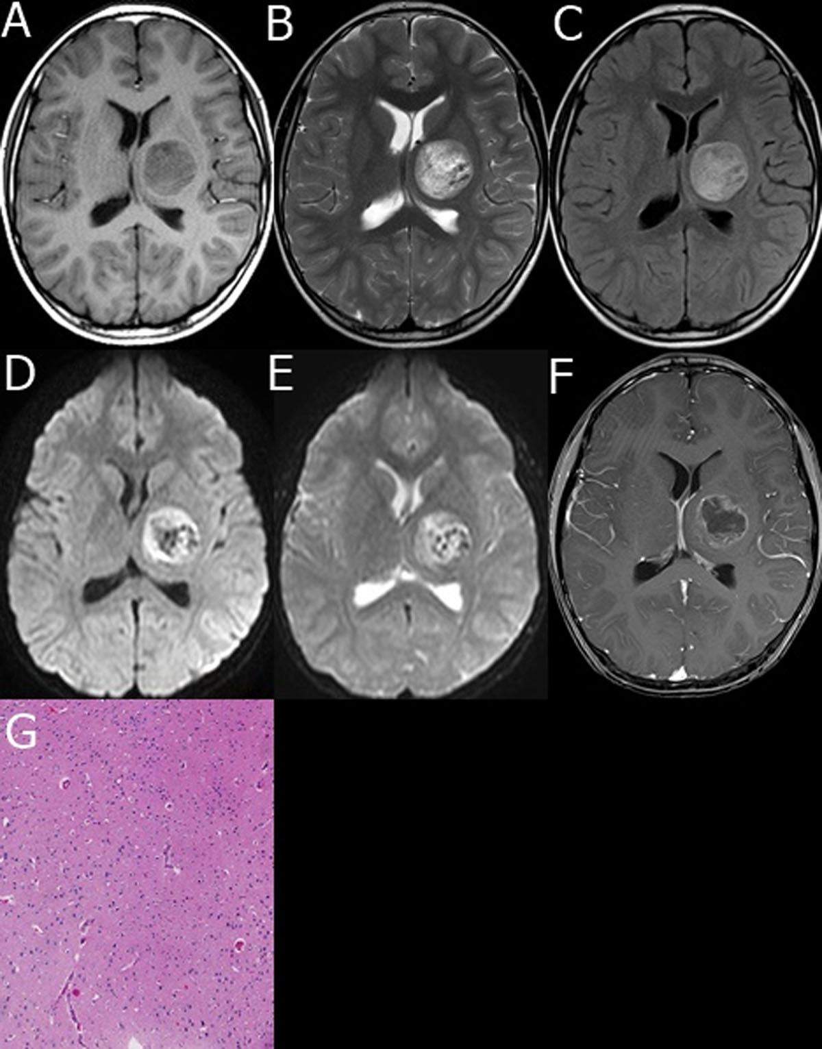
Look For First
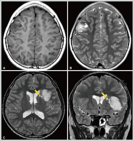
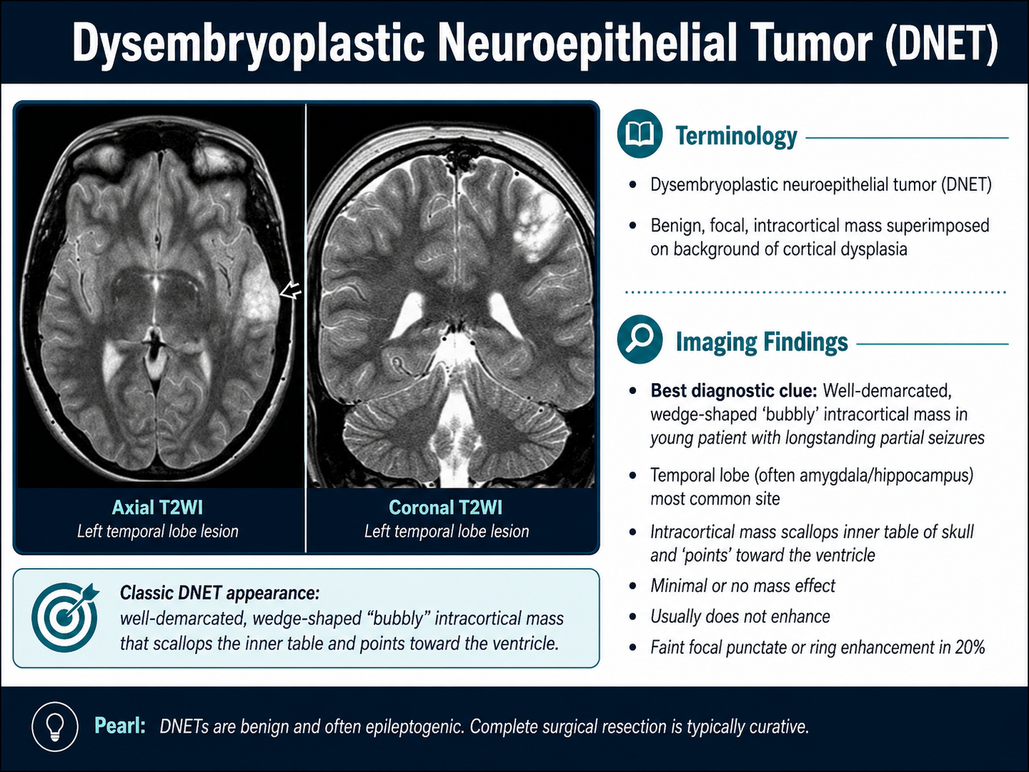
- Cortical-based multinodular or 'bubbly' appearance in a young patient with epilepsy
- T2/FLAIR hyperintense lesion with little to no gadolinium enhancement
- Temporal lobe location (most common), but can occur anywhere in cerebral cortex
Key Image Findings
- MRI is primary modality; CT shows hypoattenuating cortical and subcortical lesion with indolent imaging behavior.
- Characteristic multinodular or multicystic 'bubble-like' or 'popcorn-like' appearance reflecting the underlying nodular intracortical architecture.
- T1 hypointensity and T2/FLAIR hyperintensity, typically with absent or minimal gadolinium enhancement; patchy or partial enhancement can occur in some cases.
- Cortical and subcortical white matter involvement, often extending along an entire gyrus or wedge-shaped portion of a lobe, with variable morphology across multiple gyri.
- Chassoux classification recognizes three imaging patterns: Type 1 (polycystic), Type 2 (nodular), and Type 3 (diffuse/dysplastic), which may correlate with epileptogenic zone and seizure outcome.
- Satellite lesions present in approximately 50% of cases, typically appearing as small separate nodules located on the medial/deep aspect of the primary tumor separated by thin white matter, potentially extending toward caudate, internal capsule, or ventricular margin.
- Associated focal cortical dysplasia may surround the tumor, particularly in complex-type DNET, extending the epileptogenic zone beyond tumor boundaries.
- Poorly defined margins on T2/FLAIR imaging can make complete surgical assessment challenging, especially when satellite lesions are adjacent to eloquent cortex or white-matter tracts.
Differential Diagnosis
- Ganglioglioma: usually shows small cystic lesion with mural/nodular component rather than multinodular/multicystic pattern; demonstrates strong gadolinium enhancement in ~50% of cases (vs. minimal enhancement in DNET); ¹¹C-methionine PET shows increased uptake in ganglioglioma but low metabolic activity in DNET.
- Focal cortical dysplasia: lacks the distinct glioneuronal columns and floating neurons; imaging alone may not definitively distinguish from DNET, particularly complex-type DNET with surrounding dysplasia.
- Low-grade glioma: typically less cortically based and less epileptogenic; imaging pattern and clinical presentation favor DNET.
- Cavernous malformation: shows characteristic 'popcorn' calcification pattern and hemosiderin ring; lacks multinodular glioneuronal architecture.
- Cystic neoplasm with enhancement: strong enhancement and cystic-nodular morphology favor ganglioglioma over non-enhancing DNET.
Discussion
DNET represents ~1.5% of all childhood brain tumors and ~30% of mixed glioneuronal tumors; it is one of the classic long-term epilepsy-associated tumors (LEATs) with pronounced epileptogenicity reflecting both cortical location and developmental/dysplastic nature.
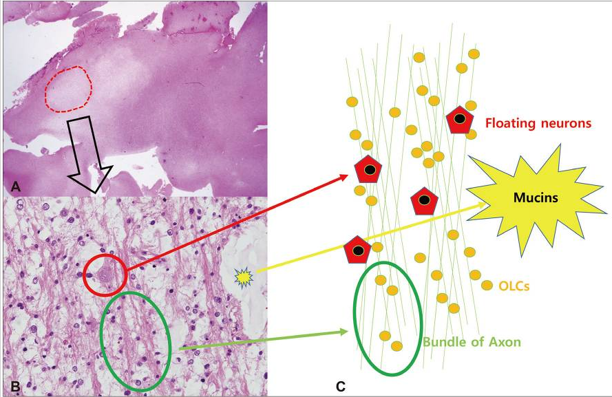
Pathologically, DNET is characterized by multinodular intracortical architecture with glioneuronal columns (axons perpendicular to cortex lined by oligodendroglia-like cells) and floating neurons suspended in a mucin-rich matrix; lacks aggressive features such as mitoses, necrosis, or high cellularity.
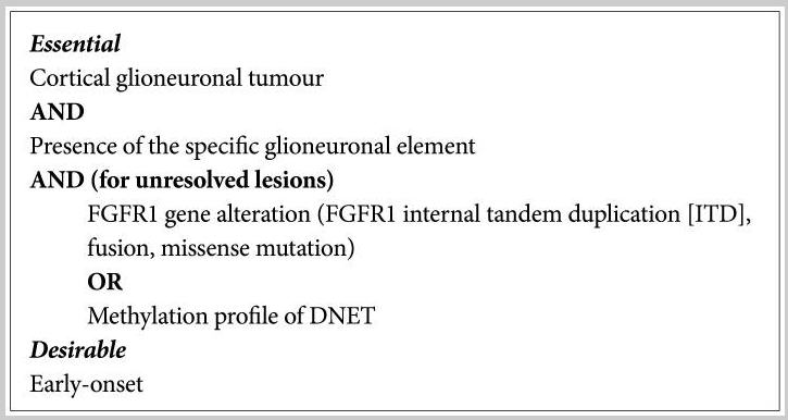
Molecularly distinct entity with characteristic FGFR1 alterations (internal tandem duplications, fusions, missense mutations), particularly those affecting the tyrosine kinase domain; modern DNA methylation and transcriptional profiling provide a distinct DNET signature useful for resolving diagnostically difficult cases.
Historically considered benign with acceptable incomplete resection, but recent evidence shows residual tumor—especially satellite nodules—can slowly enlarge; approximately 36% recurrence/progression rates reported in some series, higher than historically believed.
Gross total resection is the major determinant of both long-term tumor control and seizure freedom (~60–80% seizure-free after complete resection), making satellite nodule identification and complete removal critical for optimal outcomes.
Breakthrough seizures after initial control may signal tumor progression or satellite lesion enlargement; surgical planning must balance complete removal against avoiding injury to eloquent cortex, corticospinal tract, and optic radiations.
Reporting Pearls
Describe the lesion as a 'cortical-based multinodular/multicystic lesion with T2/FLAIR hyperintensity and minimal enhancement, consistent with DNET'; always specifically search for and comment on the presence or absence of medial/deep satellite nodules, as their identification significantly impacts surgical planning and long-term outcome—document their number, location relative to eloquent cortex, and relation to white-matter tracts.
Pitfalls
- Overlooking satellite lesions on initial imaging review; systematically evaluate the medial/deep margin and white matter adjacent to the primary tumor, as satellite nodules are present in ~50% of cases and are crucial for surgical planning.
- Overestimating the enhancement pattern; minimal or patchy enhancement is typical for DNET—do not be misled into favoring ganglioglioma if focal enhancement is present.
- Underestimating the significance of residual tumor; historical literature emphasized acceptability of incomplete resection, but modern evidence shows residual lesions including satellites can enlarge and compromise seizure freedom; always emphasize completeness of resection in reporting.
- Confusing DNET with focal cortical dysplasia based on imaging alone, particularly in complex-type DNET with surrounding dysplastic cortex; integration of imaging features (multinodular pattern, glioneuronal architecture on pathology, FGFR1 molecular signature) and clinical context is necessary for accurate diagnosis.