Gangliocytoma
Key Images
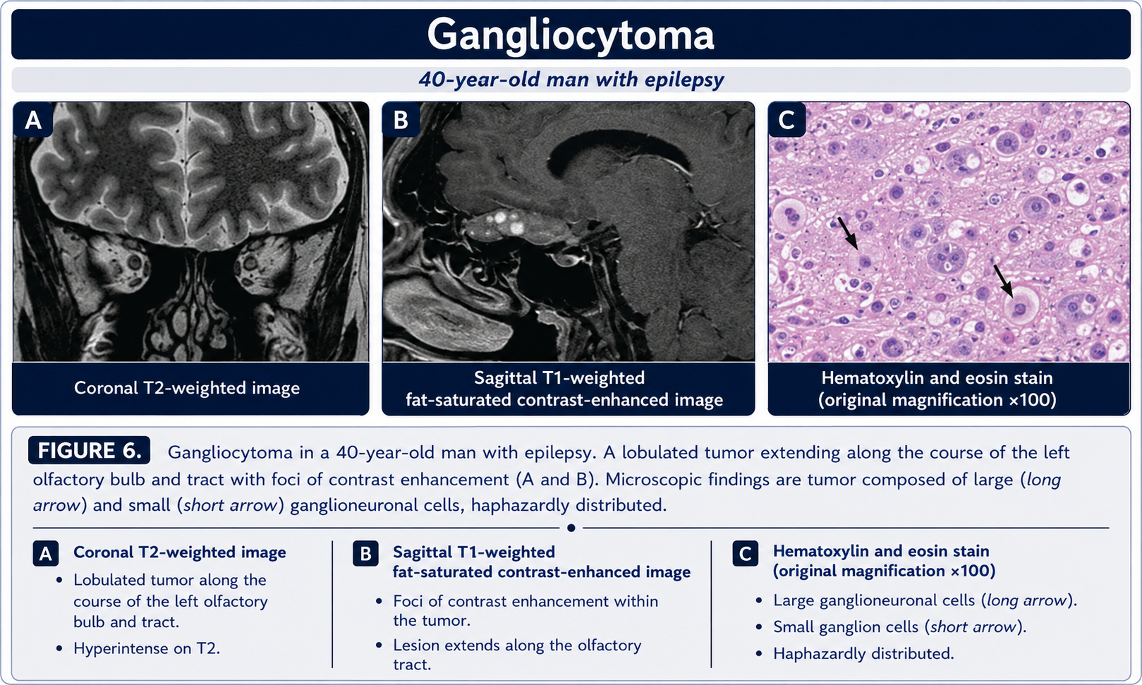
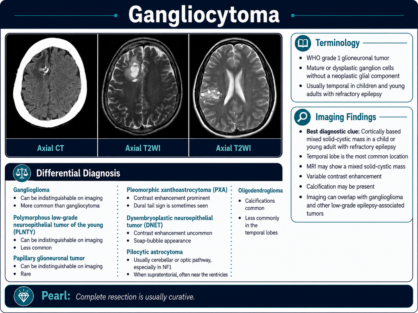
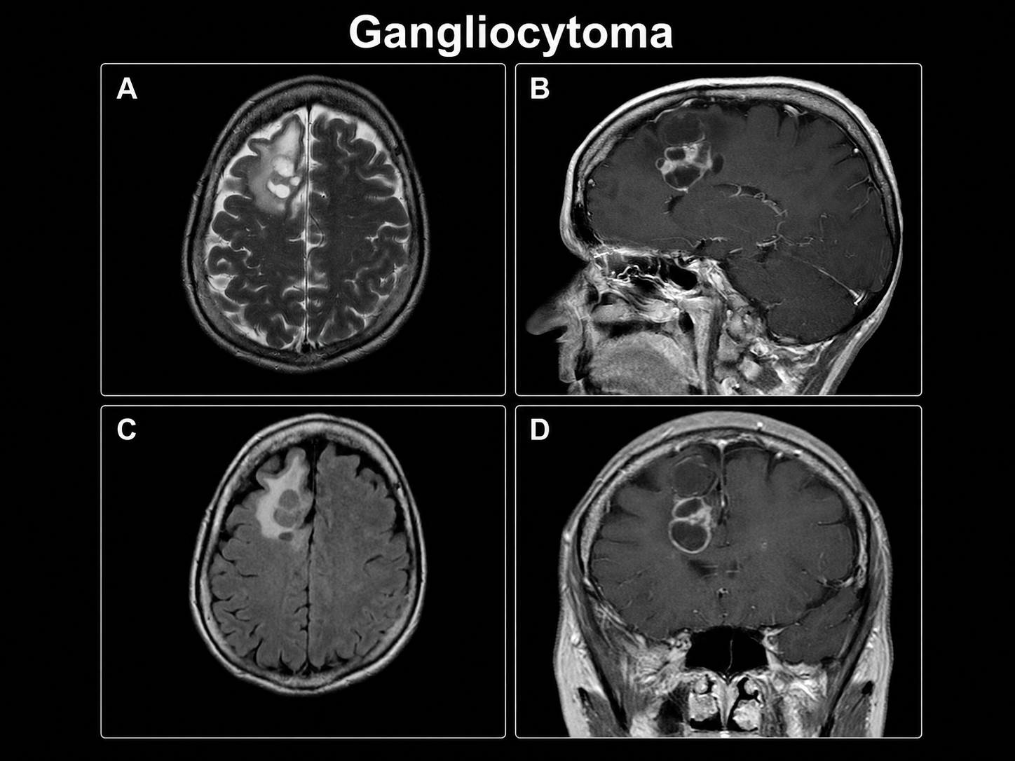
Look For First
- Superficial cortical lesion in temporal lobe with chronic seizure history in young patient
- Well-circumscribed, slowly growing mass with minimal mass effect and little vasogenic edema
- Mixed solid-cystic morphology with possible calcification and calvarial remodeling from chronic growth
- Lesion that appears indolent on imaging—this is a WHO grade 1 tumor
Key Image Findings
- On noncontrast CT, gangliocytoma often appears hyperattenuating with frequent calcification and possible cystic change reflecting the dysplastic mature ganglion cell composition.
- On T1 MRI, the solid component is typically hypo- or isointense to gray matter, reflecting the neuronal cellular density without significant T1-shortening blood products or lipid.
- On T2/FLAIR MRI, signal is variable but often isointense or mildly hyperintense depending on cystic/solid proportions and calcification burden.
- Enhancement pattern is variable but often present; tumor shows mild to moderate contrast uptake without aggressive enhancement patterns expected in high-grade tumors.
- Chronic indolent growth may produce calvarial remodeling and cortical thickening in superficial temporal lesions, reflecting slow long-term expansion.
- Mass effect is characteristically limited with minimal vasogenic edema despite often being a cortically based lesion, reflecting the slow, non-infiltrative growth pattern.
- Mixed solid-cystic architecture is common, with intratumoral cystic foci or cavitation contributing to heterogeneous signal on all sequences.
- Over 80% of gangliocytomas are temporal lobe in location, though rare cases involve spinal cord, medulla, hypothalamus, third ventricle, pineal region, or other deep gray matter.
Differential Diagnosis
- Ganglioglioma: histologic distinction (ganglioglioma contains neoplastic glial component with neoplastic neurons; gangliocytoma has only dysplastic neurons without glial component), but imaging overlap is substantial—biopsy/pathology is definitive.
- Low-grade cortical glioma: focal cortical dysplasia or low-grade astrocytoma may appear similar, but lack the pure neuronal differentiation and neuronal marker positivity (synaptophysin, neurofilament, chromogranin A, MAP2).
- Dysplastic cerebellar gangliocytoma (Lhermitte-Duclos disease): a separate cerebellar entity with characteristic striated appearance; differs in location and histology.
- Neuroblastoma or embryonal tumors: presence of small-round-blue-cell or neuroblastic component would indicate more aggressive tumor despite initial neuronal appearance.
- Cortical developmental lesion (focal cortical dysplasia, heterotopia): may mimic chronic indolent appearance but lacks mass effect and enhancement pattern; developmental lesions do not enhance.
- Cavernoma or vascular malformation: calcification and mixed signal can superficially resemble gangliocytoma, but cavernomas have hemosiderin rim and different enhancement pattern.
Discussion
Gangliocytoma is a WHO grade 1 indolent neuronal tumor fundamentally defined by dysplastic mature ganglion cells WITHOUT a neoplastic glial component; the distinction from ganglioglioma is histopathologic, not radiologic, requiring biopsy confirmation.
These tumors are significantly enriched in surgical epilepsy series (1-3% of cases) compared to general brain tumor populations (0.1-0.5%), reflecting their strong association with refractory focal cortical seizures.
Histologically, gangliocytoma cells are abnormal mature ganglion cells—often multipolar, binucleated, vacuolated, or ballooned—that express neuronal markers (synaptophysin, neurofilament, chromogranin A, MAP2) but GFAP should not label the tumor cells themselves.
The chronic, indolent natural history is reflected radiologically by circumscribed margins, minimal mass effect, sparse vasogenic edema, and chronic osseous/calvarial remodeling in superficial lesions.
Imaging alone cannot distinguish gangliocytoma from ganglioglioma due to substantial overlap; pathology, particularly GFAP immunostaining to exclude neoplastic glial components, is required for definitive diagnosis.
Atypical presentations involving deep gray matter, multifocal appearance, or non-temporal locations are rare and radiologically challenging; these may require careful correlation with immunophenotyping and negative molecular testing (IDH1, BRAF, TERT, H3.3) to establish diagnosis as 'suspicious gangliocytoma.'
Reporting Pearls
Describe a temporal cortical mass as a "circumscribed, slowly growing lesion with minimal mass effect and variable enhancement, compatible with long-term epilepsy-associated tumor (LEAT) such as gangliocytoma or ganglioglioma; histopathologic correlation is needed to distinguish neuronal from glioneuronal histology."
Pitfalls
- Assuming imaging appearance definitively distinguishes gangliocytoma from ganglioglioma—substantial imaging overlap requires pathologic confirmation with GFAP immunostaining and neuronal marker panel.
- Missing the diagnosis in atypical presentations: deep gray matter, multifocal, or non-temporal lesions are rare but may not trigger consideration of gangliocytoma, leading to diagnostic delay or misclassification.
- Overestimating aggressiveness: the indolent WHO grade 1 status and limited mass effect/edema may be underrecognized if the solid component or calcification is mistaken for higher-grade tumor features.
- Biopsy sampling error: limited sampling in mixed solid-cystic lesions risks missing heterogeneous glial components or unsampled tumor regions, necessitating the cautious qualifier 'suspicious gangliocytoma' rather than definitive diagnosis.