Ganglioglioma
Key Images
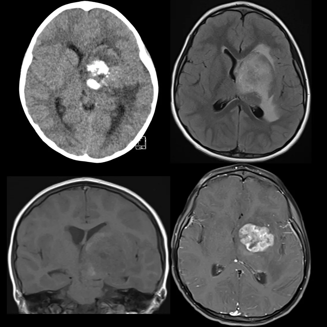
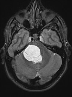
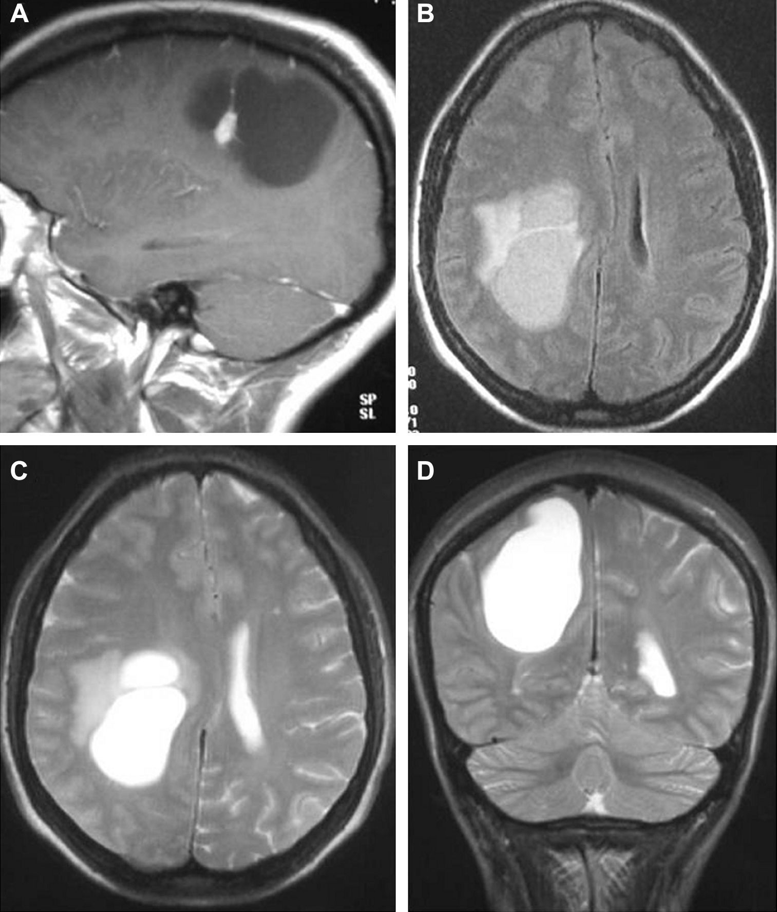
Look For First
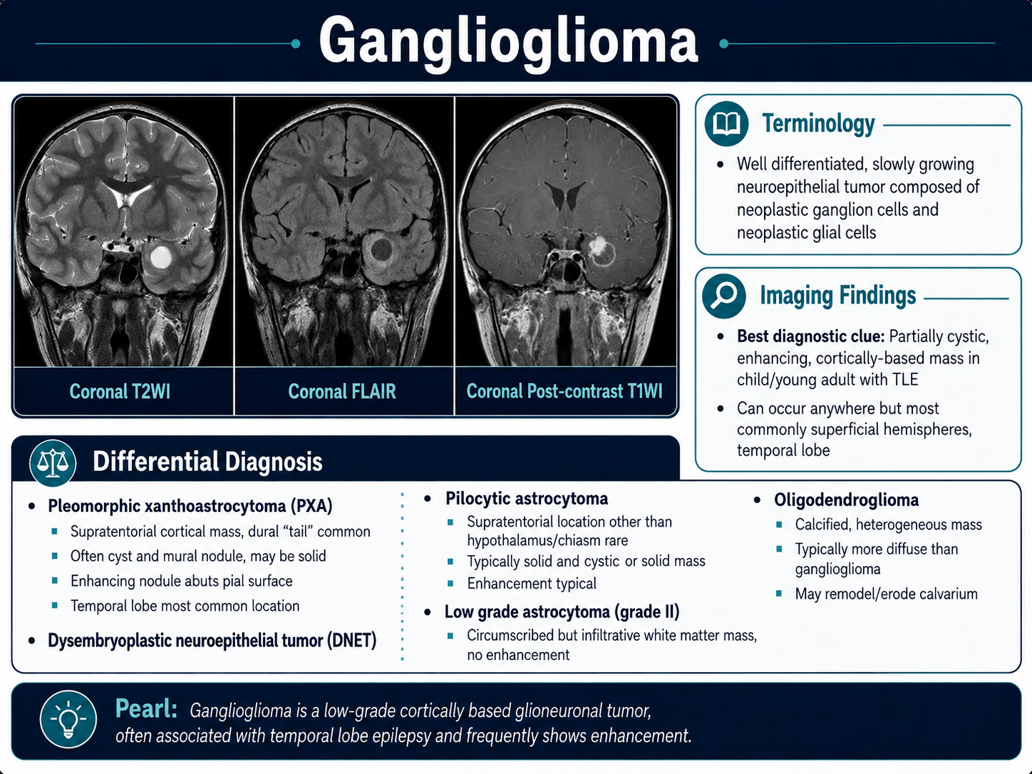
- Partially cystic mass with enhancing mural nodule in the temporal lobe (~45% of cases)
- Solid mass expanding the overlying cortical gyrus
- Calcifications present in approximately 35% of cases
Key Image Findings
- CT: iso- or hypodense mass with frequent calcifications (~35%); bony remodeling or thinning indicates slow growth; enhancement in ~50% of cases involving the solid component
- MRI T1: solid component iso- to hypointense; T1 post-gadolinium shows variable contrast enhancement of the solid component
- MRI T2: hyperintense solid component with variable signal in the cystic component depending on proteinaceous material or blood products
- Peritumoral FLAIR/T2 edema is distinctly uncommon, which helps distinguish ganglioglioma from other infiltrative gliomas
- T2* GRE/SWI imaging shows characteristic blooming signal loss from calcified areas, highlighting the calcified component
- The variable appearance reflects the mixed neuronal and glial cell populations, with tumor biology determined primarily by the grade of the glial component
- Cystic tumors are typically WHO grade 1 and have excellent prognosis following complete resection (up to 97% recurrence-free survival at 7.5 years)
- Anaplastic gangliogliomas (higher-grade lesions in ~5% of cases) may show more infiltrative growth pattern and more prominent enhancement
Differential Diagnosis
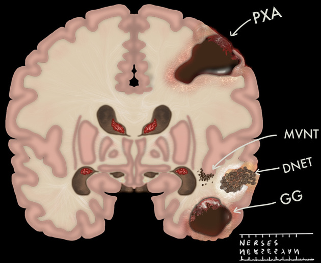
- DNET: typically shows soap bubble appearance and has uncommon contrast enhancement, unlike gangliogliomas which may show variable but more common enhancement
- Pleomorphic xanthoastrocytoma (PXA): shows more prominent contrast enhancement and sometimes dural tail sign, distinguishing from variable enhancement pattern in gangliogliomas
- Pilocytic astrocytoma: typically located in cerebellum or optic pathway (especially in NF1) or near ventricles supratentorially, whereas gangliogliomas favor the temporal lobes
- Oligodendroglioma: calcifications are common but typically less common in temporal lobes compared to gangliogliomas
- Gangliocytoma and PLNTY: can be indistinguishable on imaging but are less common; composed only of neuronal elements or have different cellular components
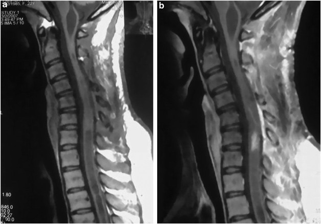
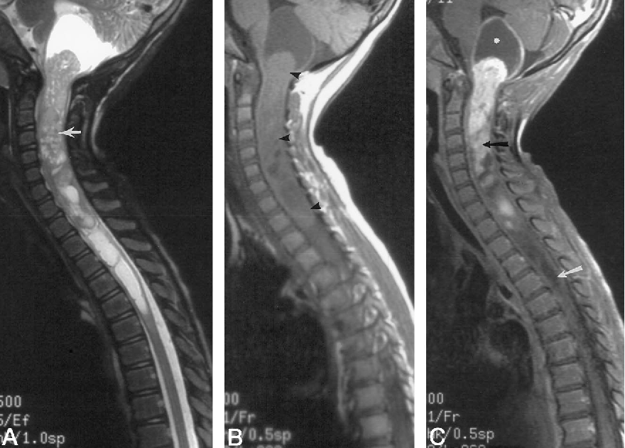
- Spinal cord location: consider astrocytoma or ependymoma, though spinal gangliogliomas are rare due to lower incidence outside the brain
Discussion
Gangliogliomas are composed of two distinct cell populations—mature neuronal ganglion cells and neoplastic glial elements (primarily astrocytic)—with the grade of the glial component determining biological behavior
The classic partial cystic presentation with enhancing mural nodule results from increased blood-brain barrier permeability allowing plasma protein accumulation and vasogenic edema, with slow clearance leading to cyst formation
WHO grade 1 (indolent) cystic gangliogliomas have excellent prognosis with complete resection, but location determines outcome; spinal gangliogliomas often progress due to difficulty achieving adequate resection margins
BRAF V600E mutations are present in 20-60% of gangliogliomas, representing a potential molecular target; IDH is negative and if positive suggests a diffuse glioma instead
Dedifferentiation to high-grade tumors is rare (~5%) and usually involves the glial component transforming into glioblastoma, while neuronal dedifferentiation into neuroblastoma is exceptionally rare
Medically refractory temporal lobe epilepsy is the most common clinical presentation, making imaging with seizure protocol MRI essential for tumor identification and surgical planning
Reporting Pearls
Clearly describe whether the lesion is cystic versus solid, document the location within the temporal lobe or other brain region, characterize enhancement pattern (minimal, variable, or prominent), note the presence of calcifications and peritumoral edema, and specifically comment on the absence of significant peritumoral FLAIR/T2 hyperintensity as a reassuring feature suggesting low-grade biology.
Pitfalls
- Mistaking a ganglioglioma for a more aggressive glioma when peritumoral edema is prominent; remember that significant peritumoral edema is distinctly uncommon in gangliogliomas and suggests higher grade or alternative diagnosis
- Confusing ganglioglioma with DNET based on appearance alone; look for soap bubble morphology in DNET and more variable enhancement in gangliogliomas
- Underestimating the importance of complete resection for prognosis; incomplete resection or residual tumor may require additional radiotherapy despite low-grade appearance
- Forgetting that location impacts prognosis significantly—spinal cord gangliogliomas have poor outcomes due to inability to achieve adequate resection margins, contrasting with excellent brain prognosis