High-grade astrocytoma with piloid features (HGAP)
Key Images
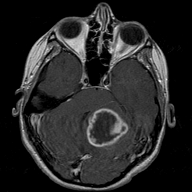
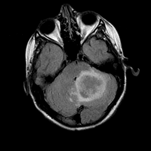
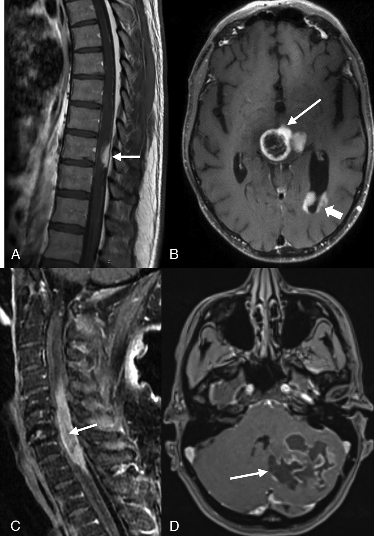
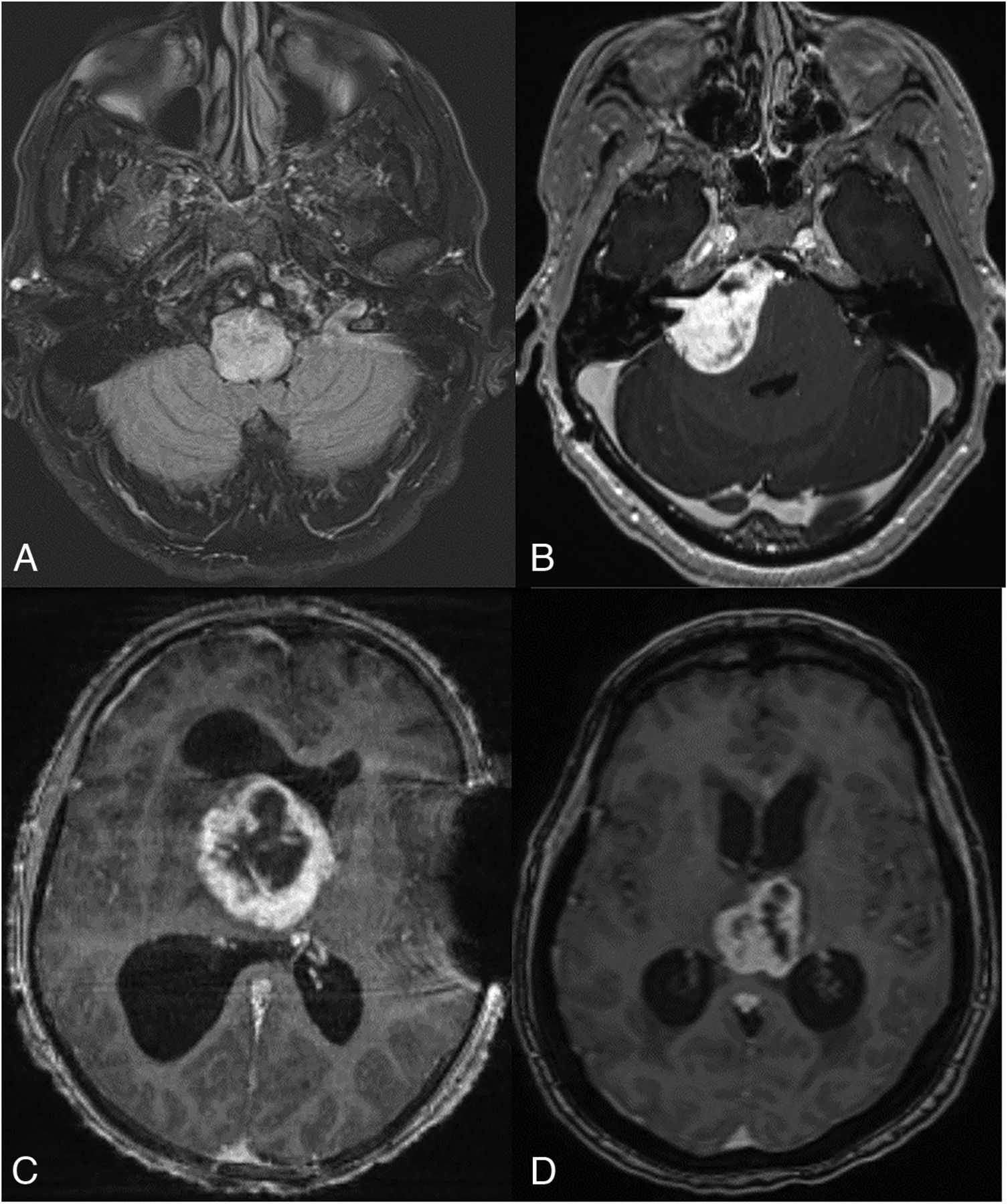
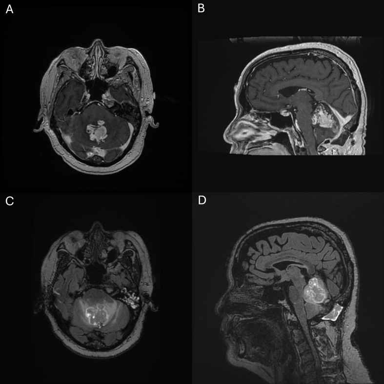
Look For First
- Heterogeneously or peripherally enhancing posterior fossa/cerebellar mass in an adult, particularly with NF1 history
- T1 hypo-/isointense and T2/FLAIR hyperintense signal with relatively circumscribed appearance
- Absence of restricted diffusion (unusual for a high-grade glioma) is a key distinguishing feature
Key Image Findings
- MRI signal: T1 hypo-/isointense, T2/FLAIR hyperintense with heterogeneous appearance and peripheral or heterogeneous enhancement pattern
- Cystic and necrotic areas may be present with variable surrounding edema, but unlike most high-grade tumors, restricted diffusion is often absent or not dominant
- Location predilection: 74% posterior fossa (especially cerebellum), with NF1-associated cases favoring midline structures including thalami
- Spinal cord involvement (when present): intramedullary tumors often with exophytic components, typically affecting dorsal cord over long segments (>3 vertebral bodies)
- Leptomeningeal and intraventricular dissemination can occur
- No specific radiographic signature distinguishes HGAP from glioblastoma, high-grade pilocytic astrocytoma transformation, or diffuse midline glioma on imaging alone
Differential Diagnosis
- Glioblastoma IDH-wildtype: typically shows more restricted diffusion and more aggressive enhancement; diagnosis confirmed by absence of HGAP methylation profile
- Diffuse midline glioma: location and molecular profile differ; methylation class testing is mandatory to distinguish
- High-grade transformation of pilocytic astrocytoma: prior benign pilocytic astrocytoma may precede HGAP, but HGAP defined by specific DNA-methylation class
- Posterior fossa metastasis: clinical history and imaging location help distinguish; lack of restriction may be atypical for metastasis
- Anaplastic pilocytic astrocytoma: histologic overlap is substantial; requires methylation profiling to confirm HGAP classification
Discussion
HGAP is defined not by imaging or histology alone but by characteristic DNA-methylation profile (WHO 2021 CNS classification), making integrated molecular workup mandatory for definitive diagnosis
Molecular hallmarks include MAPK-pathway activation (NF1, BRAF, FGFR1 alterations in ~75%), CDKN2A/B homozygous deletion (~80%), and ATRX mutation/loss (~45%), with IDH-wildtype status typical
The combination of loss of ATRX staining and CDKN2A/B homozygous deletion in a high-grade piloid tumor should strongly prompt methylation-class testing
Prognosis is poor with ~50% 5-year survival, intermediate between glioblastoma IDH-wildtype (worse) and IDH-mutant gliomas (better)
MAPK-pathway alterations create a rationale for molecularly directed therapy (mTOR and MAPK inhibitors) in selected progressive or recurrent tumors
Treatment approach: maximal safe surgical resection followed by concurrent chemoradiotherapy (e.g., temozolomide); no tumor-specific standard therapy currently exists
Reporting Pearls
For an adult posterior-fossa mass, report as: "Heterogeneously enhancing cerebellar/posterior-fossa astrocytic tumor with circumscribed appearance [and/or piloid morphology on preliminary pathology if available]. Given appearance and/or NF1 association, recommend integrated molecular workup including CDKN2A/B, ATRX, MAPK-pathway alterations, and DNA-methylation profiling to assess for high-grade astrocytoma with piloid features (HGAP)."
Pitfalls
- Absence of restricted diffusion can lead to underestimation of grade; HGAP often lacks the high diffusion restriction typical of glioblastoma, potentially causing diagnostic delay
- Histologic diagnosis of 'high-grade glioma with piloid features' is insufficient without methylation-class profiling; molecular confirmation is mandatory for HGAP classification
- Confusing HGAP with recurrent or transformed pilocytic astrocytoma; prior benign pilocytic astrocytoma may precede HGAP, requiring methylation profiling to confirm aggressive transformation
- Imaging appearance is nonspecific and overlaps substantially with glioblastoma, diffuse midline glioma, and metastasis; clinical context (NF1 history, posterior fossa location, age) should prompt suspicion and molecular workup