Adult-onset leukodystrophies
Key Images
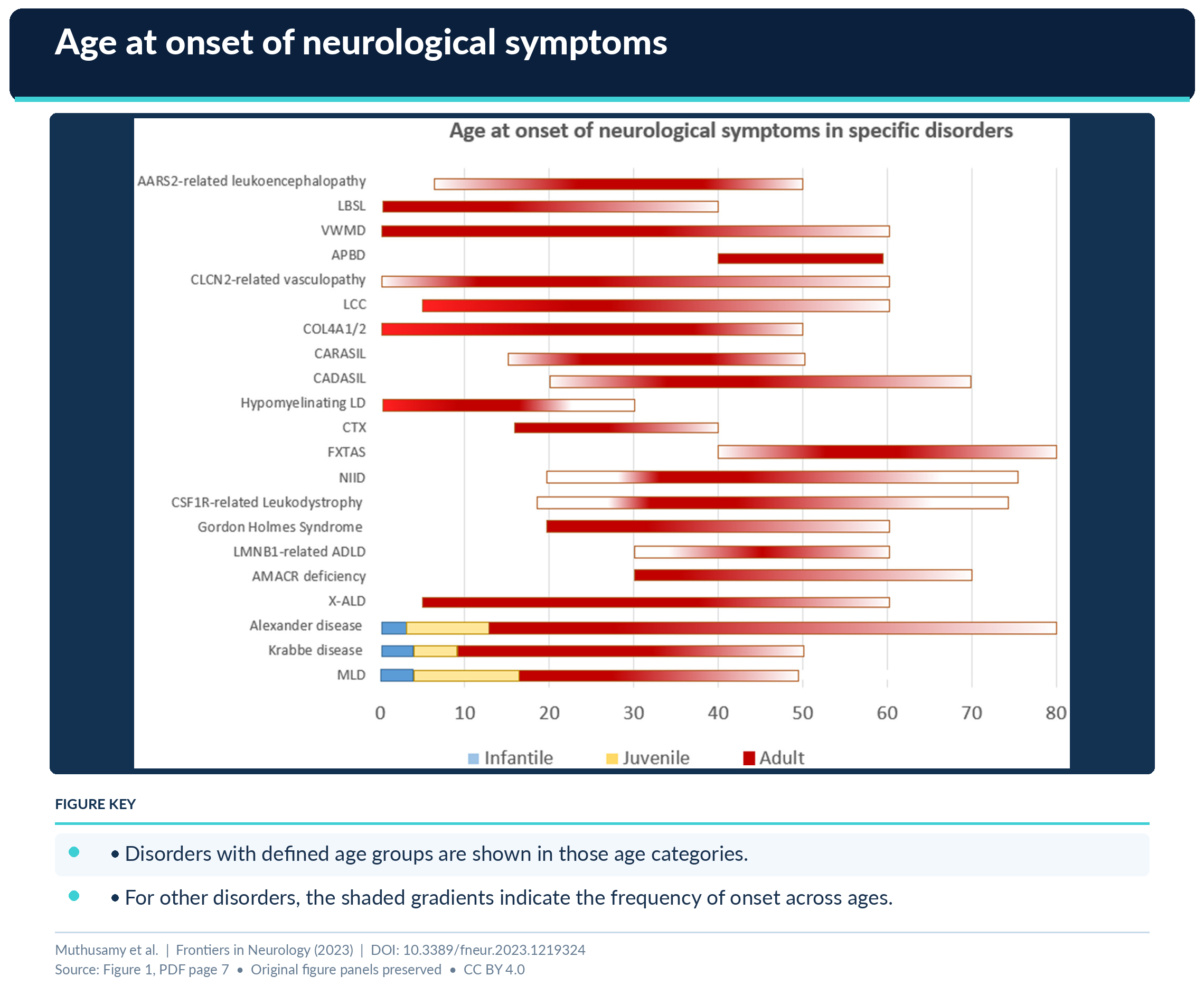
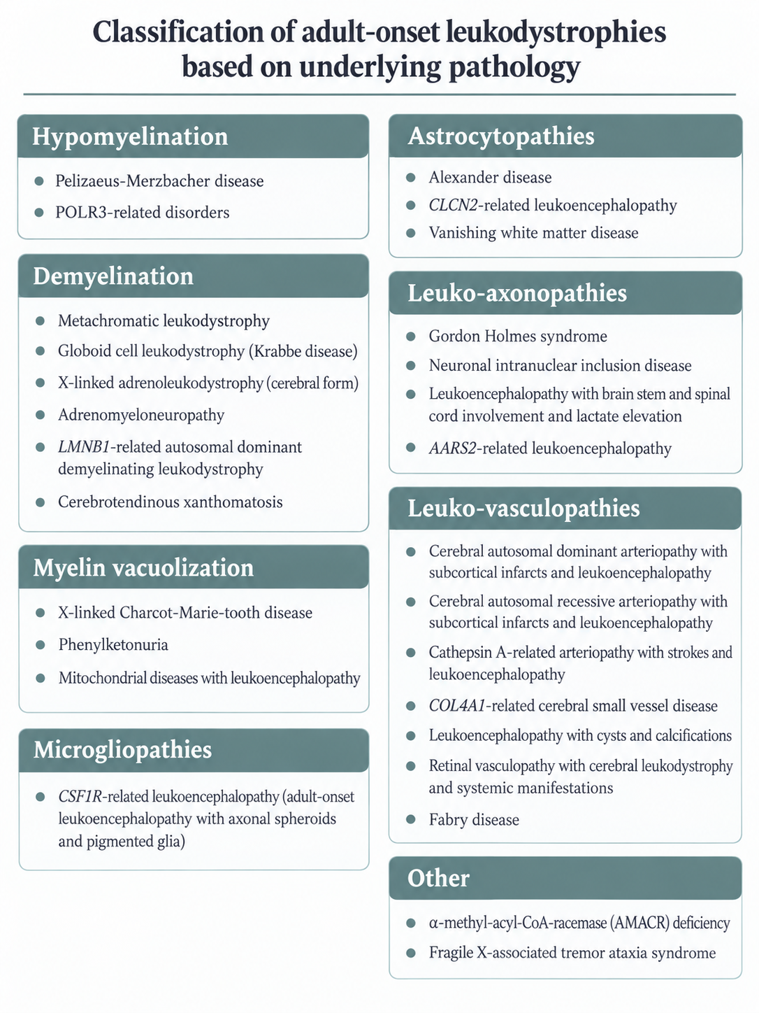
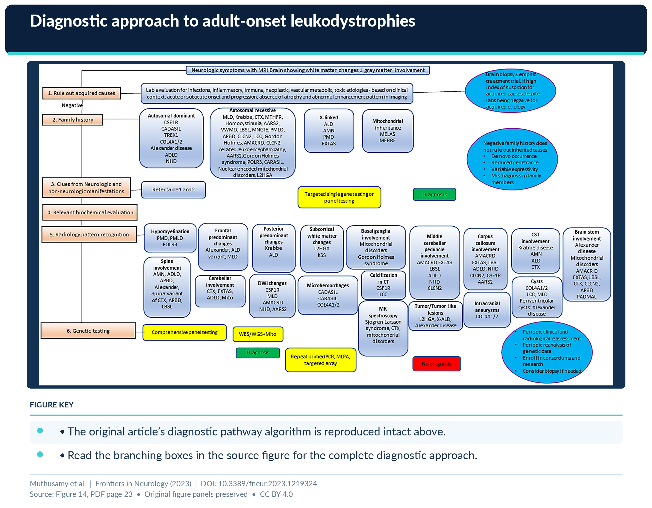
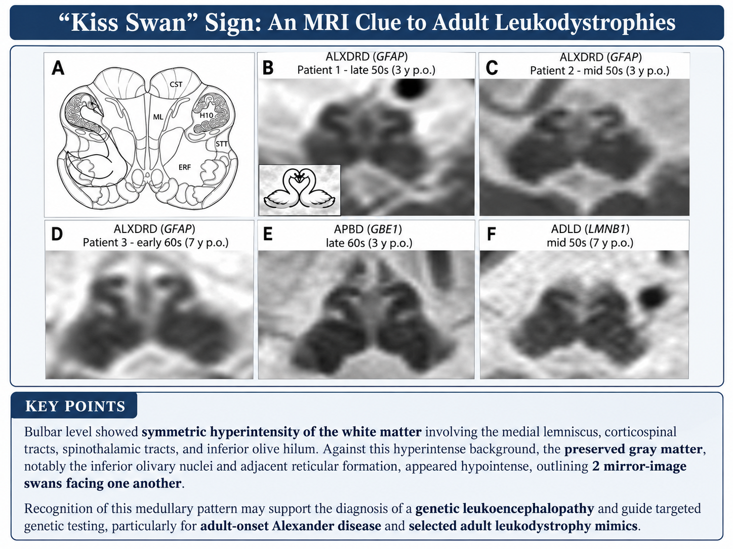
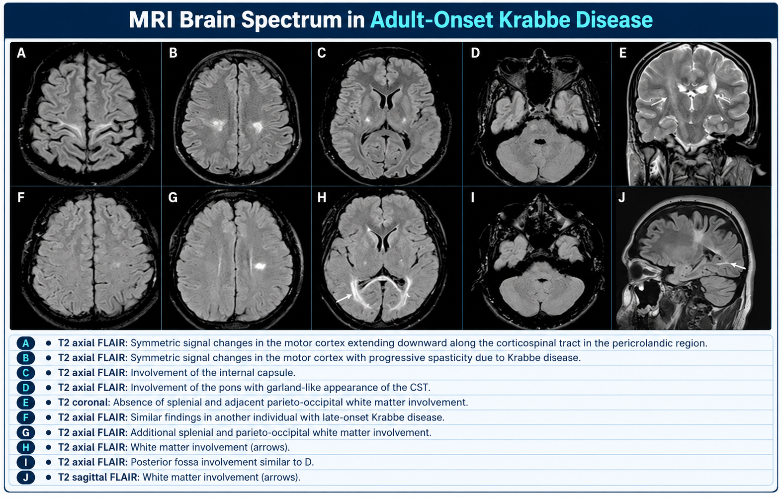
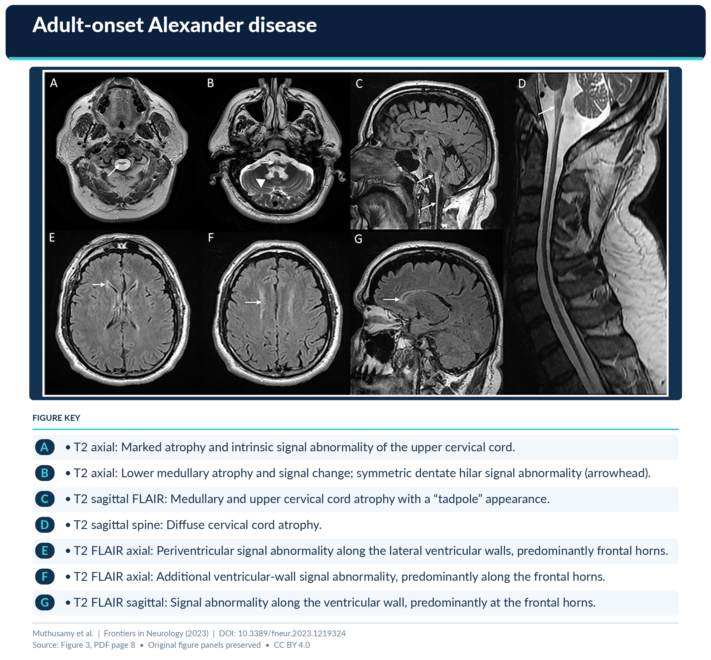
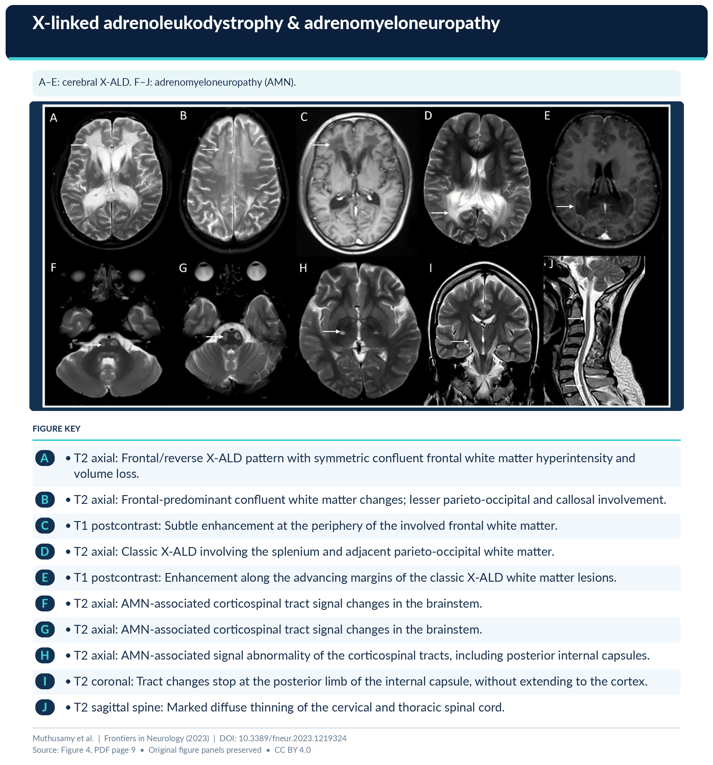
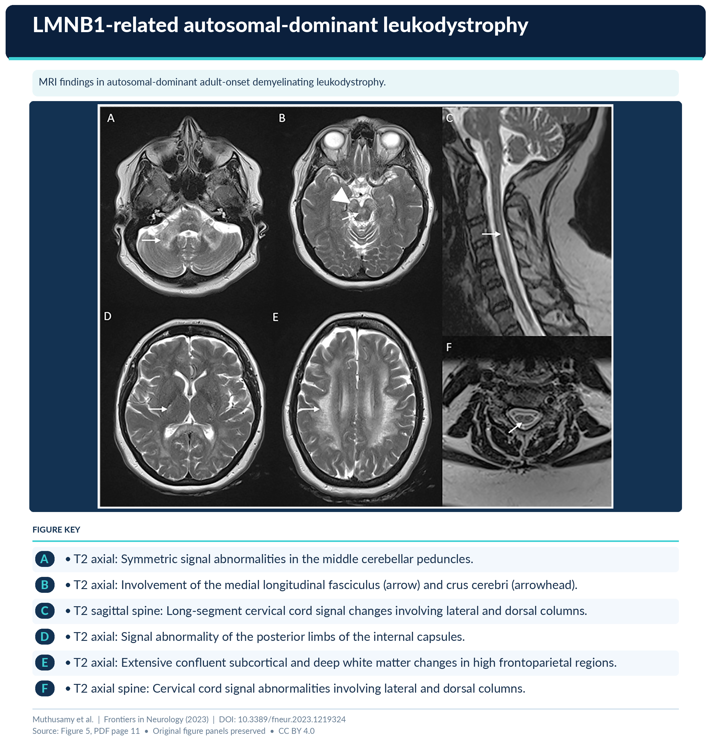
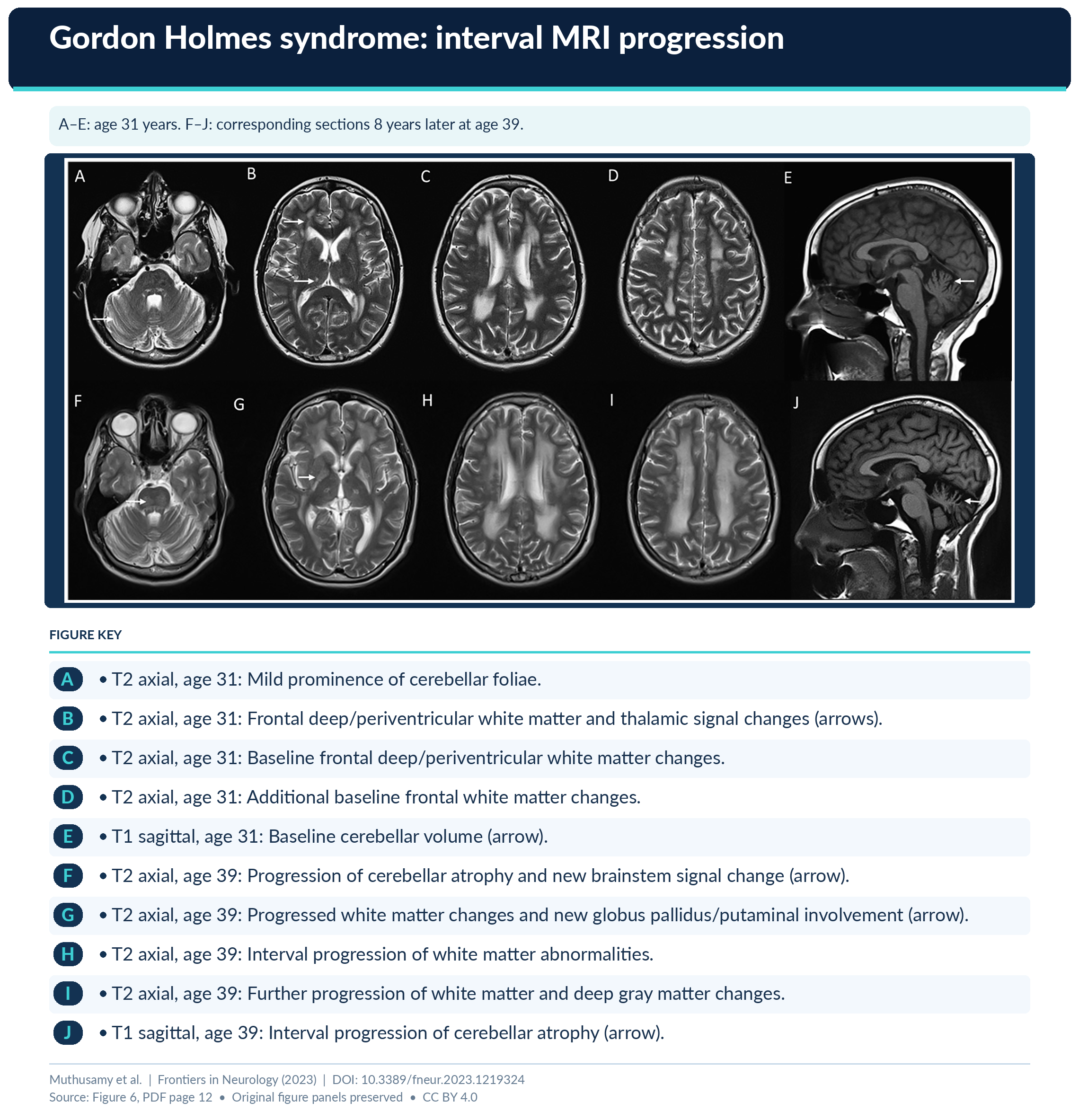
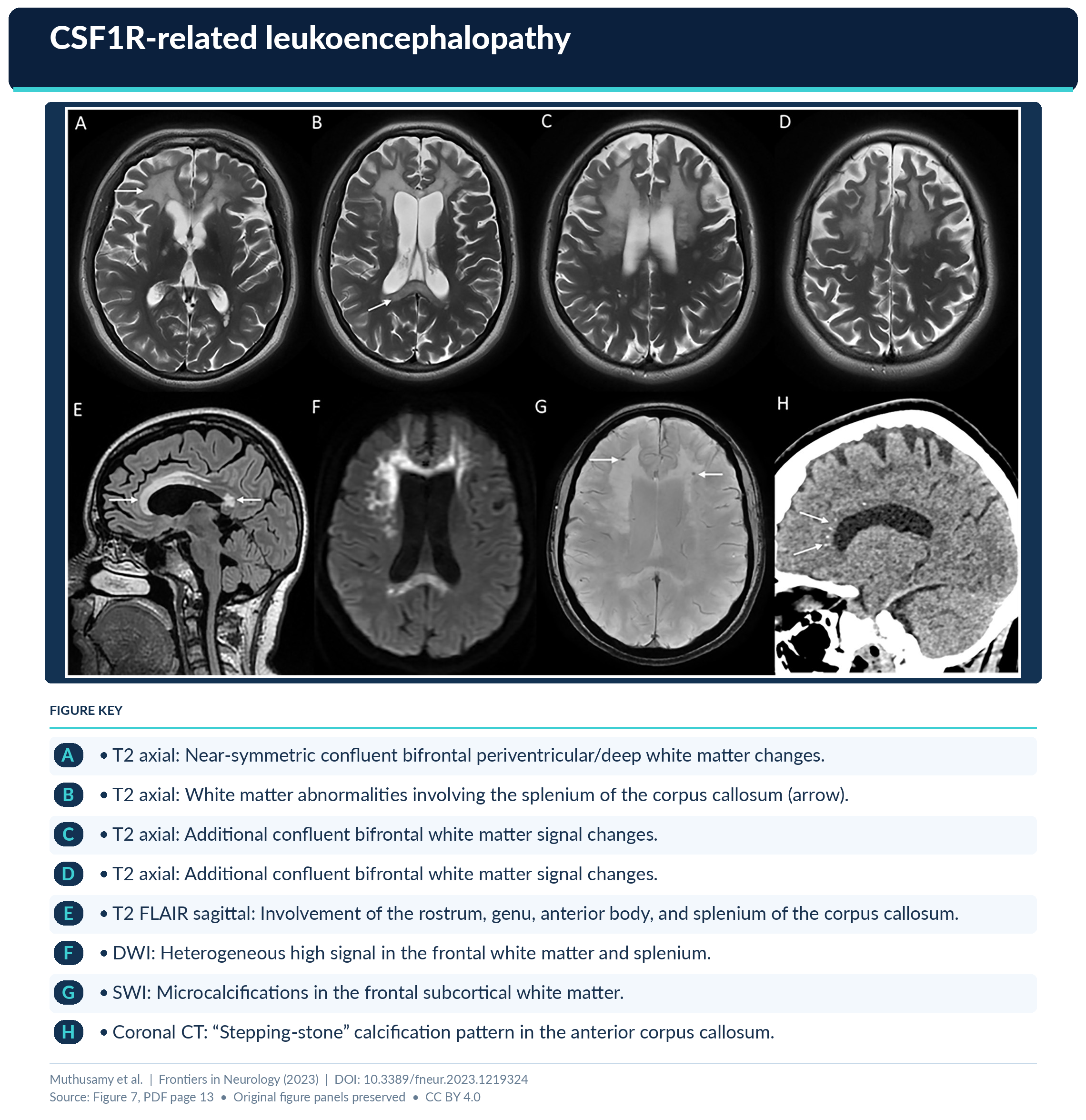
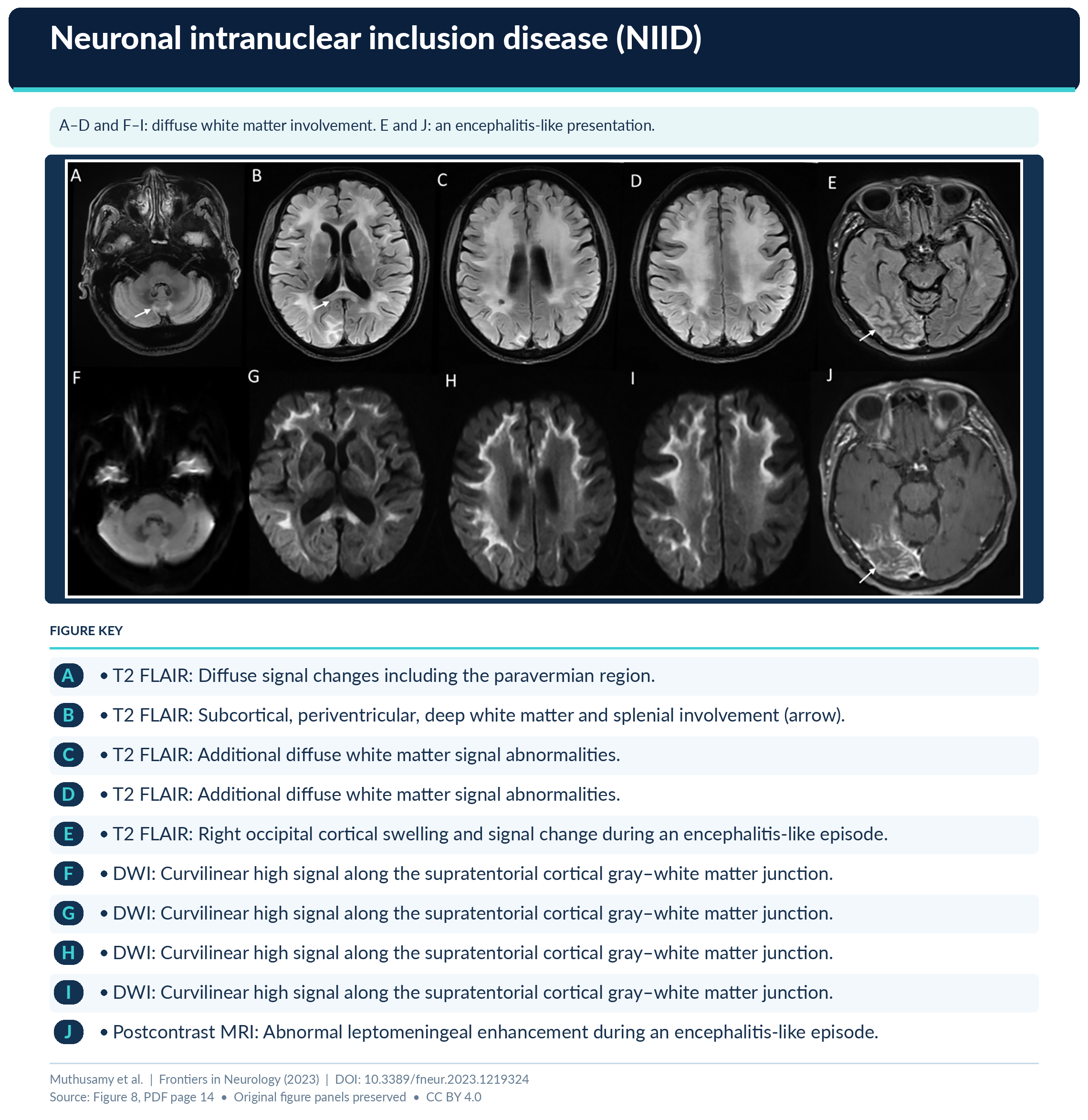
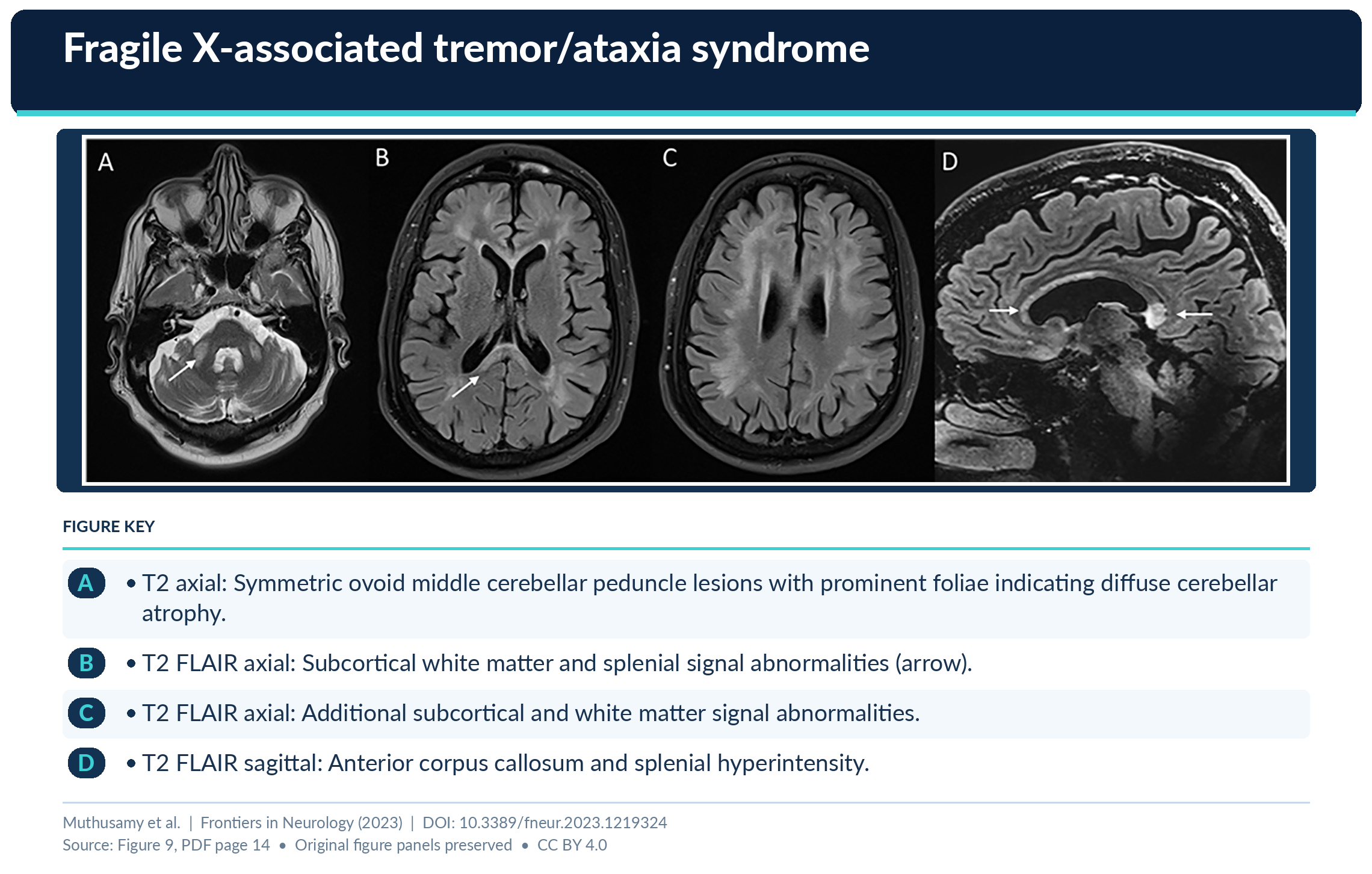
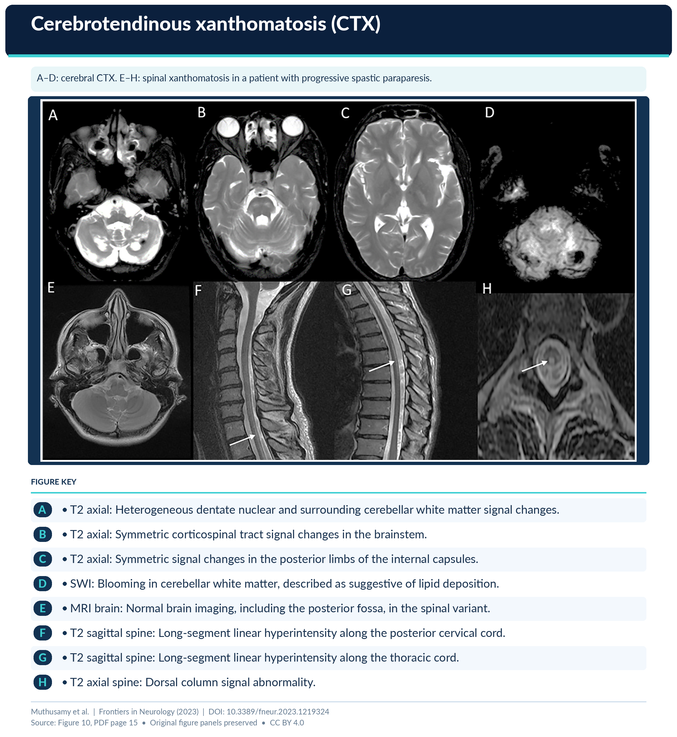
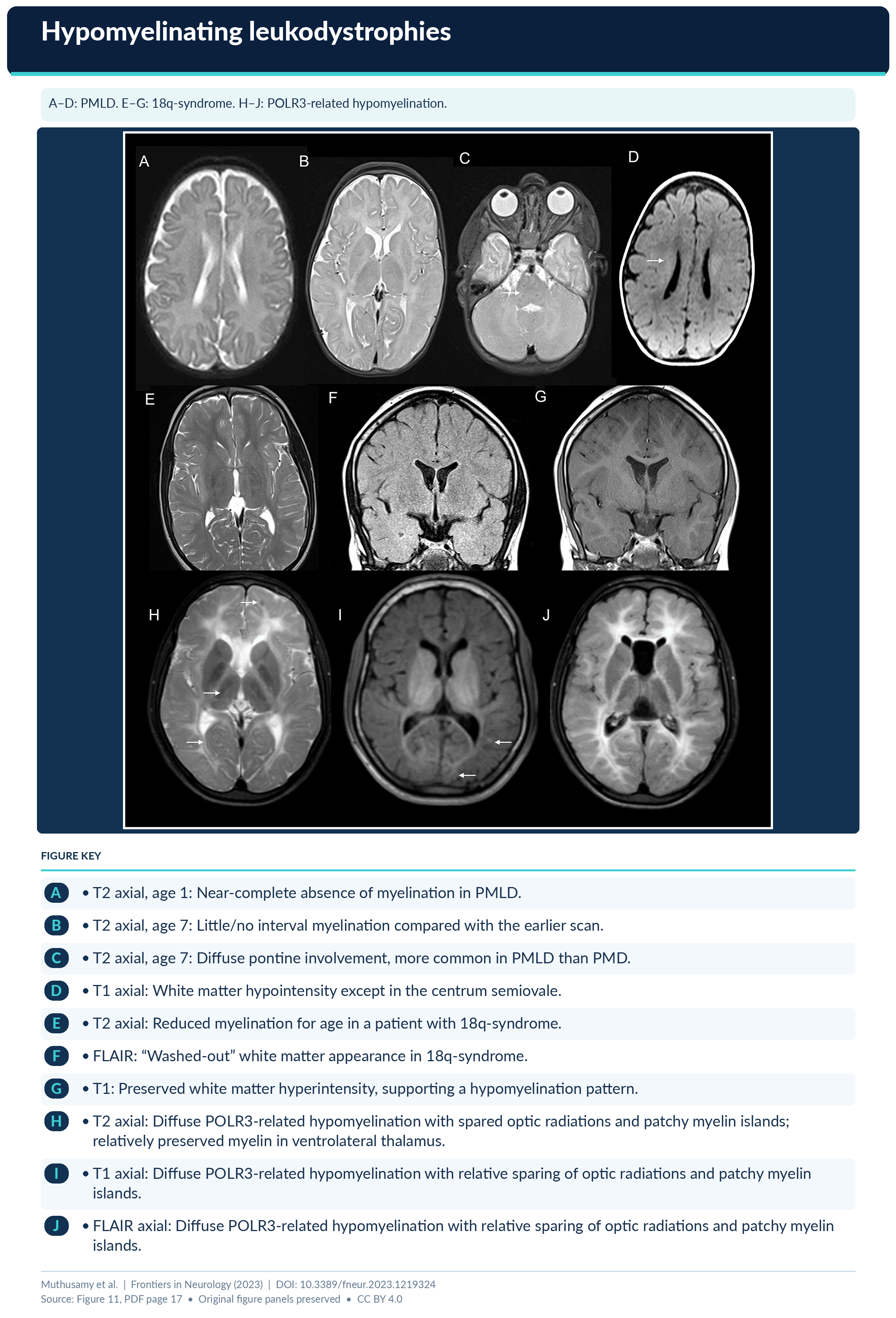
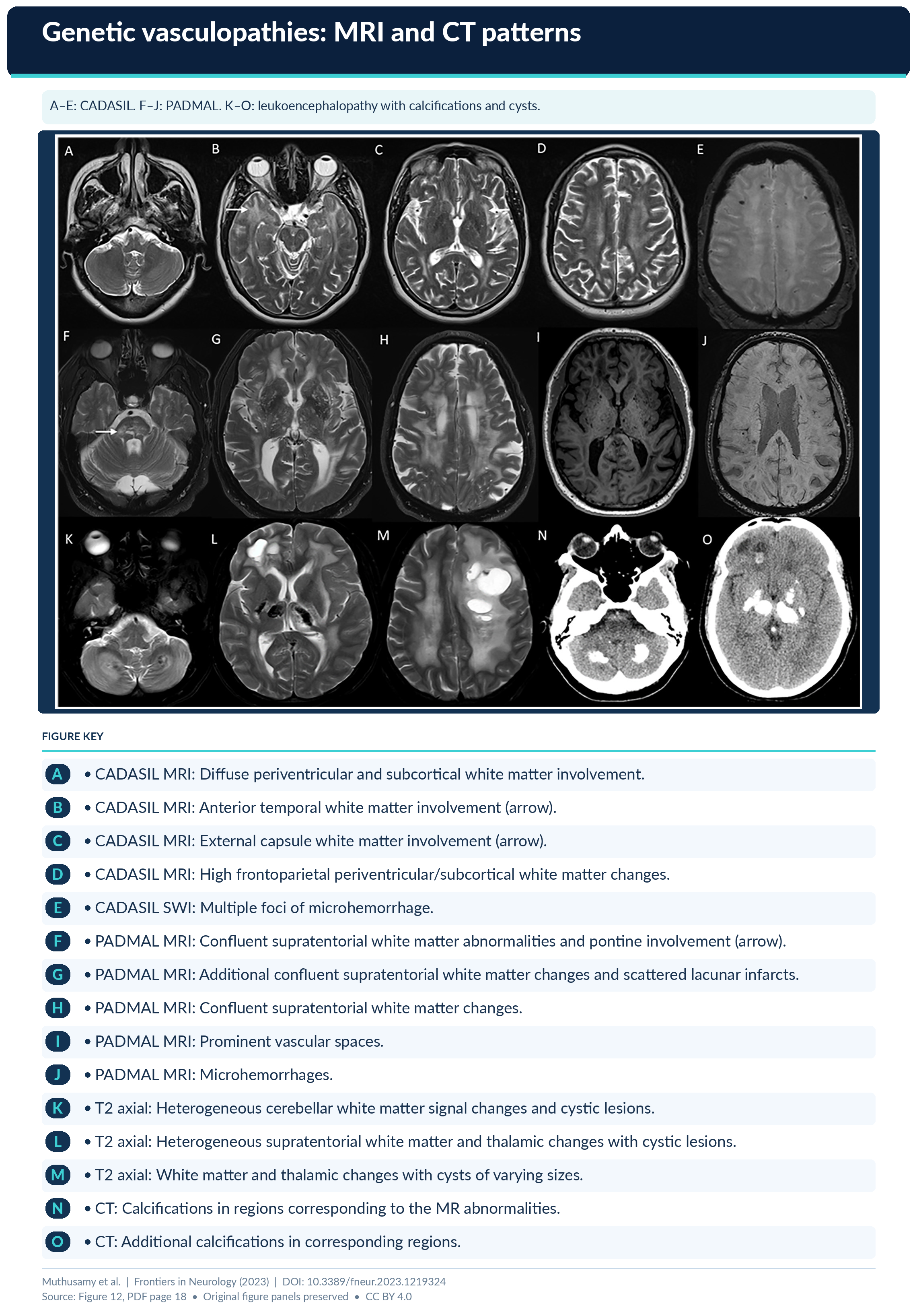
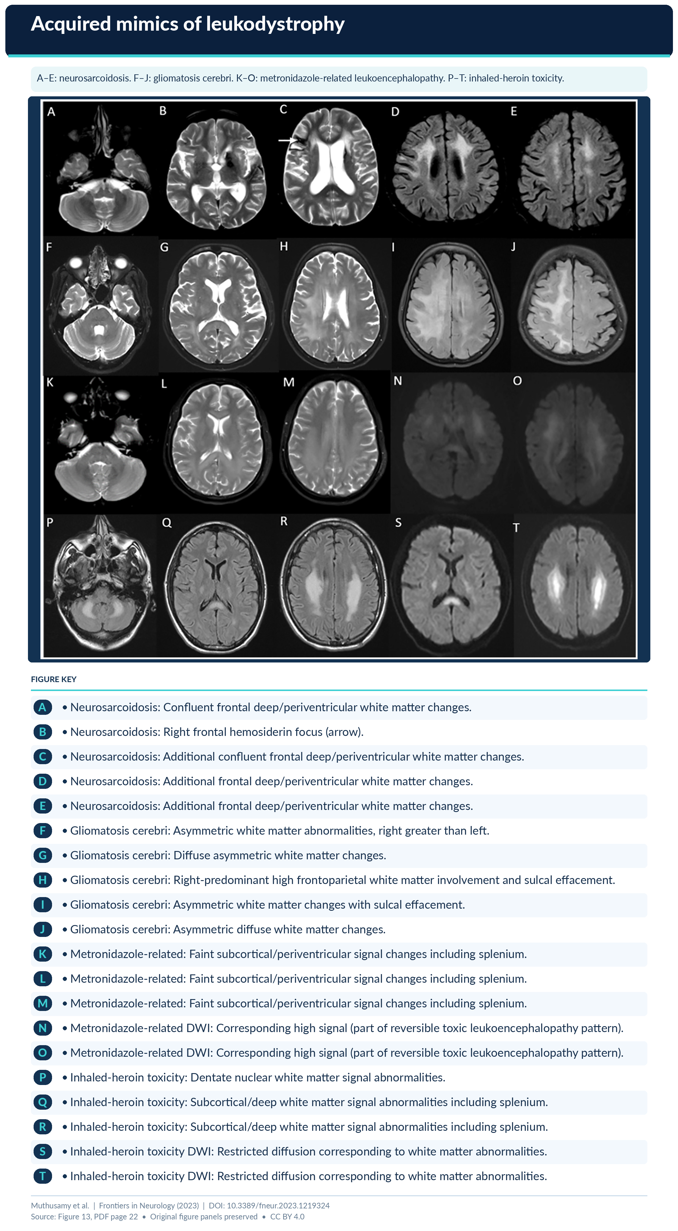
Look For First
- Abnormal white matter signal on T2/FLAIR MRI with specific pattern distribution (periventricular, subcortical, infratentorial)
- Presence of non-enhancing white matter changes to exclude demyelinating or inflammatory mimics
- Specific morphologic clues matching disease category: leukovasculopathies show subcortical infarcts, leuko-axonopathies show characteristic brainstem involvement, astrocytopathies show vanishing white matter pattern
Key Image Findings
- Leukodystrophies are characterized by bilateral and generally symmetrical white matter signal abnormalities on T2-weighted and FLAIR sequences, though specific distributions vary by subtype.
- Leukovasculopathies such as CADASIL show T2 hyperintense white matter changes predominantly in the periventricular and subcortical regions with associated lacunar infarcts, while COL4A1-related disease shows small vessel changes with microhemorrhages.
- Leuko-axonopathies including NIID present with periventricular and subcortical white matter changes with potential brainstem involvement and characteristic axonal spheroids on neuropathology.
- Astrocytopathies demonstrate distinctive patterns: vanishing white matter disease shows progressive white matter rarefaction with cystic changes; Alexander disease shows anterior predominant white matter involvement with characteristic frontal lobe changes.
- Myelin disorders like X-ALD show adductor-predominant demyelination in cerebral form with progressive white matter involvement, while Krabbe disease shows demyelinating changes with relative preservation of U-fibers.
- Microgliopathies such as CSF1R-related leukoencephalopathy show white matter changes with microglial inclusions and axonal spheroids pathologically, appearing as progressive leukoencephalopathy on MRI.
- Hypomyelinating leukodystrophies show reduced white matter volume with prolonged T1 and T2 relaxation times, reflecting inadequate myelin formation rather than demyelination.
- Brain MRI remains the primary imaging modality for detecting and characterizing white matter abnormalities; advanced sequences such as DTI, spectroscopy, and susceptibility-weighted imaging may provide additional diagnostic specificity for certain disorders.
Differential Diagnosis
- Multiple sclerosis versus demyelinating leukodystrophies: MS typically shows enhancing lesions with periventricular predilection and dissemination in space and time, while leukodystrophies show non-enhancing, symmetric, progressive white matter changes without inflammation.
- Small vessel disease and age-related white matter changes versus leukovasculopathies: Sporadic age-related changes lack family history and genetic confirmation; leukodystrophies show earlier onset, family clustering, and specific genetic mutations.
- Acquired leukoencephalopathies from infection, toxic exposure, or metabolic derangement versus inherited leukodystrophies: Acquired causes show acute presentation, systemic signs, and reversibility with treatment; inherited leukodystrophies show progressive course and genetic confirmation.
- CADASIL versus other cerebral small vessel diseases: CADASIL shows characteristic NOTCH3 mutation with migraines and early-onset strokes; COL4A1-related disease presents with congenital anomalies or renal involvement; CARASIL shows early alopecia and systemic features.
- Vanishing white matter disease versus progressive encephalopathy: VWMD shows characteristic cystic rarefaction of white matter with EIF2B gene mutations; other progressive encephalopathies lack this distinctive imaging and genetic profile.
- X-ALD cerebral form versus adrenomyeloneuropathy: Cerebral form shows progressive demyelination with rapid progression; AMN shows primarily spinal cord involvement with slower progression and peripheral neuropathy features.
Discussion
Adult-onset leukodystrophies have milder, stuttering clinical courses compared to childhood presentations, with psychiatric symptoms and cognitive decline often preceding motor signs by years, making early recognition challenging.
The epidemiology of adult leukodystrophies varies geographically with CADASIL and NIID being most common in recent cohorts, while meningeal involvement, childhood leukodystrophies with attenuated phenotypes, and new gene discoveries continue to expand the diagnostic spectrum.
Pattern recognition in MRI findings combined with characteristic clinical clues (neurologic, systemic, and family history features) is essential because many adult leukodystrophies are treatable when diagnosed early, including X-ALD with hematopoietic stem cell therapy, cerebrotendinous xanthomatosis with chenodeoxycholic acid, and others.
The classification of leukodystrophies by pathology (myelin disorders, astrocytopathies, microgliopathies, leuko-axonopathies, leukovasculopathies) provides a framework for understanding imaging patterns and helps guide genetic testing strategies.
Molecular confirmation through genetic testing (WES, WGS) is essential not only for definitive diagnosis and early treatment initiation but also for family screening and reproductive counseling in carriers and at-risk relatives.
Acquired mimics including infectious, immune-mediated, vascular, metabolic, and toxic leukoencephalopathies must be systematically excluded during initial evaluation because they can overlap clinically and radiologically with inherited leukodystrophies.
Reporting Pearls
When reporting suspected adult-onset leukodystrophy, describe the specific pattern, distribution (periventricular vs. subcortical vs. infratentorial), symmetry, and whether changes are enhancing or non-enhancing; correlate with clinical presentation and family history to guide differential diagnosis between specific leukodystrophy subtypes and acquired mimics such as demyelinating disease or small vessel disease.
Pitfalls
- Overdiagnosis of age-related or small vessel disease changes in older adults with adult-onset leukodystrophies; careful attention to family history, clinical presentation, and symmetry of changes helps distinguish inherited leukodystrophies from common acquired white matter changes.
- Misclassification as multiple sclerosis when encountering demyelinating leukodystrophies; MS typically shows enhancing lesions with dissemination in space and time, while leukodystrophies show non-enhancing symmetric progressive changes.
- Failure to recognize attenuated phenotypes of childhood leukodystrophies presenting in adulthood; reduced penetrance and variable expressivity can obscure family history and delayed diagnosis if genetic testing is not pursued.
- Relying solely on MRI imaging without clinical-radiological correlation and genetic testing; definitive diagnosis requires integration of clinical features, neuroimaging pattern, systemic manifestations, family history, and molecular confirmation.