Rare primary CNS neoplasms with neuronal elements alone or combined neuronal and glial components.
Overview
Neuronal and glioneuronal tumors are a rare, heterogeneous group of primary CNS neoplasms composed of neuronal elements alone or a mixture of neuronal and glial elements. They account for less than 2–5% of primary CNS tumors, predominantly affect children and young adults, and are strongly associated with drug-resistant epilepsy, particularly in the temporal lobe.
Most are WHO grade 1 or 2, slow-growing, and carry an excellent prognosis after gross total resection. The 2021 WHO classification incorporated molecular and DNA methylation data alongside histology, refining diagnosis and recognizing entities including MVNT, myxoid glioneuronal tumor, and DGONC.
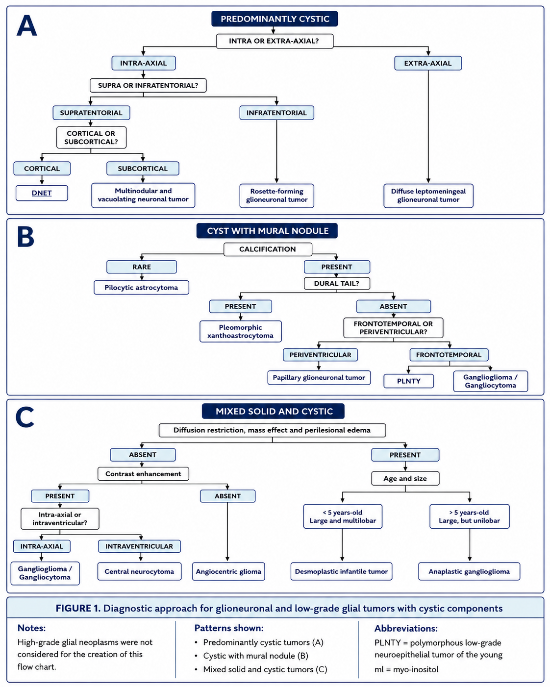
Seizures are the most frequent presentation. Other symptoms include headache, hydrocephalus with intraventricular tumors, and occasional psychiatric manifestations. MRI is the diagnostic standard; calcification, absent edema, hemosiderin, and cystic morphology can help distinguish these tumors from more common gliomas.
Purely Neuronal Tumors
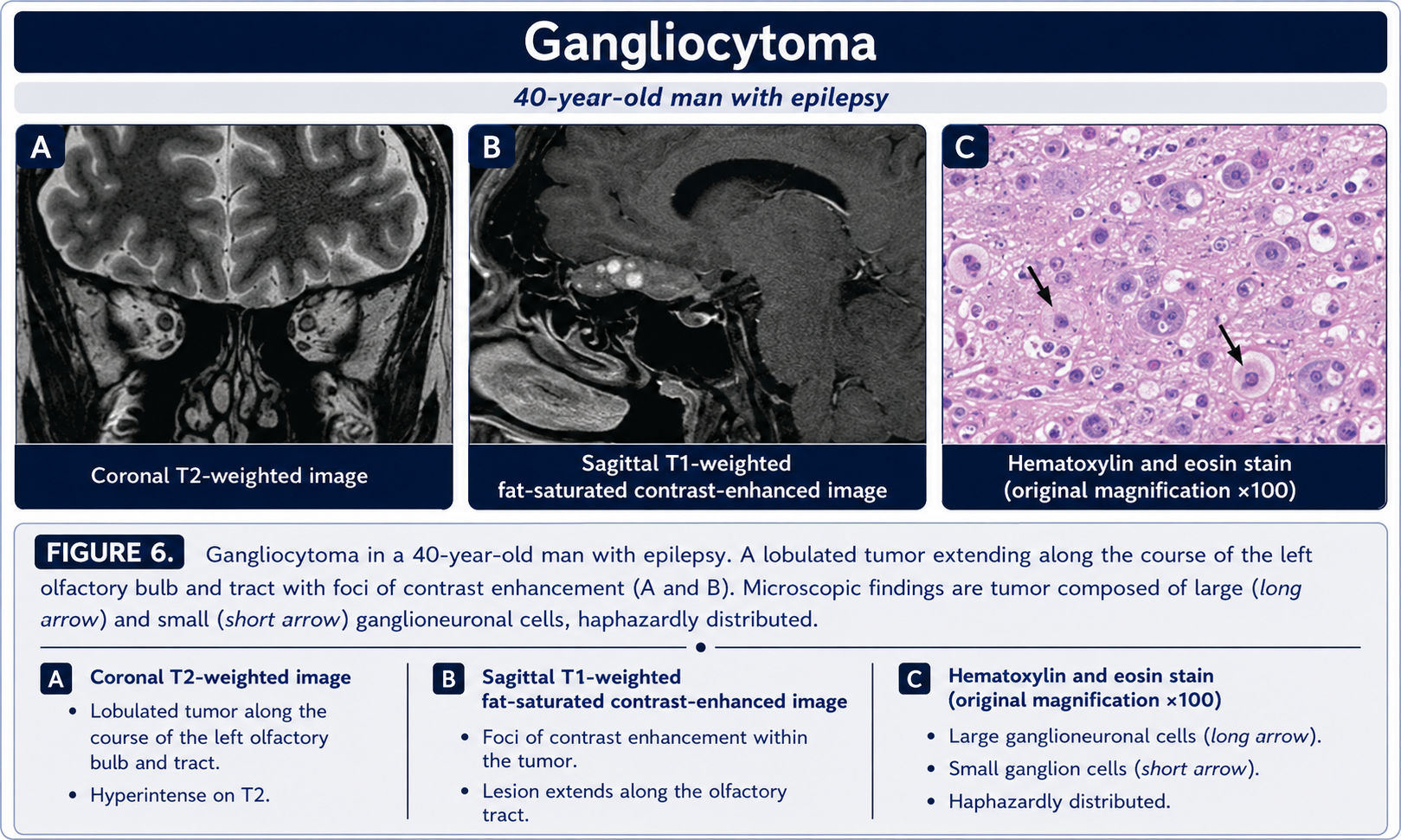
Gangliocytoma
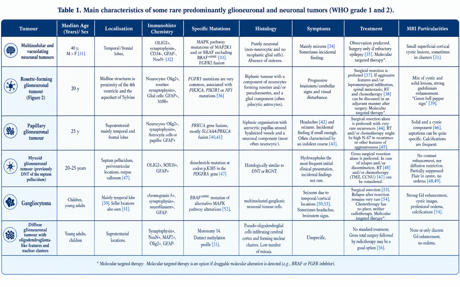
WHO grade 1; mature or dysplastic ganglion cells without a neoplastic glial component. Usually temporal in children and young adults with refractory epilepsy. MRI may show a mixed solid-cystic mass, variable enhancement, and calcification. Complete resection is usually curative.
Dysplastic cerebellar gangliocytoma
Lhermitte-Duclos disease occurs in adults and is associated with Cowden syndrome/PTEN mutation. Thickened cerebellar folia create the characteristic tigroid or “tiger-striped” MRI pattern. Elevated ADC reflects T2 shine-through. Observe small asymptomatic lesions; resect symptomatic lesions.
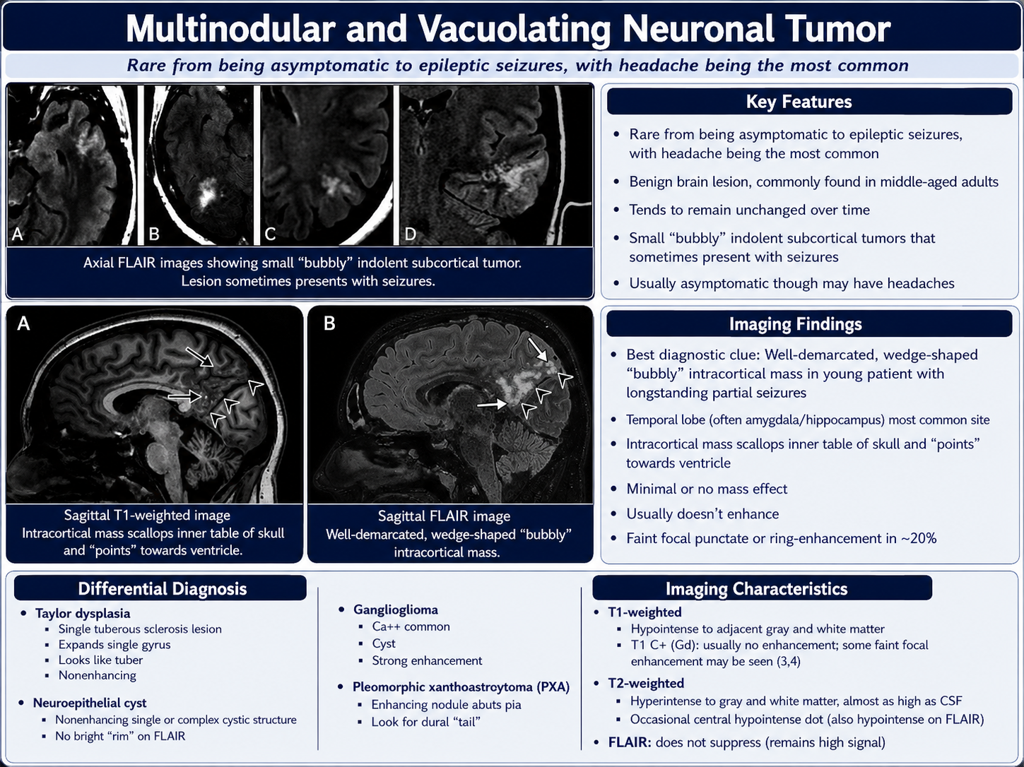
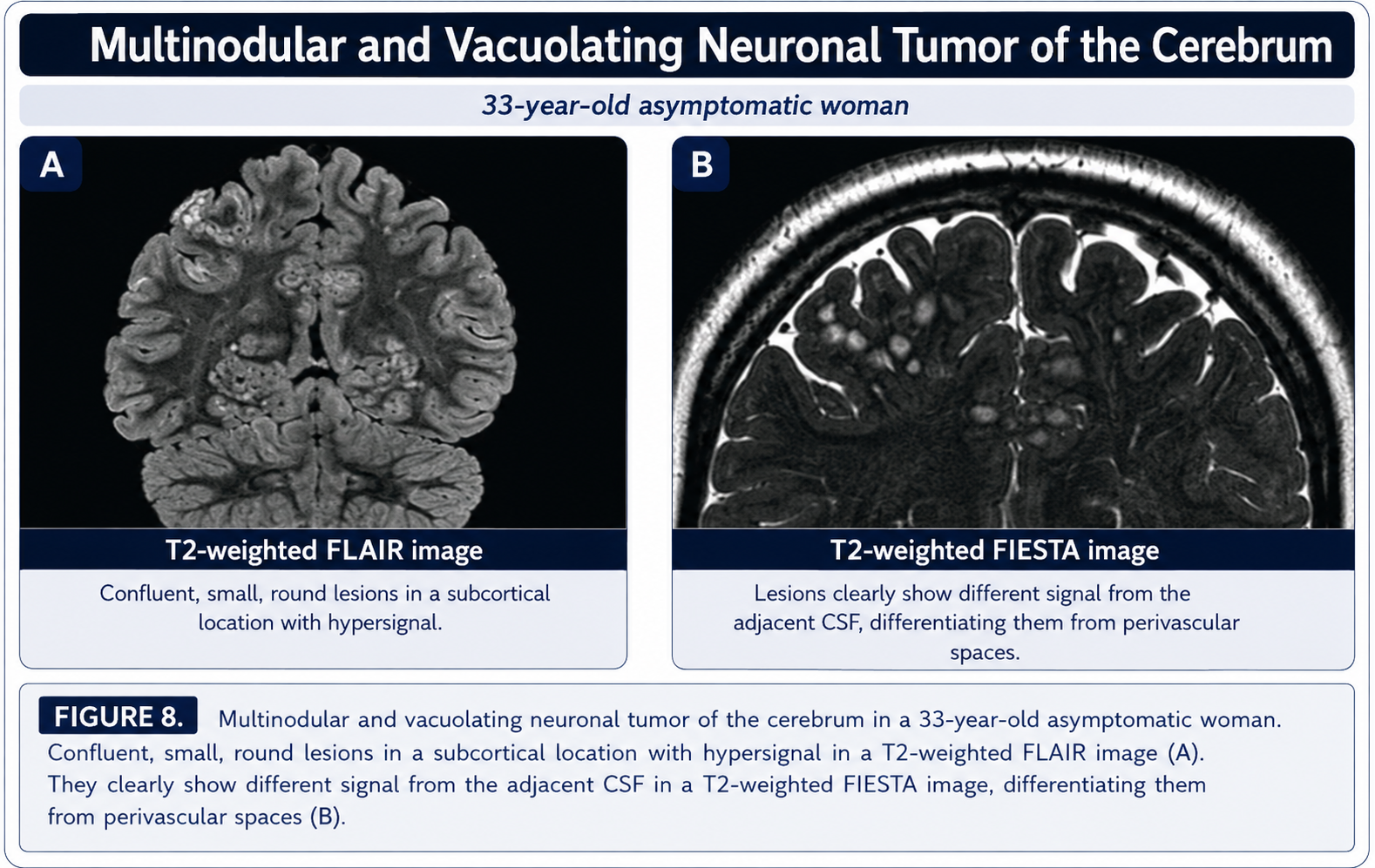
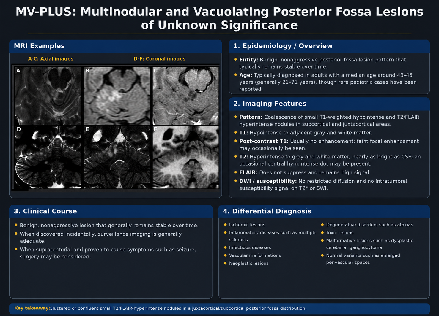
Multinodular and vacuolating neuronal tumor
Usually incidental or seizure-associated in adults. MRI shows clustered superficial cortical or subcortical nodules without enhancement, edema, or restriction. MAPK-pathway alterations are typical. Observation is preferred; surgery is reserved for refractory epilepsy.
Neurocytic Tumors
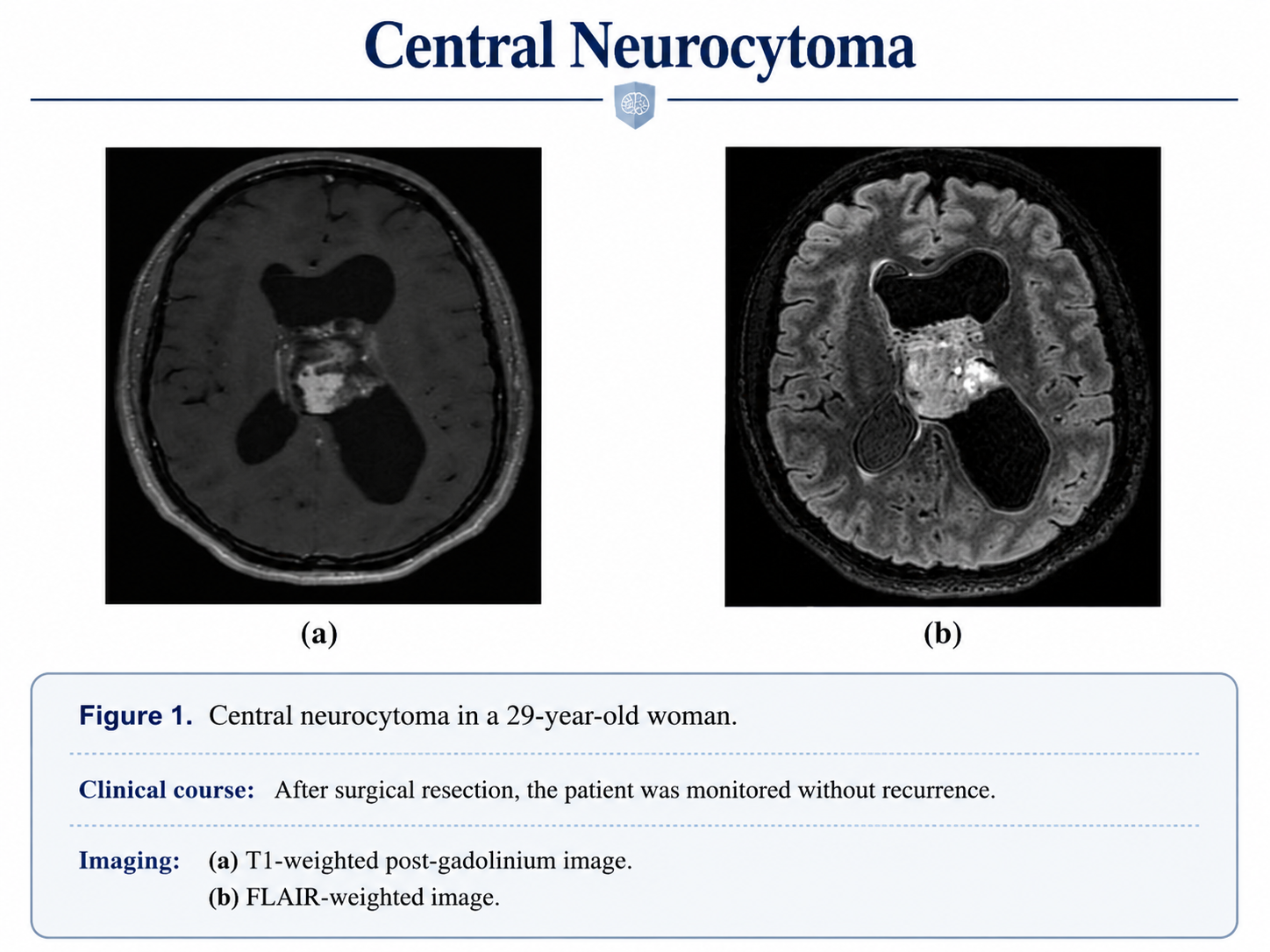
Central neurocytoma
WHO grade 2 tumor near the foramen of Monro or lateral ventricle in adults aged 20–50. A bubbly heterogeneous intraventricular mass, scalloping, punctate calcifications, and flow voids are characteristic. Resection is first-line; radiotherapy may follow incomplete resection or relapse.
Extraventricular neurocytoma
Usually temporal or frontal in young adults and often mixed solid-cystic without peritumoral edema. FGFR1::TACC1 fusion is frequent. Complete resection is often curative, while atypical forms recur more often.
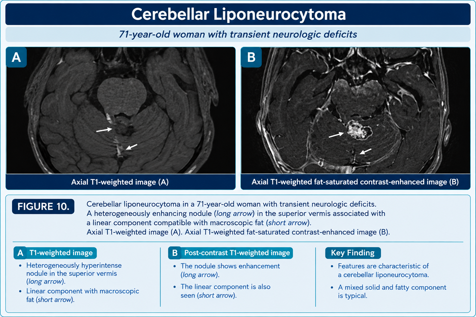
Cerebellar liponeurocytoma
Rare WHO grade 2 posterior fossa tumor of older adults. Macroscopic fat produces T1-hyperintense, fat-suppressible areas. Resection is first-line, although approximately one-third recur.
Mixed Glioneuronal Tumors
Ganglioglioma
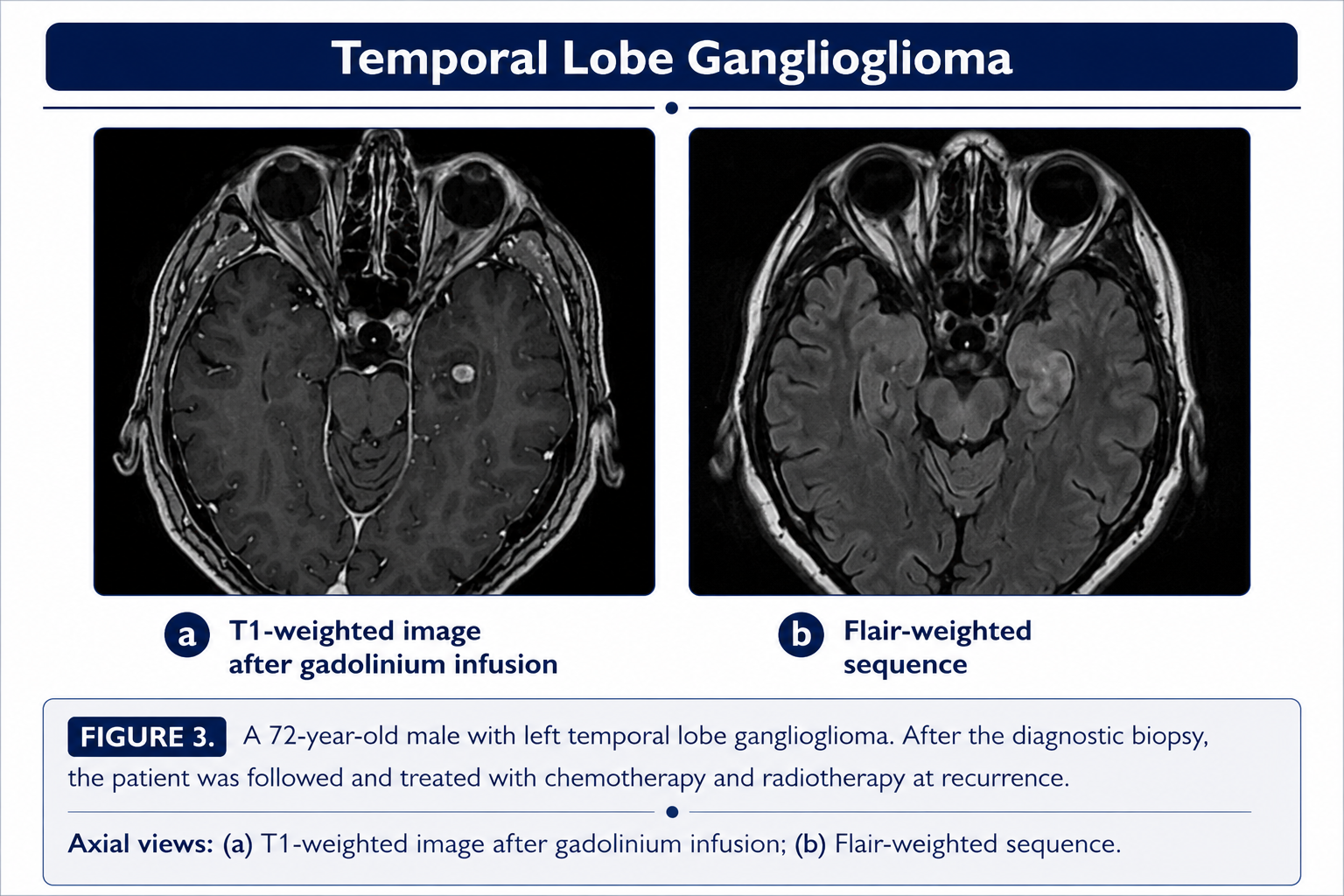
The most common glioneuronal tumor; usually temporal and strongly epileptogenic. It often has solid, cystic, or calcified components with variable enhancement. BRAF V600E occurs in about 30% and may be targetable. Complete resection has excellent long-term survival.
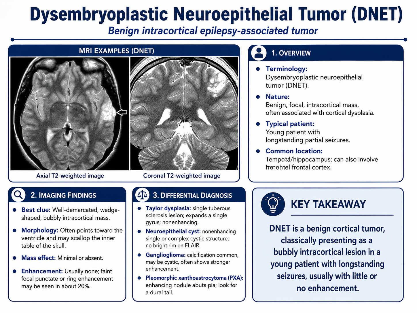
Dysembryoplastic neuroepithelial tumor
WHO grade 1 epilepsy-associated tumor, typically in patients aged 10–25. MRI shows a well-demarcated multicystic “bubbly,” wedge-shaped cortical lesion with high ADC, minimal edema, and infrequent enhancement. FGFR1 alterations are common. Resection produces seizure freedom in most patients.
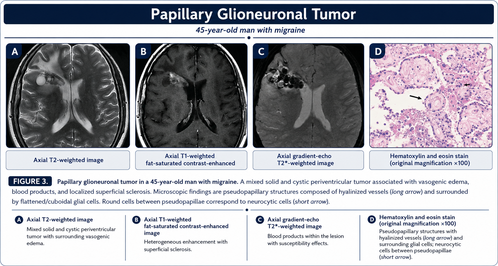
Papillary glioneuronal tumor
Rare WHO grade 1 tumor of young adults with SLC44A1::PRKCA fusion. Imaging ranges from cystic to solid, commonly a circumscribed cyst with an enhancing mural nodule. Calcification or superficial siderosis may occur. Complete resection is generally curative.
Additional Glioneuronal Tumors
Rosette-forming glioneuronal tumor
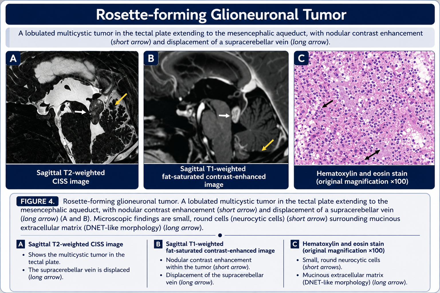
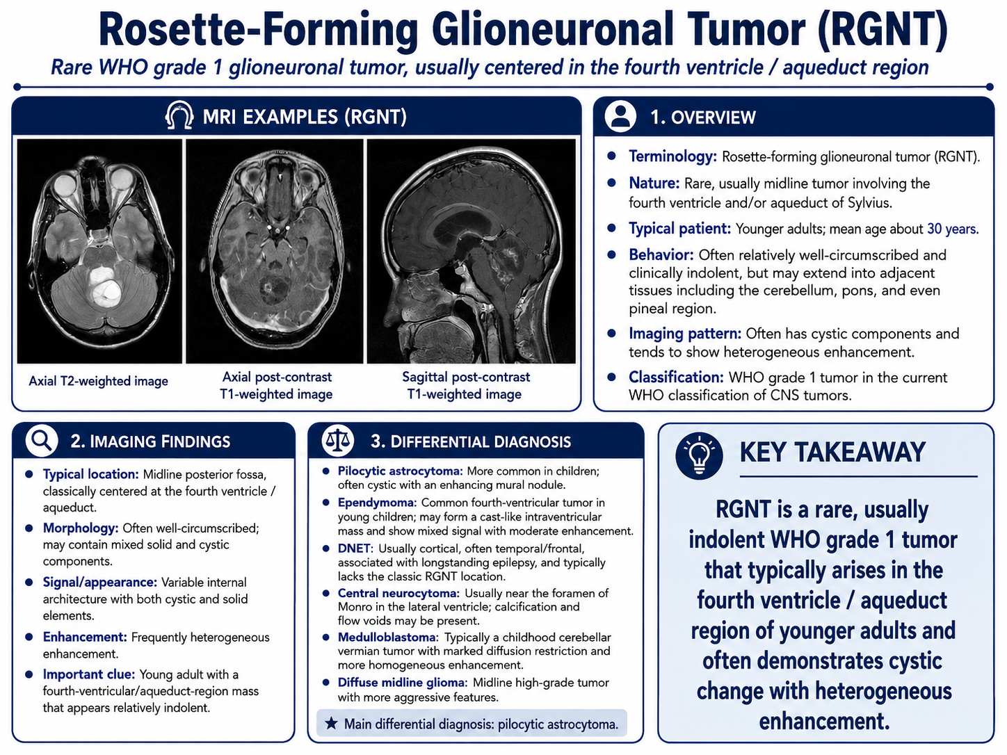
WHO grade 1 biphasic tumor, classically near the fourth ventricle or aqueduct. Mixed cystic-solid architecture and strong enhancement produce the “green bell pepper” appearance. FGFR1 alterations are common. Surgery is preferred.
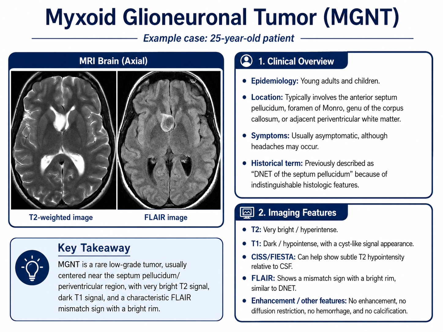
Myxoid glioneuronal tumor
Usually centered in the septum pellucidum or periventricular corpus callosum region in children or young adults. PDGFRA p.K385 mutation is characteristic. MRI typically lacks enhancement, restriction, and edema, with partial FLAIR suppression. Resection is preferred.
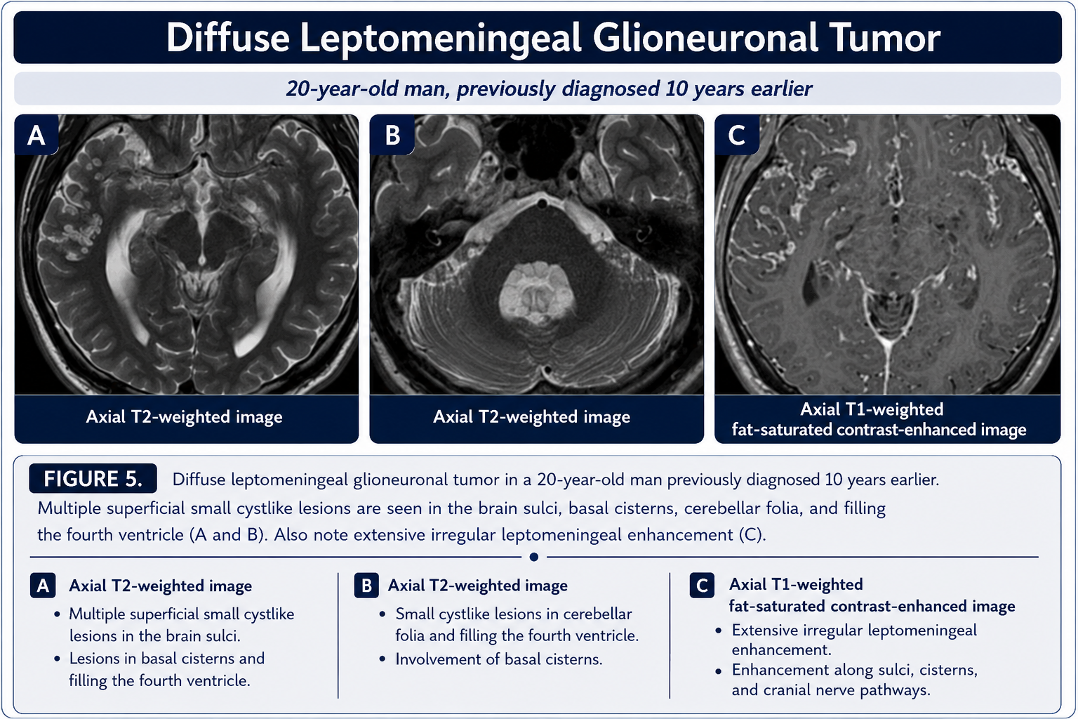
Diffuse leptomeningeal glioneuronal tumor
WHO grade 1–2 tumor, mainly in children and adolescents. MRI shows diffuse leptomeningeal thickening and enhancement with small subpial cystic lesions. 1p deletion and BRAF fusions are characteristic. Indolent disease may be monitored; systemic therapy is used when progressive.
DGONC
Diffuse glioneuronal tumor with oligodendroglioma-like features and nuclear clusters is a very rare provisional molecular entity with monosomy 14 and a distinct methylation profile. It affects children and young adults; no standard treatment is established.
Emerging and Related Entities
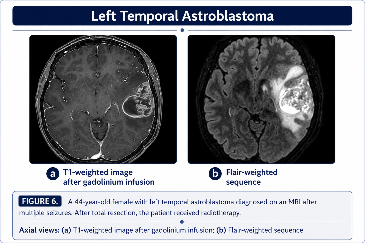
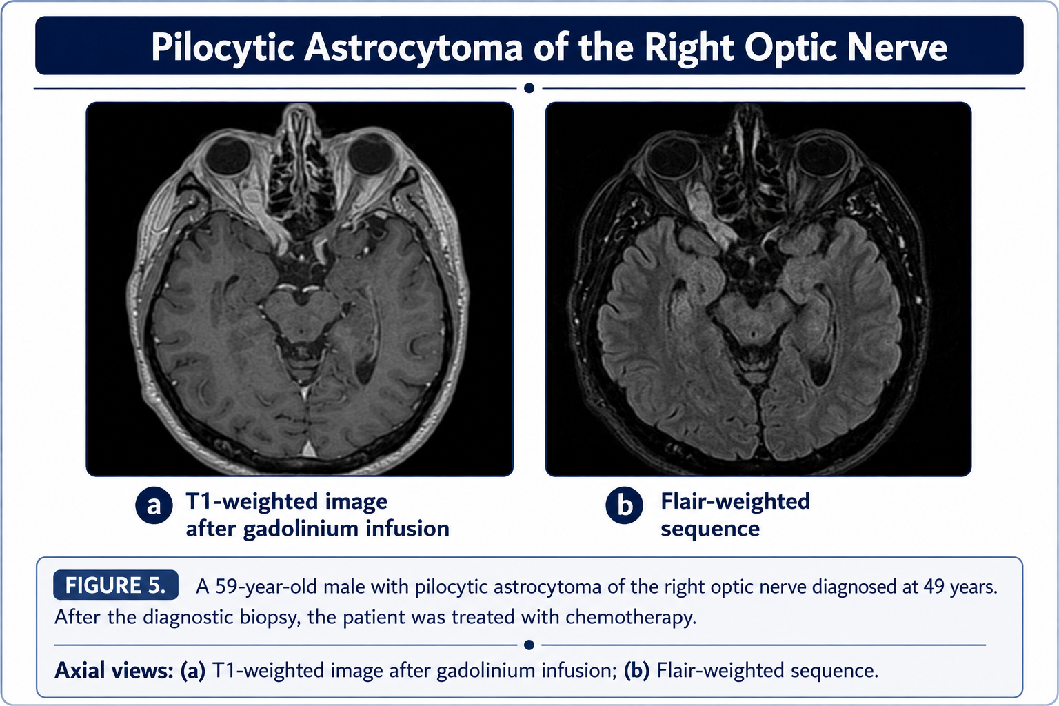
Several predominantly glial tumors overlap with glioneuronal tumors in their epilepsy association, imaging appearance, or MAPK-pathway biology.
Additional uncommon entities
- Desmoplastic infantile astrocytoma/ganglioglioma: almost exclusively under age 2; complete resection can be curative.
- PLNTY: temporal-lobe epilepsy tumor with oligodendroglioma-like cells, CD34 positivity, and BRAF V600E or FGFR2/3 fusion.
- Paraganglioma: neuroendocrine tumor that may be hereditary and can metastasize; treated with surgery and/or radiotherapy.
Treatment principles
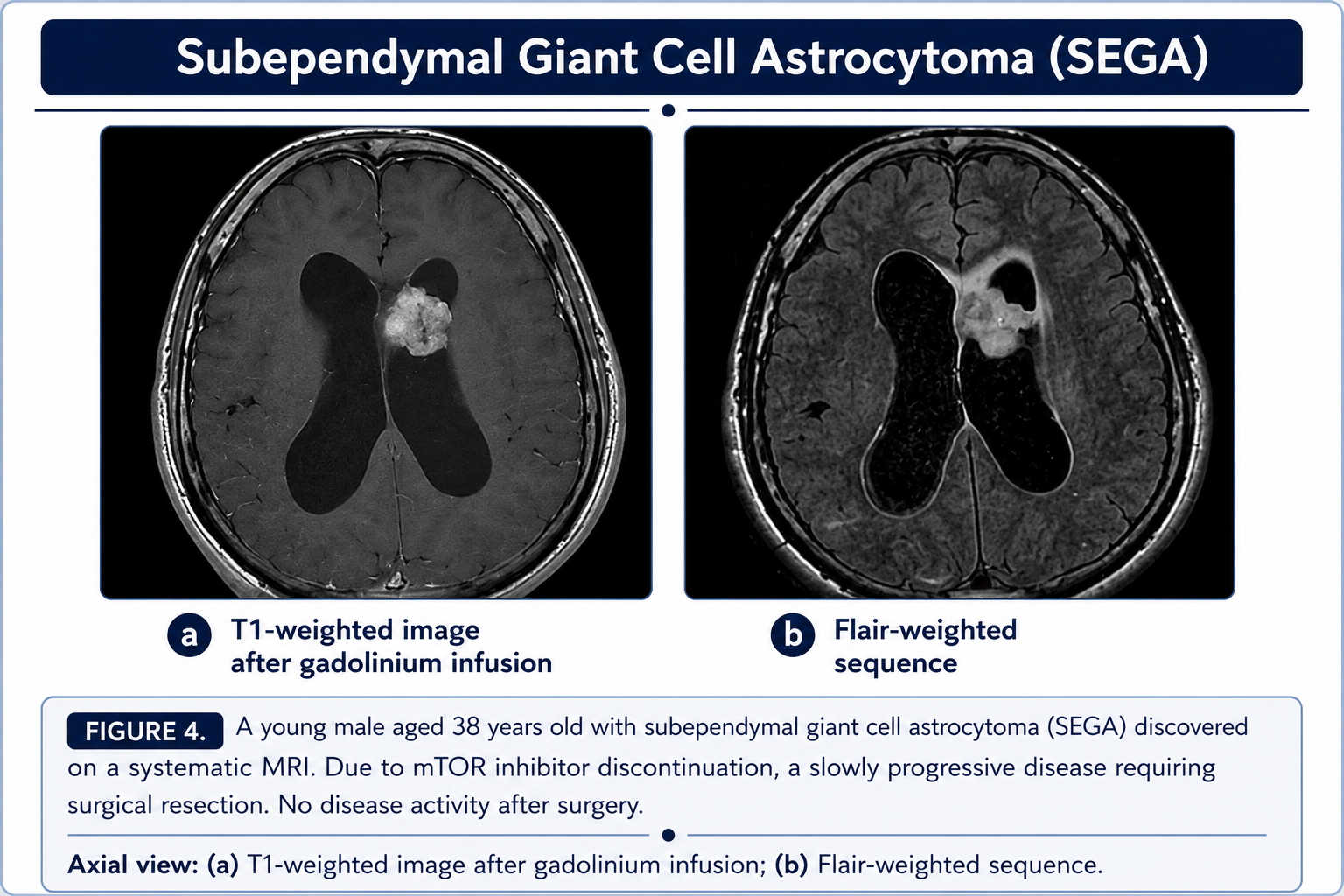
Maximal safe resection offers the best chance for seizure control and cure. Radiotherapy is generally reserved for incomplete resection, recurrence, or aggressive disease. Chemotherapy has a secondary role. Targeted BRAF, MEK, mTOR, or FGFR therapy may be useful when the corresponding alteration is present.