Bertolotti syndrome
Key Images
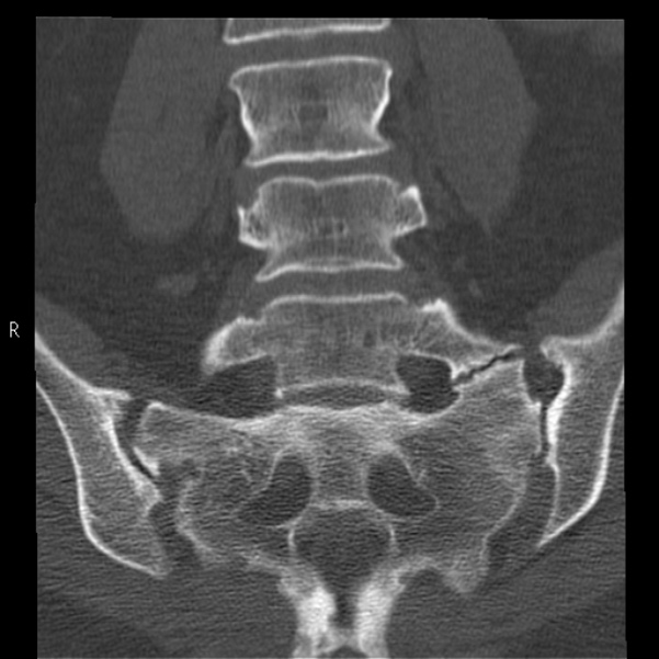
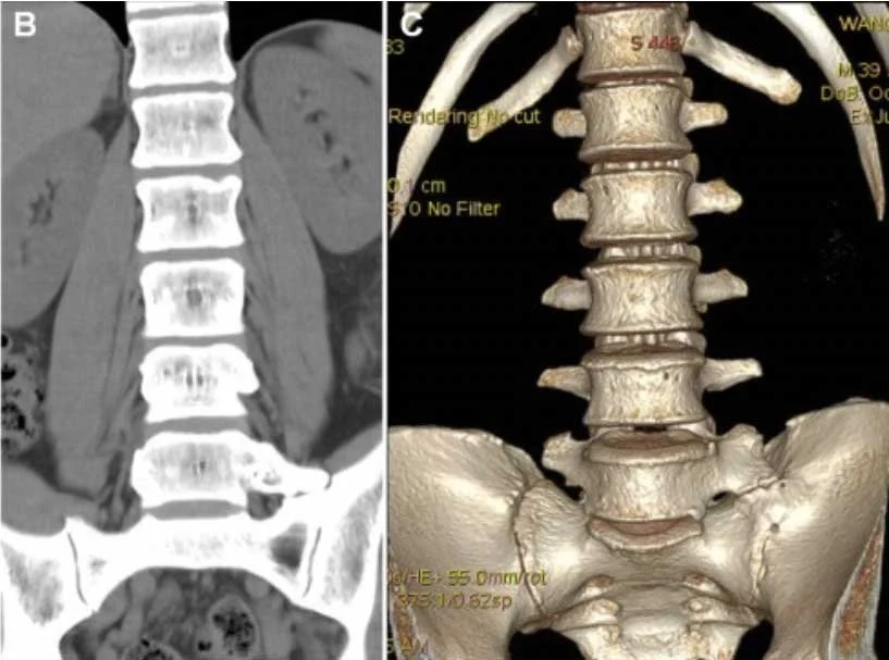
Look For First
- Partial or complete fusion of L5 vertebra to the sacrum creating a transitional anatomy between lumbar and sacral spine
- Asymmetric vertebral development with unilateral or bilateral pseudo-articulations at the L5-sacral junction
- Sagittal narrowing and facet hypertrophy at the level above the transitional vertebra (L4-L5) due to abnormal biomechanics
Key Image Findings
- On radiographs, Castellvi classification distinguishes LSTV: Type I (unilateral or bilateral enlarged transverse processes), Type II (unilateral or bilateral transverse process with incomplete fusion), Type III (unilateral or bilateral complete fusion), Type IV (complete fusion on one side with unilateral complete fusion on the contralateral side).
- Types II and IV LSTV show the strongest positive correlation with low back pain and buttock pain prevalence and severity.
- MRI T2-weighted and STIR sequences best demonstrate spinal canal narrowing and nerve root compression when radiculopathy is present, showing signal intensity changes in compressed nerve roots.
- The level above the transitional vertebra (L4-L5) typically demonstrates accelerated degenerative changes including disc herniation, facet hypertrophy, and central canal stenosis due to altered biomechanical stress.
- On sagittal MRI, the pedicles of the transitional vertebra appear broadened and the intervertebral disc above (L4-L5) shows premature degeneration relative to the patient's age.
- The transitional disc space (L5-S1) often appears preserved or normal compared to the hypermobile disc above it.
- Three-dimensional CT or advanced imaging can help confirm the anatomy and detect associated facet arthrosis and foraminal stenosis.
Differential Diagnosis
- Disc herniation at L4-L5 without LSTV: distinguished by normal lumbosacral anatomy without transitional vertebrae on radiographs.
- Facet syndrome from primary facet hypertrophy: distinguished by normal vertebral anatomy and central rather than foraminal pain pattern.
- Spondylolisthesis: distinguished by vertebral slip on radiographs with intact anterior vertebral body alignments in LSTV.
- Occult spondylolysis: distinguished by break in pars interarticularis rather than vertebral fusion anomaly.
- Idiopathic low back pain without structural cause: identified by completely normal imaging and imaging findings must correlate with clinical presentation to justify LSTV as pain source.
Discussion
Bertolotti syndrome remains controversial because many asymptomatic patients harbor LSTV at 5% population prevalence, making it difficult to establish causation without careful clinical-radiological correlation.
Types II and IV transitional vertebrae show statistically stronger association with low back pain than Types I and III, suggesting incomplete fusion creates more biomechanical instability than complete fusion.
The transitional vertebra itself is typically pain-insensitive; pain symptoms likely arise from accelerated degenerative changes at the hypermobile level above (L4-L5) and facet arthrosis.
Conservative management (physical therapy, NSAIDs, injections) remains first-line therapy and surgery is reserved for refractory cases with imaging correlation to the pain pattern.
MRI is essential only when radiculopathy or myelopathy symptoms are present; radiographs alone may miss neural compromise.
Documentation of LSTV type, presence and severity of degenerative changes at adjacent levels, and any nerve root compression is critical for communicating risk and prognosis to clinicians.
Reporting Pearls
When reporting LSTV, clearly specify the Castellvi type, describe the degree of fusion (complete vs. incomplete, unilateral vs. bilateral), note the presence and severity of degenerative changes at the L4-L5 level, and explicitly state whether foraminal or central canal narrowing correlates with the patient's pain location and radicular symptoms to justify LSTV as a pain source.
Pitfalls
- Assuming LSTV causes pain without clinical correlation: remember that 5% of asymptomatic population have LSTV, so anatomic presence alone does not prove causation.
- Misclassifying the transitional vertebra level: ensure you count correctly from L1 to identify which vertebra is transitional, as miscounting leads to incorrect surgical planning.
- Overlooking degenerative changes at L4-L5: the pain generator is often the hypermobile disc above, not the fusion itself; describe stenosis and disc herniation at the adjacent level thoroughly.
- Failing to distinguish Types II and IV (higher pain correlation) from Types I and III (lower pain correlation) in the report: this distinction influences treatment recommendations and prognosis discussion.