PE-RADS v2026
Table 5: Assessment Categories
Zoom Table 5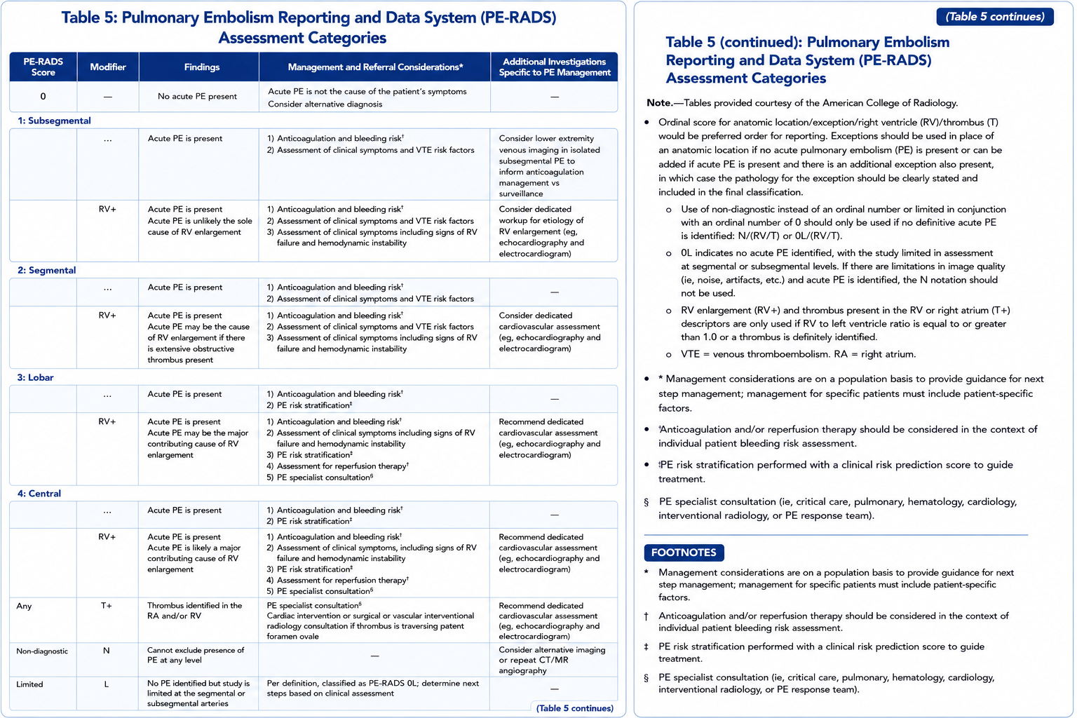
Primary management table. Use the zoom button, then use Zoom + inside the viewer for a closer read.
Reference Images
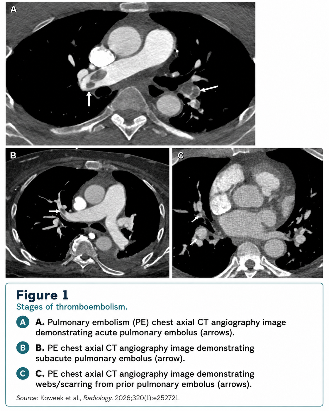
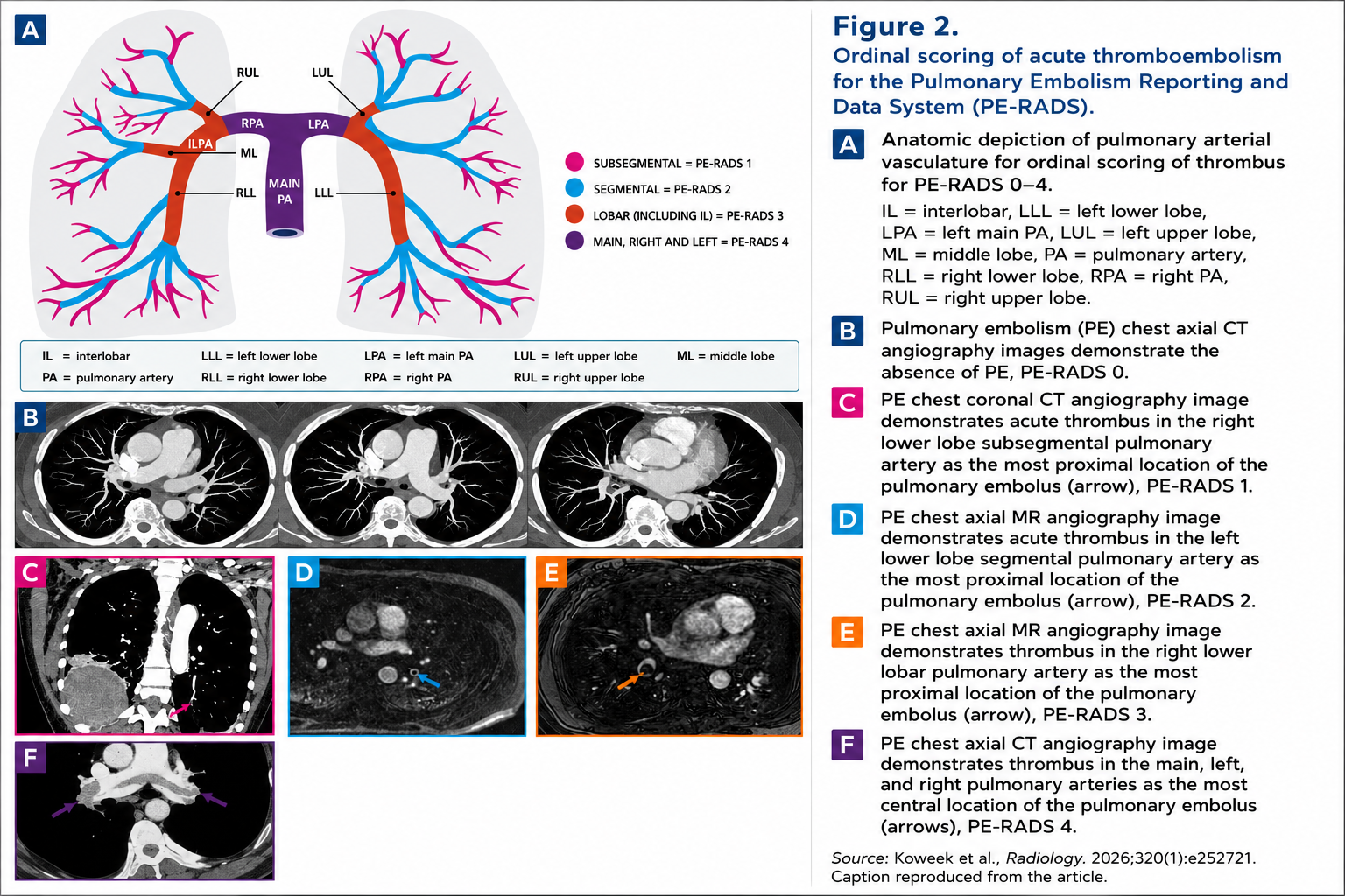
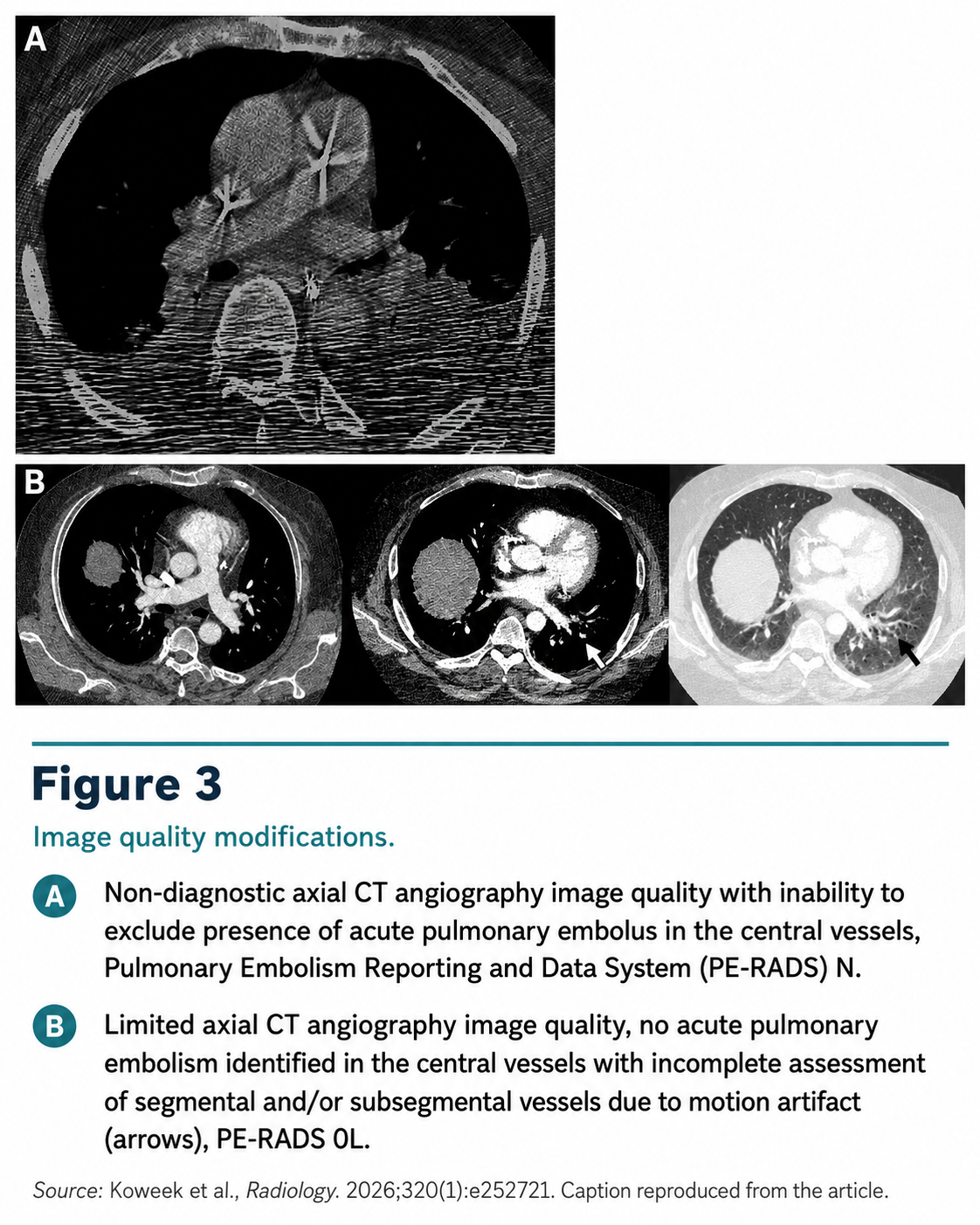
Core Concept
Use PE-RADS for
CT pulmonary angiography or MR angiography exams evaluating pulmonary embolism.
What the label communicates
- Most proximal acute PE location.
- Image quality limitations or nondiagnostic central assessment.
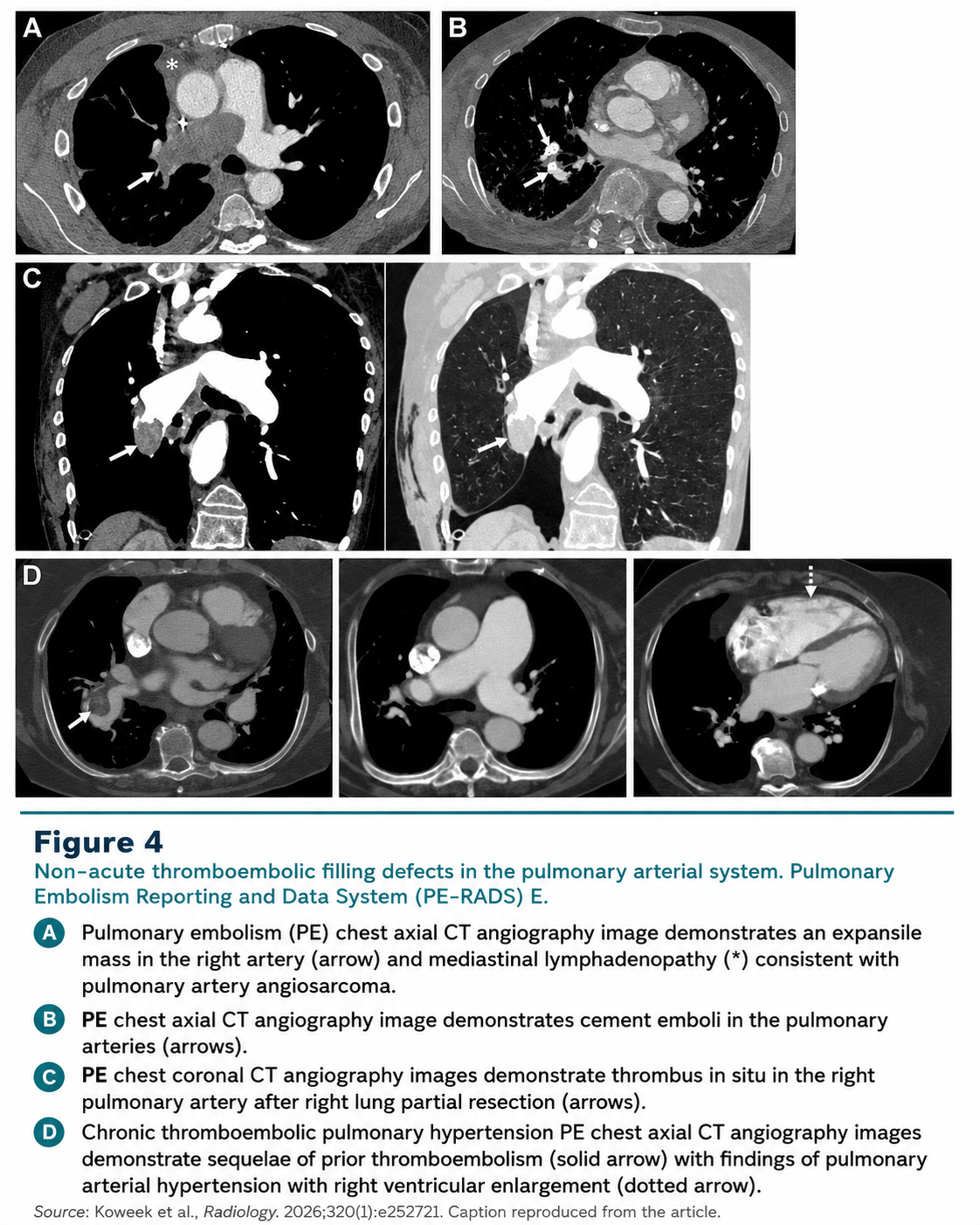
- Nonacute or non-PE intravascular filling defects.
- Right ventricular enlargement and definite right heart thrombus.
Reporting principle
- Use the most proximal acute embolus for the ordinal score.
- Do not use the score as a total clot burden estimate.
- Final shorthand examples: PE-RADS 2, PE-RADS 0L, PE-RADS E, PE-RADS 4/RV+/T+.
Ordinal Score
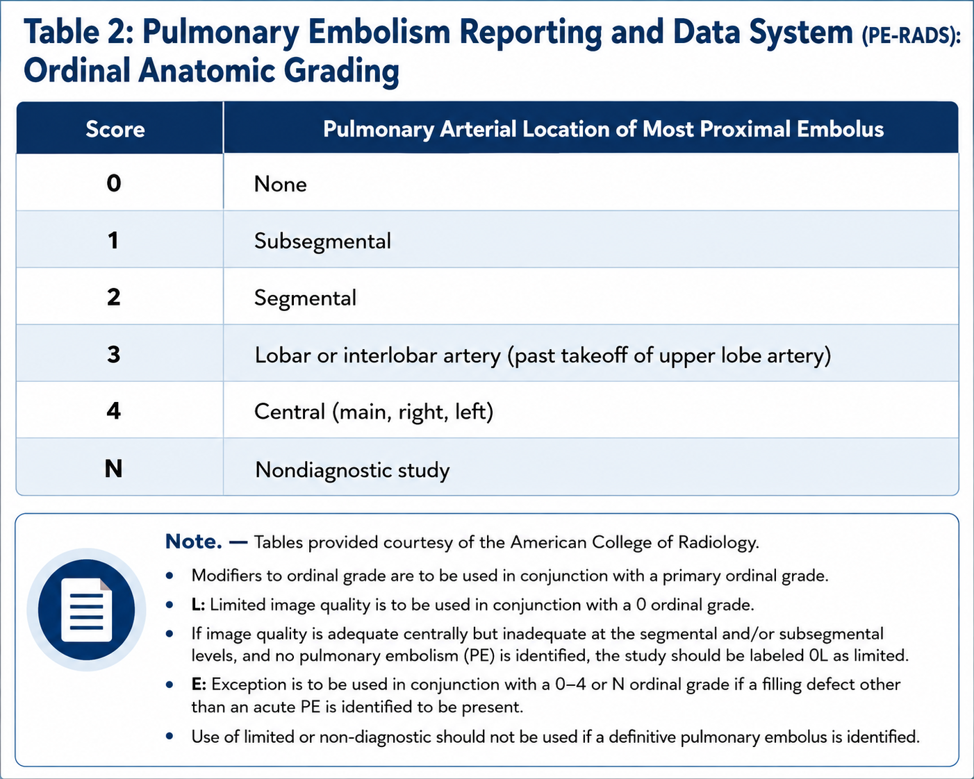
Acute PE anatomic grade
- 0: no acute pulmonary embolism.
- 1: most proximal embolus is subsegmental.
- 2: most proximal embolus is segmental.
- 3: most proximal embolus is lobar or interlobar artery.
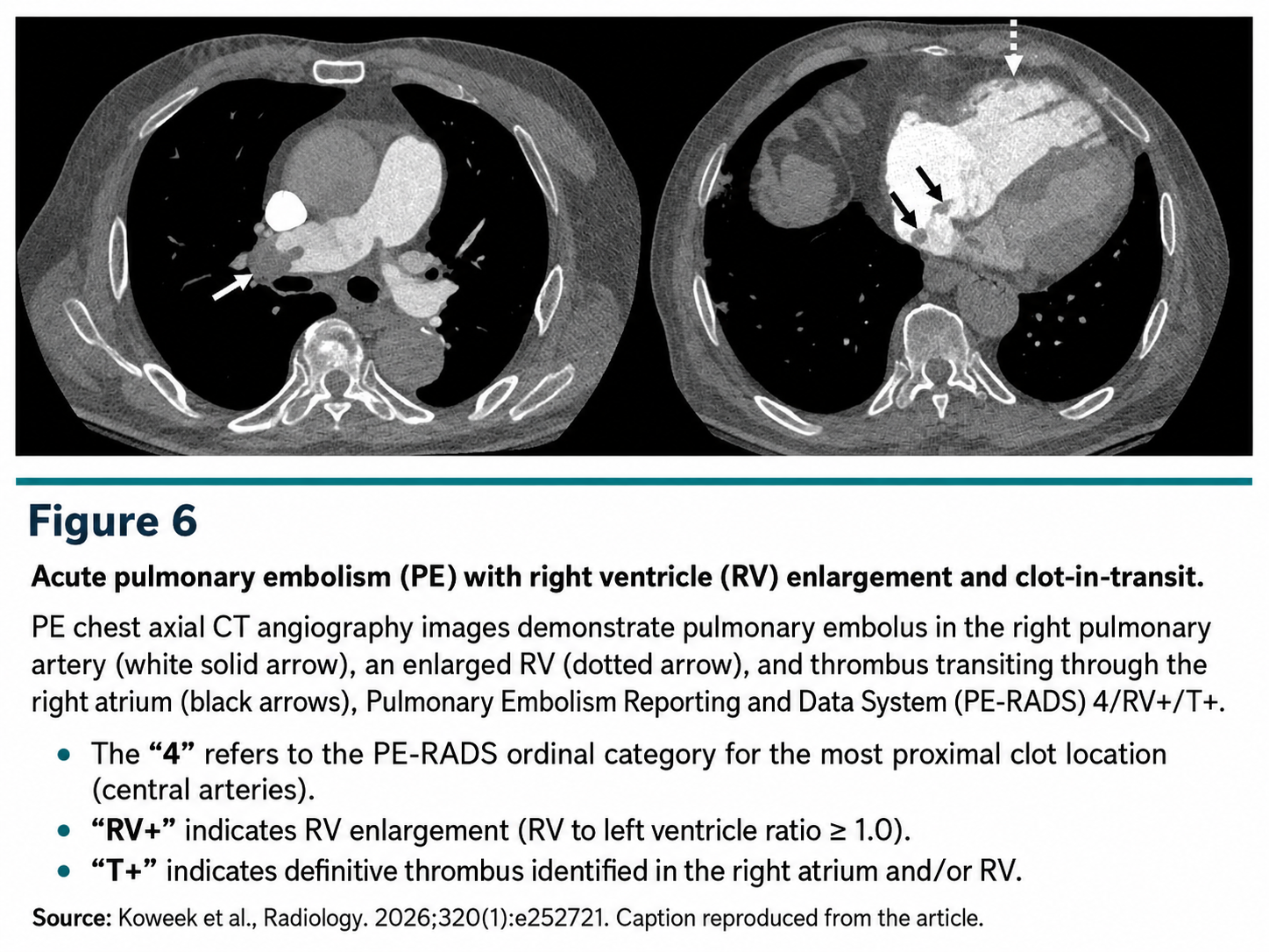
- 4: most proximal embolus is central, involving main, right, or left pulmonary arteries.
- N: nondiagnostic study; cannot exclude acute PE at main or lobar level.
Location wording
- State the most proximal level and laterality when useful.
- Describe burden separately when clinically important.
Quality / Exceptions
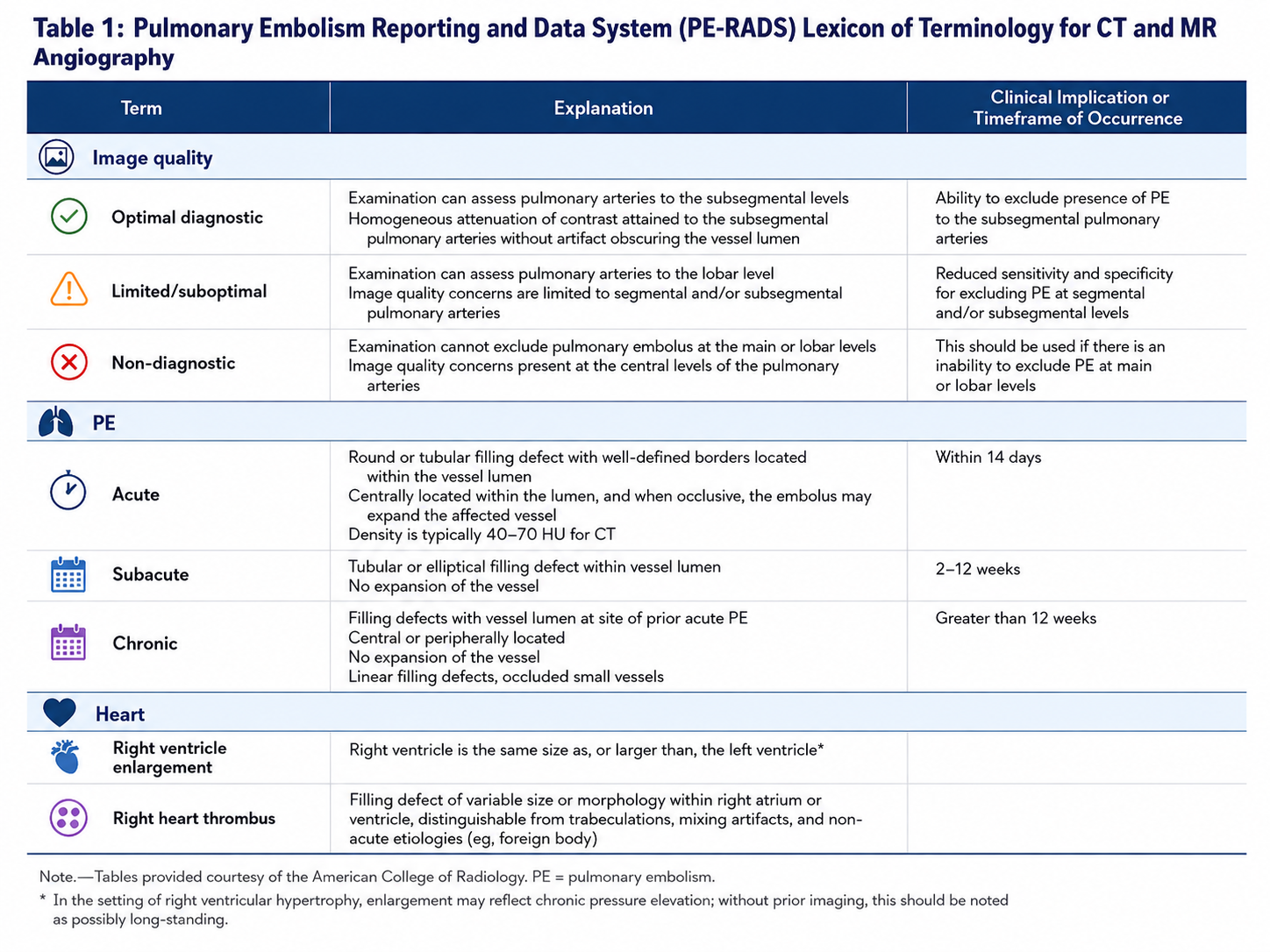
Image quality modifiers
- Optimal diagnostic: adequate to exclude PE to subsegmental level.
- Limited/suboptimal: central assessment is adequate, but segmental or subsegmental assessment is limited.
- Non-diagnostic: cannot exclude PE at the main or lobar pulmonary arteries.
How to use 0L and N
- Use 0L when no acute PE is identified but distal assessment is limited.
- Use N when central evaluation is nondiagnostic.
- Do not use limited or nondiagnostic labels when a definite acute PE is identified.
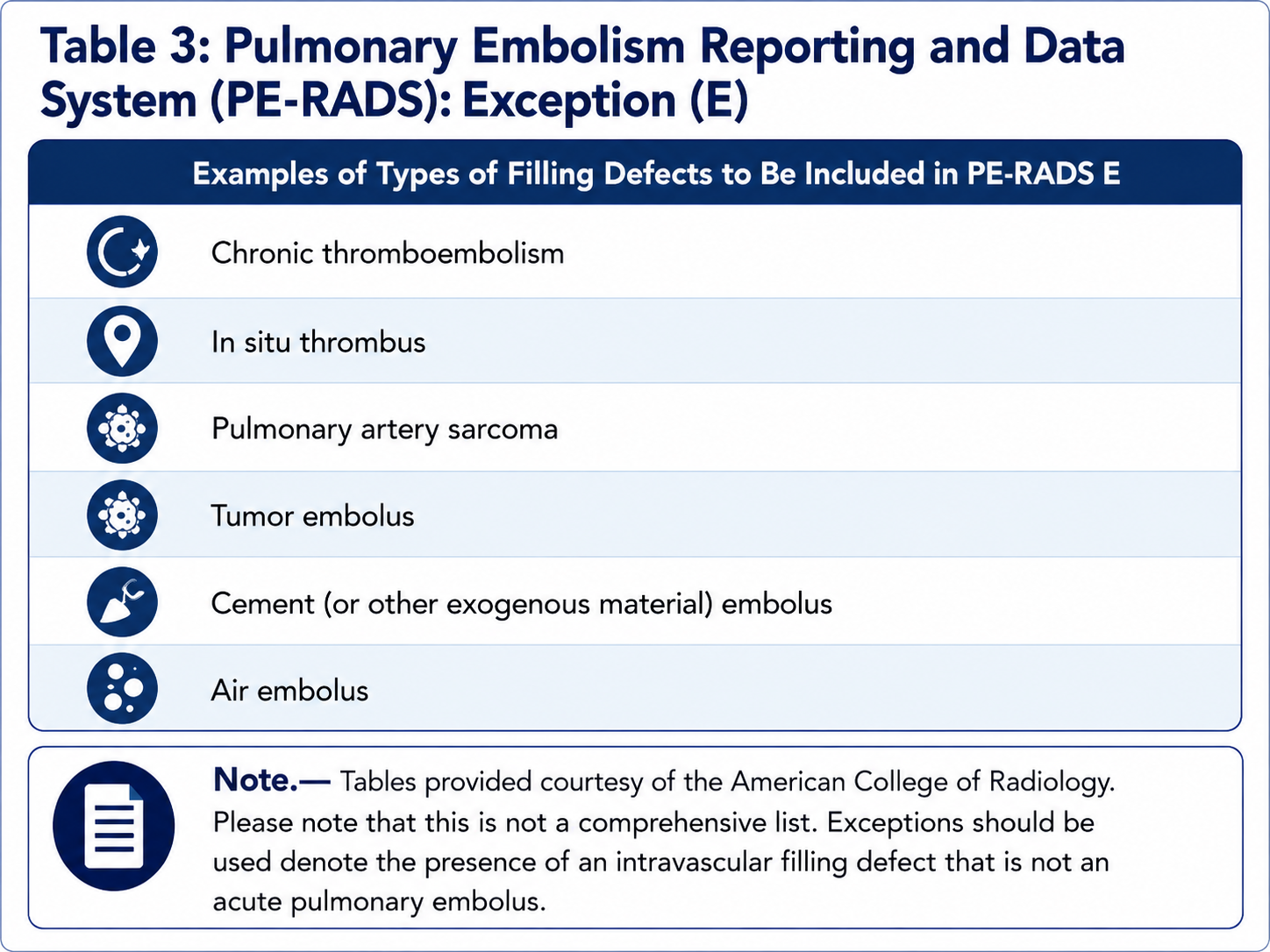
Exception E
- Use E for a filling defect that is not acute PE.
- Examples: chronic thromboembolism, in-situ thrombus, pulmonary artery sarcoma, tumor embolus, cement or other exogenous material embolus, and air embolus.
- Name the suspected process in the report when possible.
RV+ / T+
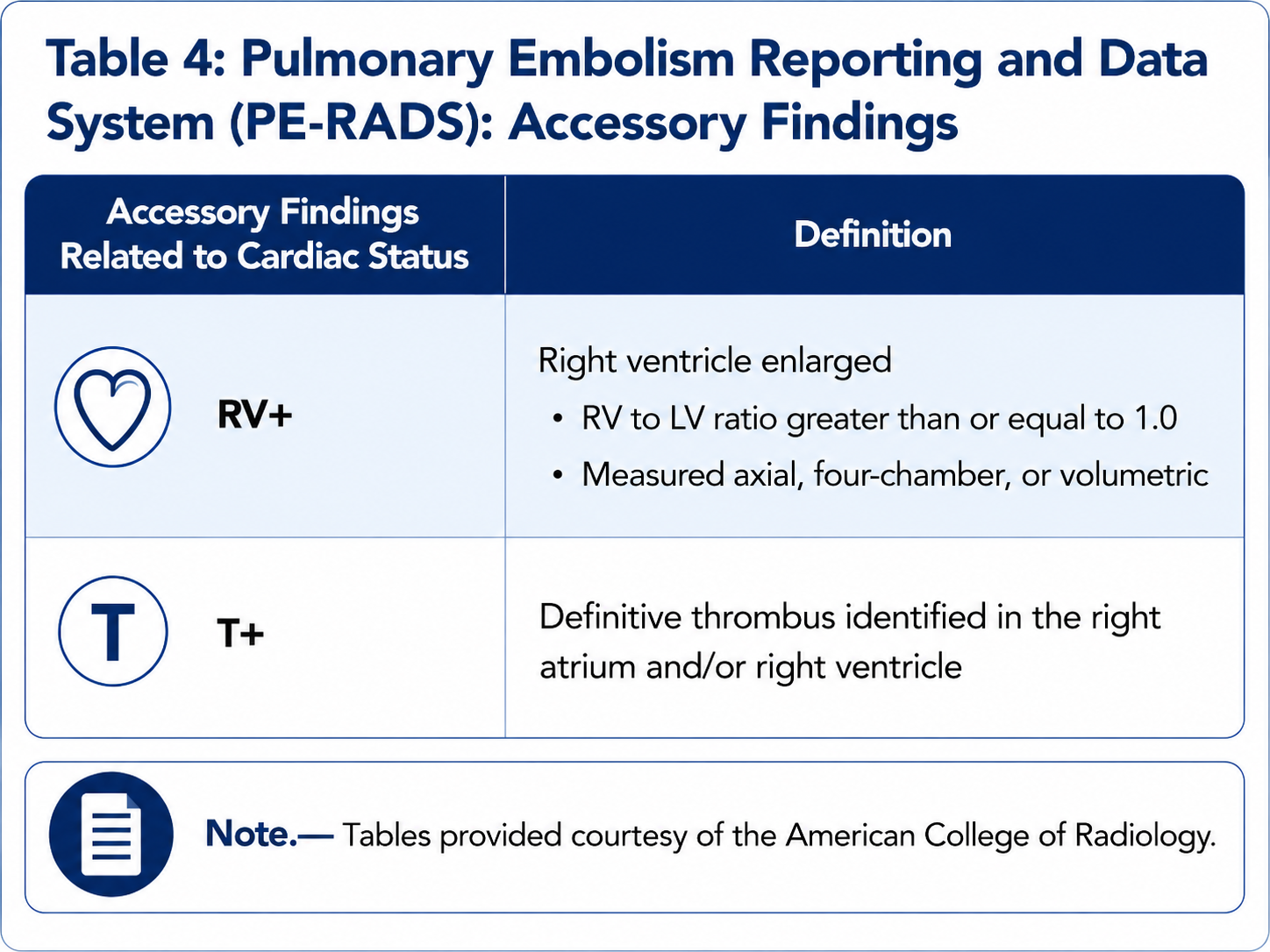
RV+ modifier
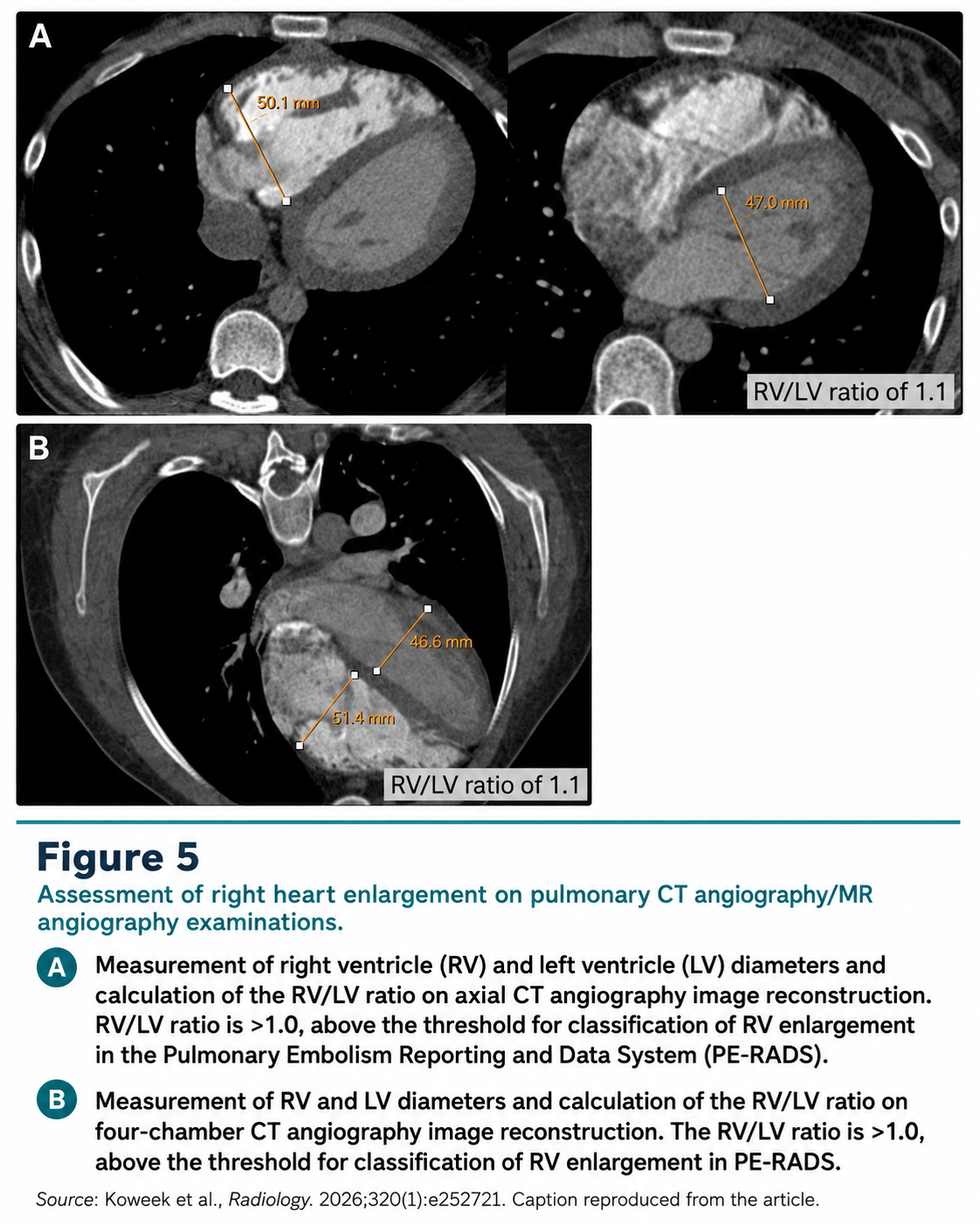
- RV+ means right ventricular enlargement.
- Use when RV/LV ratio is greater than or equal to 1.0.
- Measurement may be axial, four-chamber, or volumetric.
- Prefer "RV enlargement" over "RV strain"; chronic pressure overload can also enlarge the RV.
T+ modifier
- T+ means definite thrombus in the right atrium and/or right ventricle.
- Use only when the thrombus is confidently identified.
- Example: PE-RADS 4/RV+/T+ = central acute PE with RV enlargement and right heart thrombus.
Management Snapshot
Acute PE
- Any acute PE: consider anticoagulation in the context of bleeding risk and clinical severity.
- Isolated subsegmental PE: individualized anticoagulation versus surveillance; consider lower extremity venous imaging.
- Segmental PE: anticoagulation and clinical VTE/symptom assessment.
- Lobar or central PE: PE risk stratification and clinical severity assessment.
Escalation patterns
- RV+ may warrant dedicated cardiovascular assessment, such as echocardiography and ECG.
- Lobar/central PE with RV+ may warrant reperfusion therapy assessment and PE specialist or PERT consultation.
- T+ warrants urgent specialist input; consider cardiac surgery or vascular intervention when thrombus is traversing a patent foramen ovale.
- PE-RADS N: repeat or alternative CT/MR angiography may be needed.
Report Language
Reusable phrases
- No acute pulmonary embolism. PE-RADS 0.
- No acute PE in the central or lobar arteries; segmental/subsegmental evaluation is limited by motion artifact. PE-RADS 0L.
- Acute pulmonary embolus is present to the segmental pulmonary arteries. PE-RADS 2.
- Central acute pulmonary embolism with RV/LV ratio 1.1. PE-RADS 4/RV+.
- Central acute pulmonary embolism with RV enlargement and definite right atrial/right ventricular thrombus. PE-RADS 4/RV+/T+.
- Nonacute pulmonary arterial filling defect compatible with chronic thromboembolic disease. PE-RADS E.
Source
- Koweek et al. Radiology. 2026;320(1):e252721.
- PE-RADS is a reporting framework; management should incorporate patient-specific risk, hemodynamics, biomarkers, comorbidities, and local PE response pathways.
Source Article
Primary Reference
- PE-RADS article PDF
- Koweek et al. Radiology. 2026;320(1):e252721.