Regions
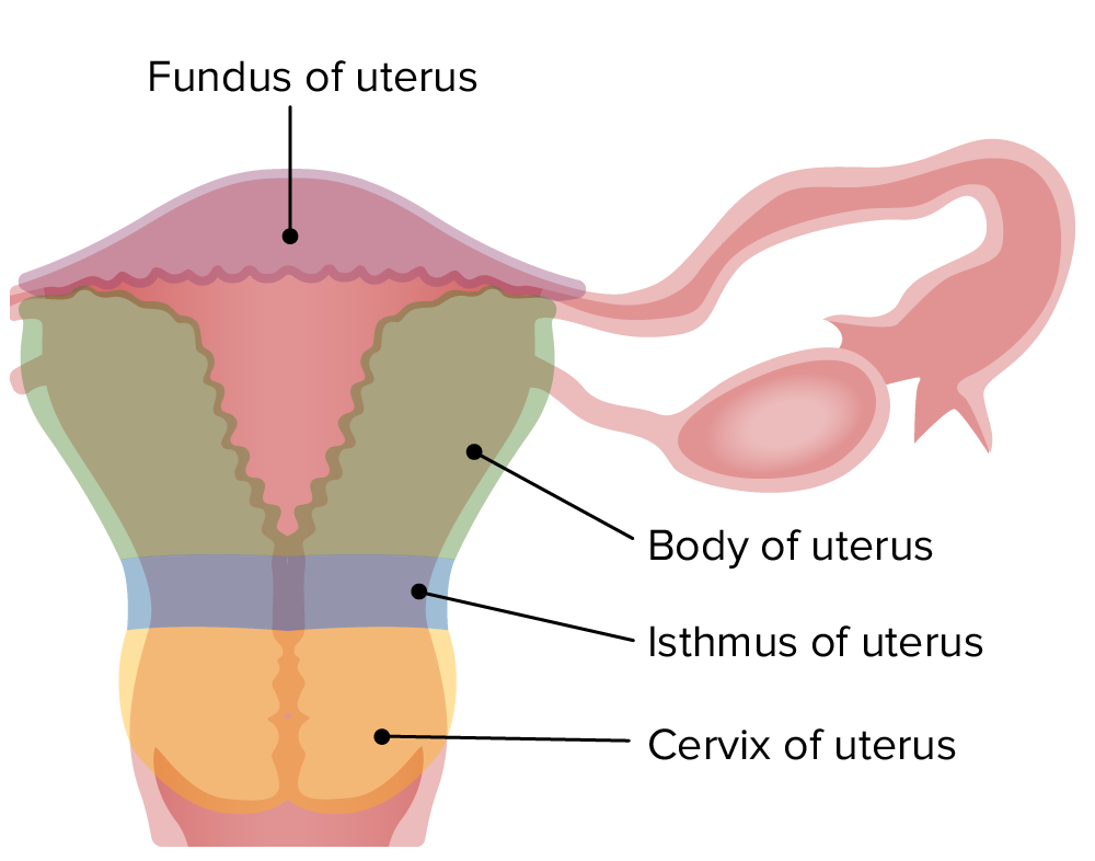
Fundus and body above the isthmus; cervix below it.
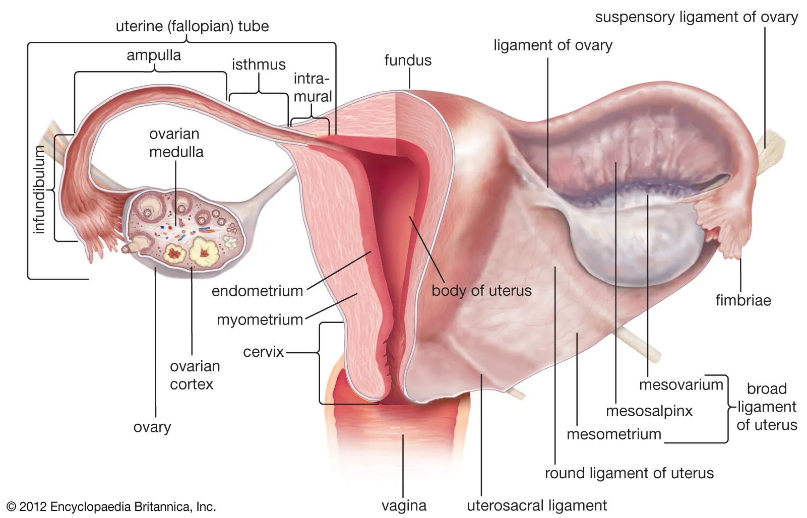
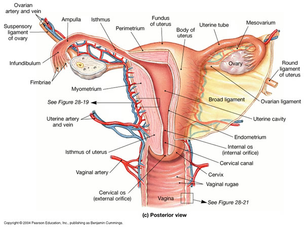
Anatomy of the uterus, uterine tubes and supporting ligaments, with imaging appearances and life-stage measurements.
 Enlarge figure 1
 Enlarge figure 2
 Enlarge figure 3
 Enlarge figure 4
 Enlarge figure 5
 Enlarge figure 6
 Enlarge figure 7
 Enlarge figure 8
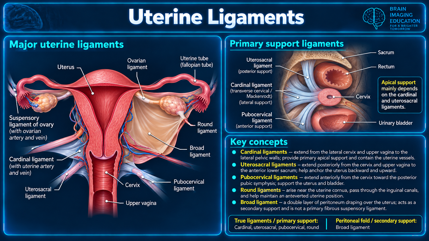 Enlarge figure 9
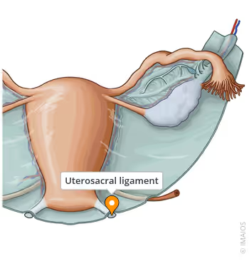 Enlarge figure 10
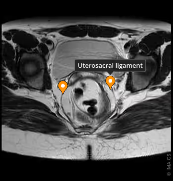 Enlarge figure 11
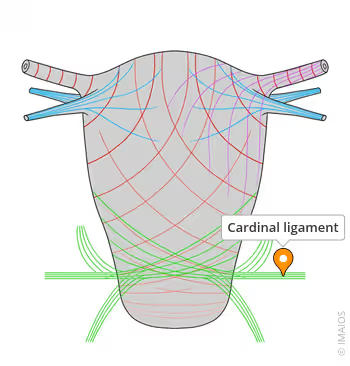 Enlarge figure 12
 Enlarge figure 13
 Enlarge figure 14
 Enlarge figure 15
 Enlarge figure 16
Source limitations: Numbered citation markers are retained from the supplied text, but no bibliography or destinations for its ?see? references and mnemonic were supplied. Figure 7 contains prevalence estimates without identified studies. The text describes a role for the round ligament in anteflexion; figure 9 describes anteversion and groups it with primary support ligaments while emphasizing cardinal and uterosacral ligaments for apical support. These source formulations are preserved without reconciliation. Adult size measurements in the gross anatomy and life-stage sections differ; the supplied values are retained. Original annotations, credits and watermarks remain visible.
Fundus and body above the isthmus; cervix below it.
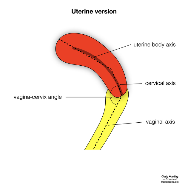
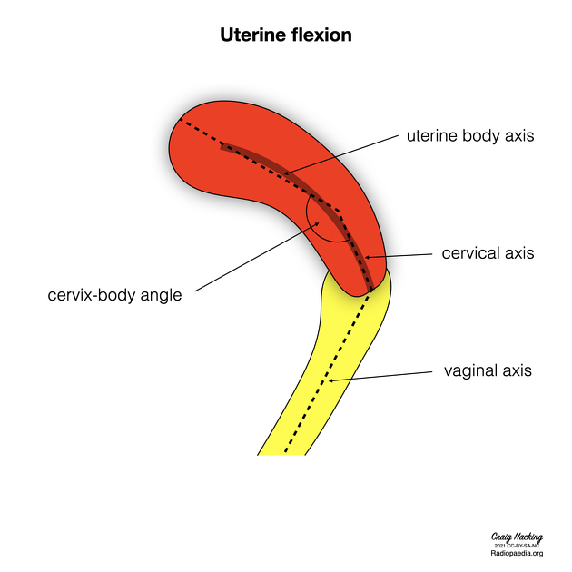
Version compares cervical and vaginal axes. Flexion compares uterine body and cervical axes.
T2: high-signal endometrium, low-signal junctional zone, intermediate-signal outer myometrium.
The uterus is a hollow, thick-walled, muscular organ of the female reproductive tract that lies in the lesser pelvis. The body of the uterus is intraperitoneal and the cervix uteri is subperitoneal.
The uterus has an inverted pear shape. It measures about 7.5 cm in length, 5 cm wide at its upper part, and nearly 2.5 cm in thickness in adults. It weighs approximately 30-40 g.
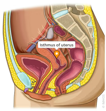
The uterus is divisible into two portions: body and cervix. About midway between the apex and base is a slight constriction known as the isthmus. The portion above the isthmus is termed the body, and that below, the cervix. The part of the body which lies above a plane passing through the points of the entrance of the uterine tubes is known as the fundus.
The body gradually narrows from the fundus to the isthmus. The cavity of the body is a mere slit, flattened anteroposteriorly. It is triangular in shape:
Although anatomically a part of the uterus, the uterine cervix has a different function and is associated with separate pathological entities. It is discussed in detail in a separate article.
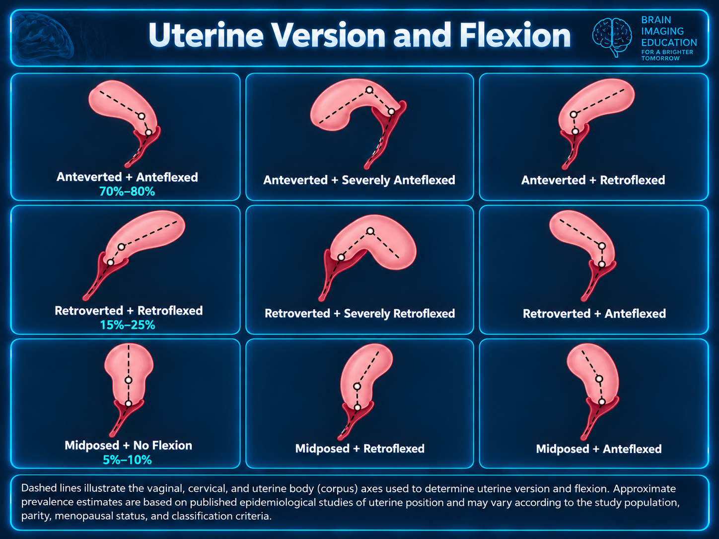
Uterine position in adults is variable and largely depends on the degree of bladder and rectal distension. When the bladder is empty, the entire uterus is directed forward and is at the same time bent on itself at the junction of the body and cervix so that the body lies upon the bladder. As the latter fills, the uterus gradually becomes more and more erect until, with a fully distended bladder, the fundus may be directed back toward the sacrum.
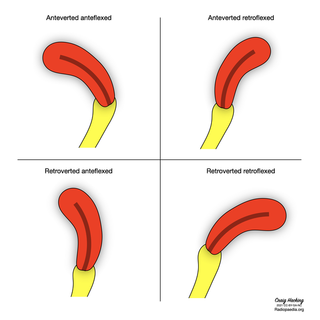
The uterine position can be described in terms of version and flexion:
This results in a number of uterine positions:
In the fetus, the uterus is contained in the abdominal cavity, projecting beyond the superior aperture of the pelvis. The cervix is considerably larger than the body.
At puberty, the uterus is pyriform in shape and weighs from 14-17 g. It has descended into the pelvis, the fundus being just below the level of the superior aperture of this cavity. The palmate folds are distinct and extend to the upper part of the cavity of the organ.
During menstruation, the uterus is enlarged, more vascular, and its surfaces rounder; the external orifice is rounded, its labia are swollen, and the lining membrane of the body thickened, softer, and of a darker color.
During pregnancy, the uterus becomes enormously enlarged, and by the eighth month, it reaches the epigastric region. The increase in size is partly due to the growth of pre-existing muscles and partly to the development of new fibers.
After parturition, the uterus nearly regains its usual size, weighing about 42 g. Its cavity is larger than in the pre-pregnancy state, its vessels are tortuous, and its muscular layers are more defined. The external orifice is more marked, and its edges present one or more fissures.
In old age, the uterus becomes atrophied and paler and denser in texture; a more distinct constriction separates the body and cervix. The internal orifice is frequently, and the external orifice is occasionally obliterated, while the lips almost entirely disappear.
The nerves are derived from the inferior hypogastric and ovarian plexuses (via the aorticorenal plexuses) from the third and fourth sacral nerves.
Ultrasound is the primary diagnostic modality in characterizing uterine lesions. Meanwhile, CT scan is normally done for other non-gynecological indications. If there is indeterminate lesion found on ultrasound or CT, MRI is helpful in characterizing the uterine lesions 7.
The transabdominal US allows evaluation of the size and position of the uterus in the pelvic cavity. The transvaginal US allows the internal structure of the uterus to be examined 2.
The normal uterus lies between the bladder and rectum and myometrial attenuation is between 30-50 HU depending on age and hormonal influences. The endometrial complex is relatively hypoattenuating to myometrium and this difference persists following IV contrast medium injection. The myometrium typically enhances earlier and more intensely 2. The enhancement pattern is variable 7:
These variable enhancement patterns could be due to differences in age, parity and menstrual status. The basal layer of endometrium adjacent to the myometrium could contribute to thin sub-endometrial enhancement. Thick sub-endometrial enhancement could be due to the junctional zone. Meanwhile, diffuse myometrial enhancement could be due to the density of smooth muscles and blood vessels in the myometrium that affects the uptake of contrast 7.
The cervix is relatively hypoattenuating and may have a target appearance after IV contrast medium with a central ring of lower enhancement. Enhancement is relatively delayed, compatible with the fibrous nature of the cervix.
MRI displays the zonal anatomy of the uterus. The myometrial layers are indistinguishable on T1 imaging. It can be divided into three zones on T2 weighted imaging 7:
On contrasted MRI study, myometrium shows homogeneous hyperintensity on arterial and venous phases. Meanwhile, endometrium enhances slowly and is more hypointense when compared to myometrium 7.
There is some physiologic variability to the myometrial zonal appearance. The junctional zone is less distinct pre-menarche and during pregnancy 4. In the postmenopausal patient, the outer myometrium is thinner and of lower signal due to reduced fluid content and therefore approximates the junctional zone, with poor delineation of the margin in some patients.
Myometrial zonal anatomy has diagnostic implications in the assessment of adenomyosis and in the staging of endometrial carcinoma, where the depth of myoinvasion is assessed in relation to the junctional zone.
Endometrium on MRI undergoes expected physiological variation in thickness, but the structural, cyclical changes seen on ultrasound are not reproduced.
The uterine length is measured in the midsagittal plane from the outer serosal surface of the fundus to the external cervical os. The uterine height (AP) is measured in the midsagittal plane anteroposteriorly from outer to outer serosal surfaces. The uterine width is measured in the transverse plane. The uterine body length is measured in the midsagittal plane from the fundal outer serosal surface to the internal os. The uterine cervix length is measured in the midsagittal plane from the internal os to the external os.
Several methods in the assessment of the uterine size are present in the literature:
According to this method, normal mean uterine volumes are as follows:
| Age | Mean uterine volume |
|---|---|
| 0-1 month | 3.4 mL |
| 3 months | 0.9 mL |
| 1-5 years | 1 mL |
| 7 years | 0.9 mL |
| 9 years | 1.3 mL |
| 11 years | 1.9 mL |
| 13 years | 11 mL |
| 15 years | 21.2 mL |
Uterine length and uterine body to cervix ratio by life stage 6 :
| Life stage | Uterine length | Body : cervix ratio |
|---|---|---|
| neonatal | 3.5 cm | 1:2 |
| pediatric | 1-3 cm | 1:1 |
| prepubertal | 3-4.5 cm | 1-1.5: 1 |
| pubertal | 5-8 cm | 1.5-2:1 |
| reproductive | 8-9 cm | 2:1 |
| postmenopausal | 3.5-7.5 cm | 1-1.5:1 |
The fallopian tube (TA: tuba uterina 8), also known as the uterine tube or, less commonly, the oviduct, is a paired hollow tube that bridges the ovary and uterus and functions to convey the mature ovum from the former to the latter. If conception occurs, it usually does so within the tube, which can be affected by a wide range of pathologies.
The fallopian tube is approximately 10-12 cm long and 1-4 mm in diameter. It courses laterally between the ovary and the uterus. Through it, the ovum passes into the uterine cavity. The peritoneal reflection draping over the salpinges forms the mesosalpinx.
The fallopian tube can be divided into five anatomic segments (from lateral to medial, the path that an ovum takes after ovulation):
The extrauterine part of the fallopian tube courses between the two folds of the broad ligament at its superior aspect 5.
The fallopian tubes are one of the components of the adnexa.
The broad ligaments (TA: ligamentum latum uteri or plica lata uteri 4) are the paired lateral folds of the parietal peritoneum which reflect over the upper genital tract in females.
The broad ligament extends from the lateral aspect of the uterus to the lateral pelvic wall and can be divided into three main components - the mesosalpinx, mesovarium and mesometrium. The broad ligament is usually considered to form part of the adnexa.
The contents of the broad ligament include:
A useful mnemonic to remember the contents can be found here.
The round ligament is one of the supporting structures of the uterus. It has a function in uterine anteflexion.
Together with the ligament of the ovary, it is one of the remnants of the gubernaculum in females.
The round ligament is a rope-like fibromuscular band that extends from the anterolateral aspect of the uterus in the region of the cornu, just below the origin of the uterine tube. It courses anterolaterally between the layers of the broad ligament to the pelvic sidewall. It then passes through the deep inguinal ring into the inguinal canal. After exiting the canal through the superficial inguinal ring, it terminates in the labia majora and mons pubis.
A branch of the inferior epigastric artery, the artery of the round ligament (also known as Sampson's artery) runs under the round ligament and provides an anastomosis between ovarian and uterine arteries.
Can be seen as a soft tissue density band between the uterus and deep inguinal ring.
The ligament is low T1 and T2 signal.
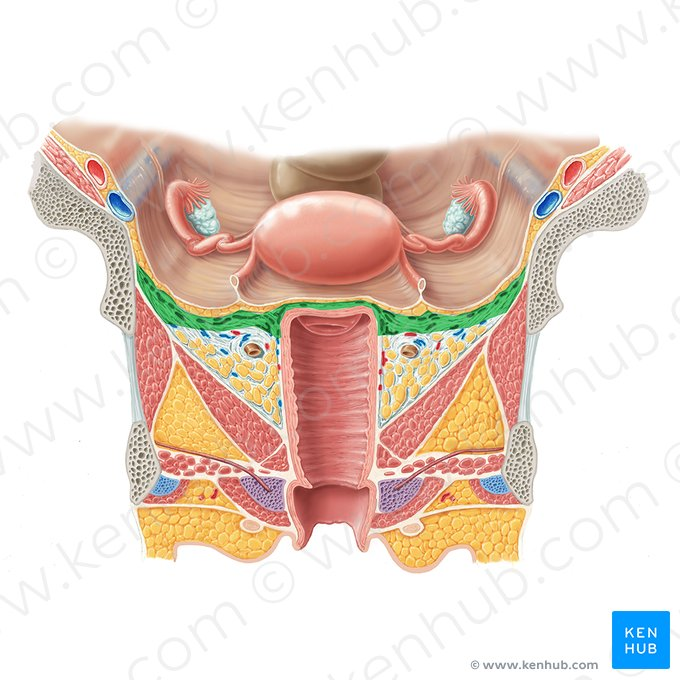
The cardinal ligaments, also known as transverse cervical ligaments or Mackenrodt ligaments, are paired structures that act to support the pelvic organs of the female pelvis. These along with the uterosacral and pubocervical ligaments, provide support to prevent pelvic organ prolapse 1.
The cardinal ligaments originate at the border of the greater sciatic foramen before “inserting” at the lateral aspect of the superior vagina and cervix 1. Along with the uterosacral ligaments, they act to suspend the uterus, cervix, and upper vagina. This can be demonstrated by the cardinal ligaments being in a relatively vertical orientation when an individual is standing upright - resisting the effect of gravity on the structures it supports 1,2.
While referred to as a "ligament", the cardinal ligament is rather a condensation of connective tissue rather than a discrete ligamentous structure in the traditional sense 1,2.
The cardinal ligament consists primarily of vascular, neural, and areolar tissue containing fat with a network of connective tissue separating the different elements 2.
They are comprised of both a vascular component (located cranially) and a neural component (located caudally) 2,4:
The ureter crosses the cardinal ligament at the point where the ligament passes inferior to the uterine vessels 4.
Cardinal ligaments are best demonstrated on coronal T2-weighted MRI imaging. Vessels appear as dot-shaped structures with low T2 signal intensity, while areas of high T2 signal intensity correlate with adipose and connective tissue 3.
Given the position of the uterine vessels and ureter relative to the cardinal ligament, manipulation of the structure during pelvic surgery can result in ureteric or uterine artery injury 4.
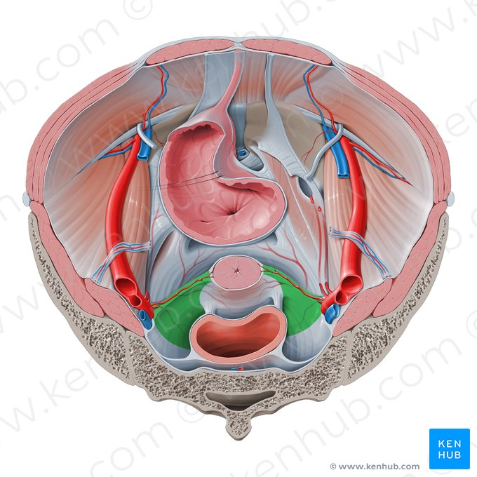
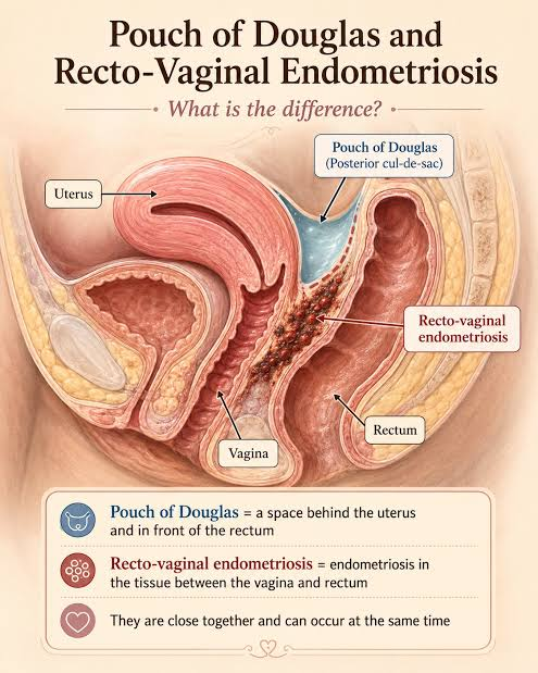
The paired uterosacral ligaments are one of the supporting structures of the uterus. Uterosacral ligaments are not infrequently affected in cases of deep infiltrating endometriosis.
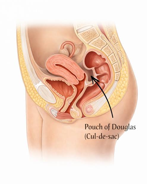
The paired uterosacral ligaments are extraperitoneal structures which extend posteriorly from the uterine cervix to sacrum, forming the lateral boundaries of the rectouterine and rectovaginal spaces.
They are composed of smooth muscle and connective tissue along with splanchnic nerve fibers.
Can be detected on CT as soft tissue attenuation bands, and on MRI as low T1 and T2 signal bands between the cervix and sacrum.