Localize
Medial anterior parahippocampal cortex, medial to the collateral sulcus. Exact boundaries are difficult to delineate and definitions vary.
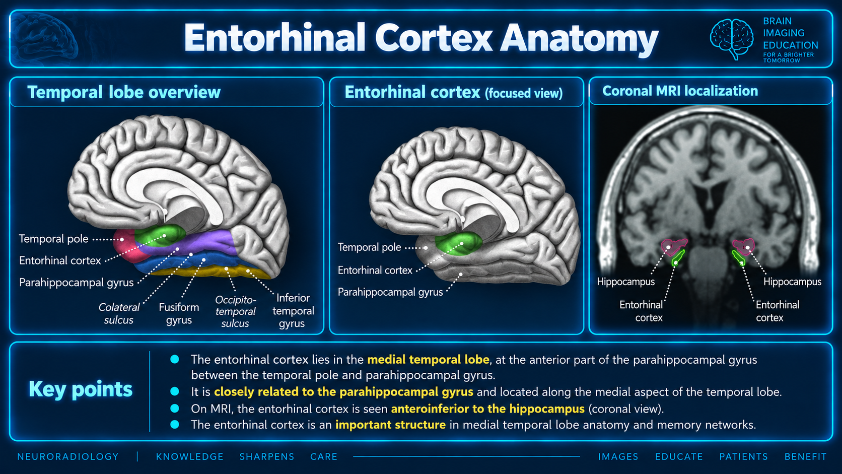
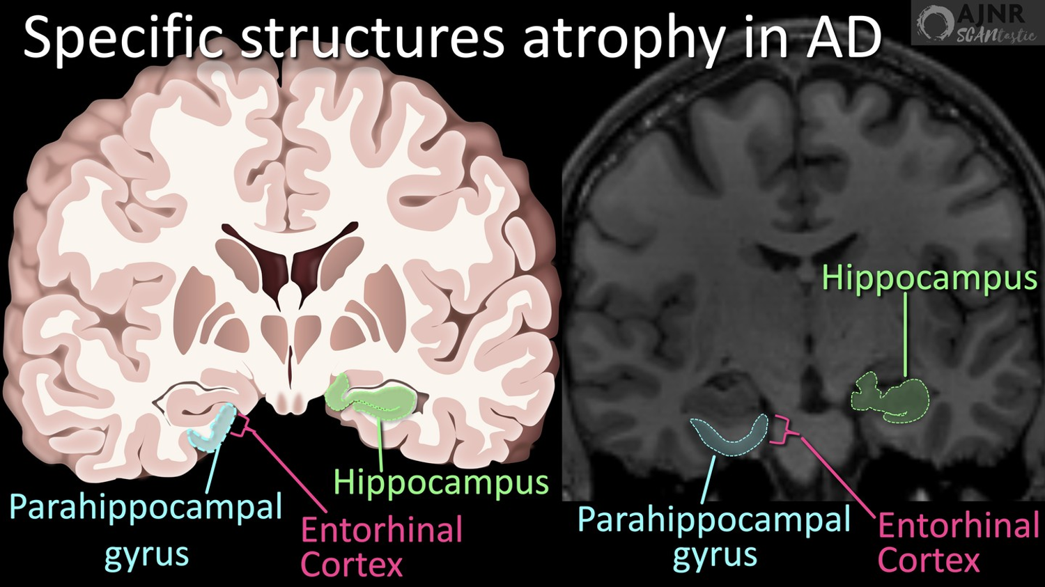
Medial anterior parahippocampal cortex, medial to the collateral sulcus. Exact boundaries are difficult to delineate and definitions vary.
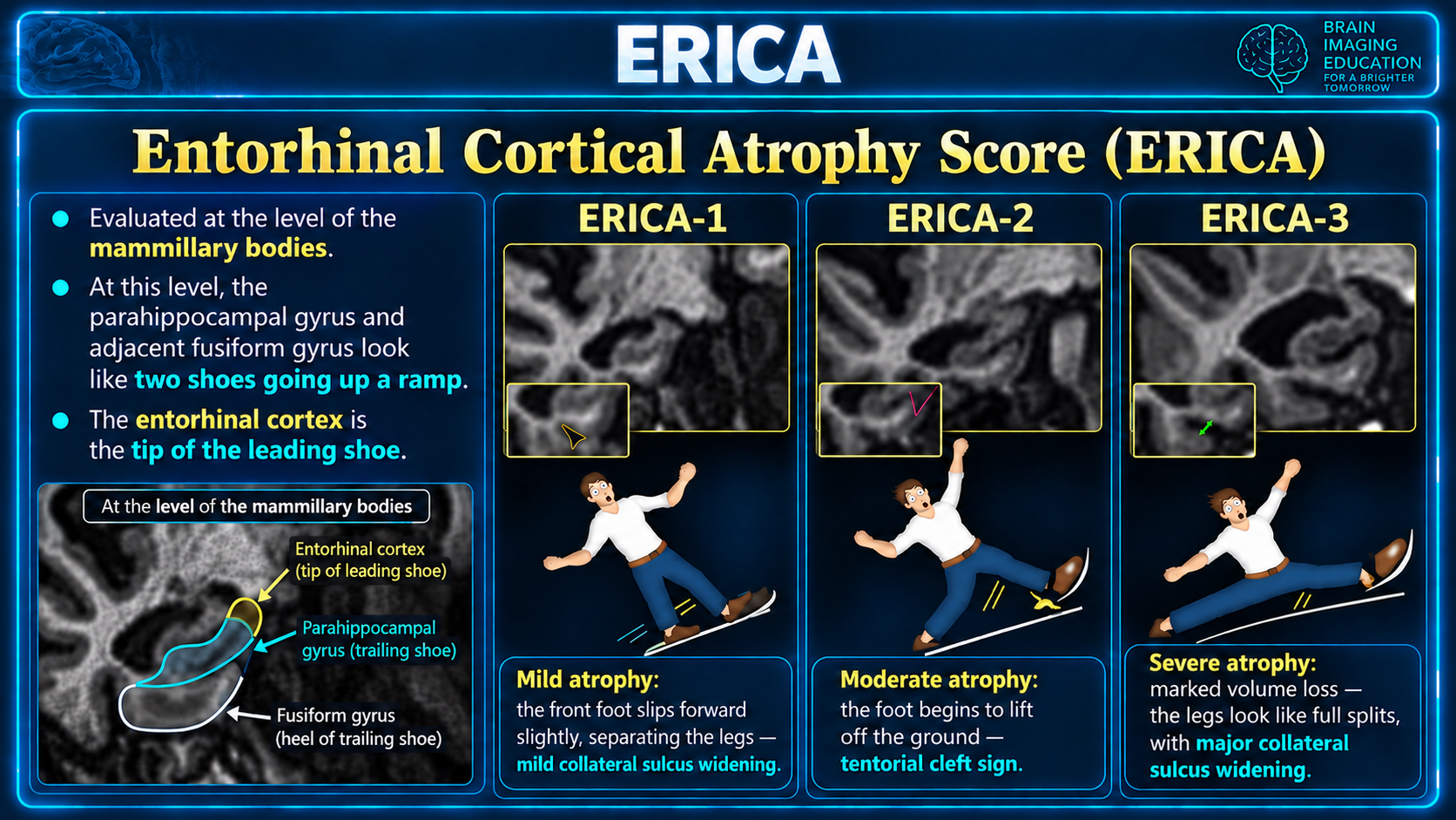
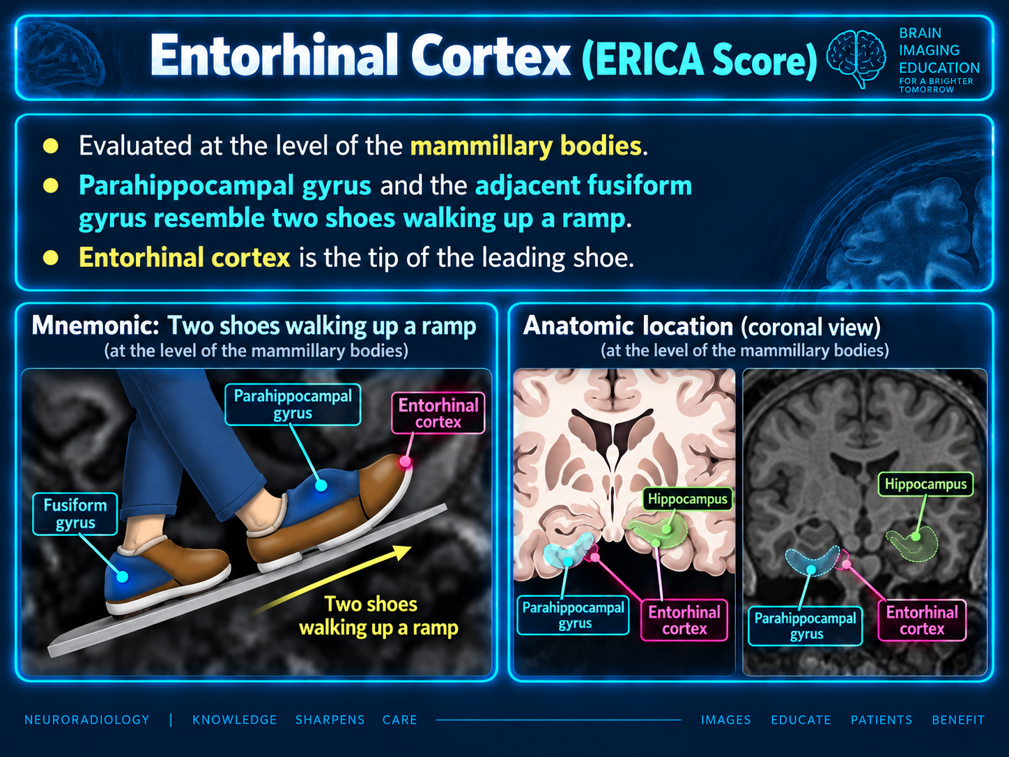
Thin coronal imaging perpendicular to the hippocampal long axis, at the level of the mammillary bodies.
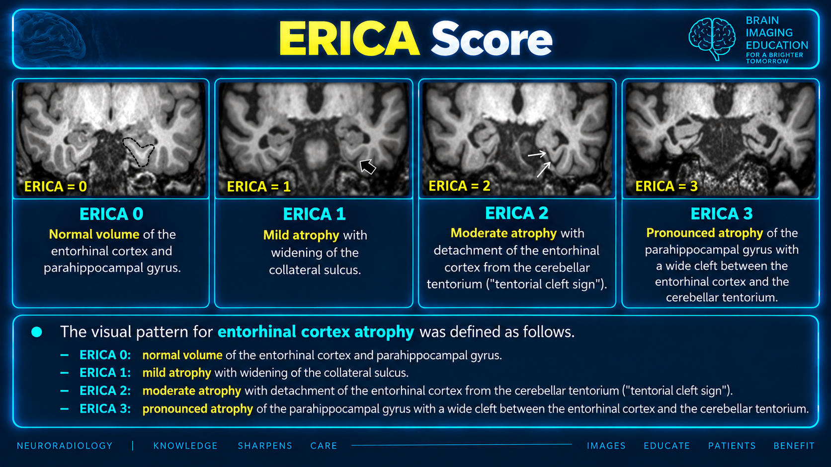
ERICA 0–3. CSF between the entorhinal cortex and tentorium at grades 2 or 3 is the tentorial cleft sign.
It occupies the anterior parahippocampal region, including portions of the parahippocampal gyrus and gyrus ambiens. Useful anatomical relationships are: radiopaedia
For imaging localization, think of the cortex along the medial anterior parahippocampal region, medial to the collateral sulcus—not the hippocampus itself. Its exact boundaries are difficult to delineate anatomically, and definitions vary. radiopaedia
The entorhinal cortex is more than a relay: it integrates information about objects, places, and time into the hippocampal memory system. pubmed.ncbi.nlm.nih
| Component | Principal role |
|---|---|
| Medial entorhinal cortex | Spatial context and navigation; contains grid, head-direction, and border cells. |
| Lateral entorhinal cortex | Information about items and the content of an experience, including relationships to local landmarks. |
These are functional tendencies rather than completely separate systems. sciencedirect
Its superficial layers provide major hippocampal inputs: layer II projects principally to the dentate gyrus and CA2/CA3, while layer III projects to CA1 and the subiculum. The perforant pathway is the major entorhinal afferent system to the hippocampal formation. frontiersin
The transentorhinal and entorhinal regions are among the earliest cortical sites affected by tau pathology in Alzheimer disease. The transentorhinal region is a neighboring transition zone, largely overlapping Brodmann area 35—not simply another name for the entorhinal cortex.
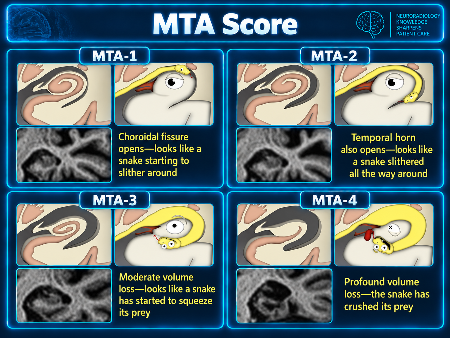
The entorhinal cortical atrophy (ERICA) score has been developed as an alternative to the medial temporal lobe atrophy (MTA) score to help visually identify patients with Alzheimer disease by evaluating the entorhinal cortex for volume loss 1.
diagnostic accuracy = 91%
sensitivity = 83%
specificity = 98%
The ERICA score ranges from 0 to 3. Higher values indicate a higher degree of atrophy and a higher likelihood of Alzheimer disease. In the supplied age-matched case-control study, a score ≥2 had 83% sensitivity and 98% specificity for probable Alzheimer disease rather than subjective cognitive decline.
| Score | Volume | Imaging criterion |
|---|---|---|
| 0 | Normal volume of the entorhinal cortex and parahippocampal gyrus | Normal volume |
| 1 | Mild atrophy of the entorhinal cortex and parahippocampal gyrus | Widening of the collateral sulcus |
| 2 | Moderate atrophy of the entorhinal cortex and parahippocampal gyrus | Elevation of the entorhinal cortex away from the adjacent cerebellar tentorium |
| 3 | Marked atrophy of the entorhinal cortex and parahippocampal gyrus | Wide cleft between the entorhinal cortex and adjacent cerebellar tentorium |
The presence of CSF between the entorhinal cortex and the tentorium (ERICA 2 or 3) has been termed the “tentorial cleft sign”
Entorhinal thinning and atrophy can precede symptomatic cognitive impairment and may occur earlier than conspicuous hippocampal atrophy. However, entorhinal atrophy is not specific for Alzheimer disease; it also occurs in other disorders, including frontotemporal dementia. frontiersin