Typical appearance
Extra-axial, well-circumscribed mass with a broad dural base, usually isointense to grey matter on T1 and T2, with vivid enhancement.
Meningiomas are extra-axial tumors and are the most common tumor of the meninges. They are non-glial neoplasms that originate from the meningocytes or arachnoid cap cells of the meninges and are located anywhere meninges are found, and in some places where only rest cells are presumed to be present.
Although they are usually easily diagnosed and are typically indolent with a low rate of recurrence following surgery, there are 15 subtypes with variable imaging features and, in some instances, more aggressive biological behavior and higher grades.
Typical meningiomas appear as dural-based masses that are isointense to grey matter on both T1- and T2-weighted imaging, and enhance vividly on both MRI and CT. Some of the subtypes can vary dramatically in their imaging appearance.
This article is a general discussion of meningioma, focusing on typical primary intradural meningiomas and the imaging findings of intracranial disease.
Spinal meningioma and primary extradural meningioma, as well as some of the various subtypes, are discussed separately.
Seven supplied teaching slides. Select an image to view the complete slide and its original annotations.
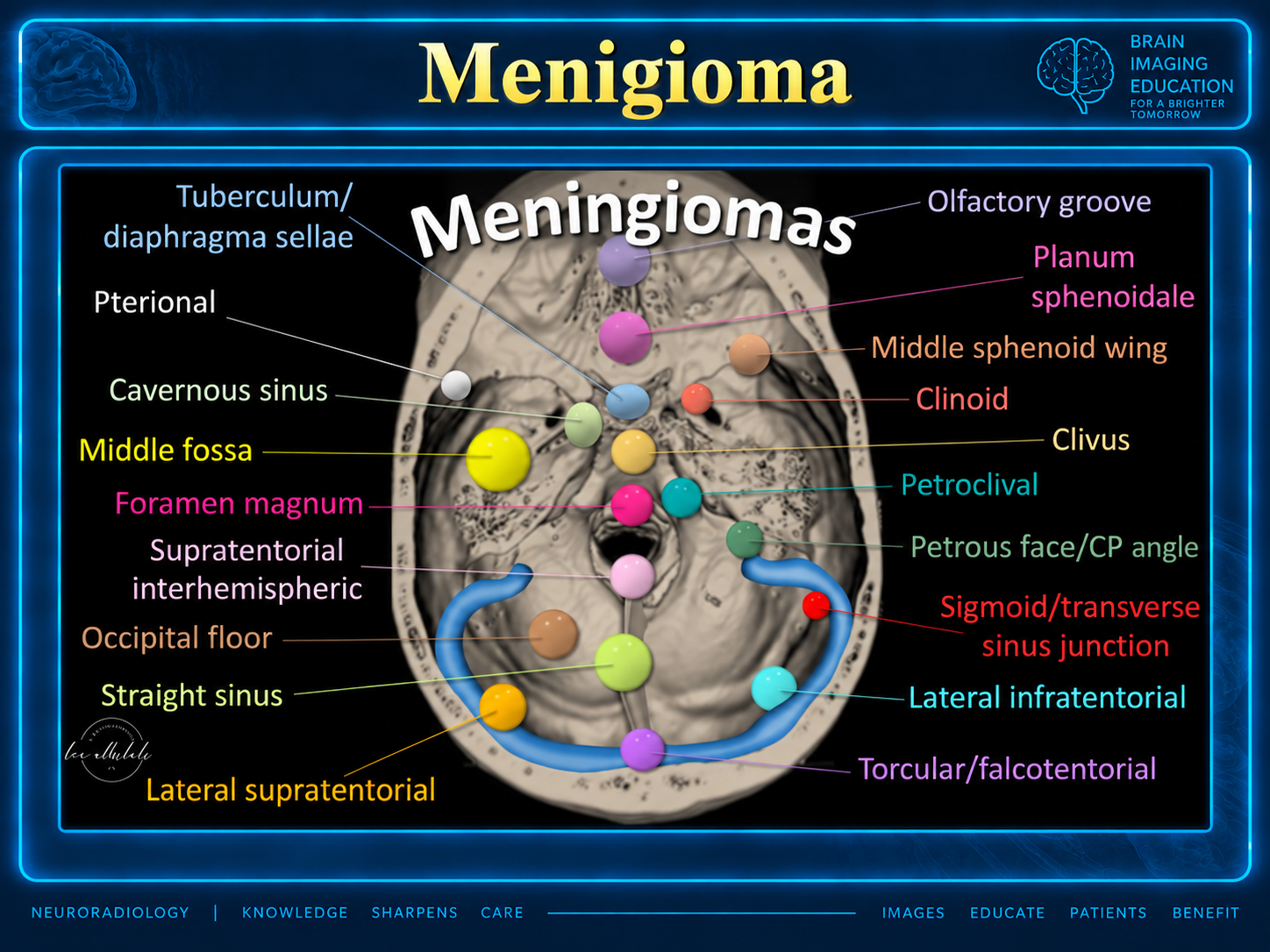
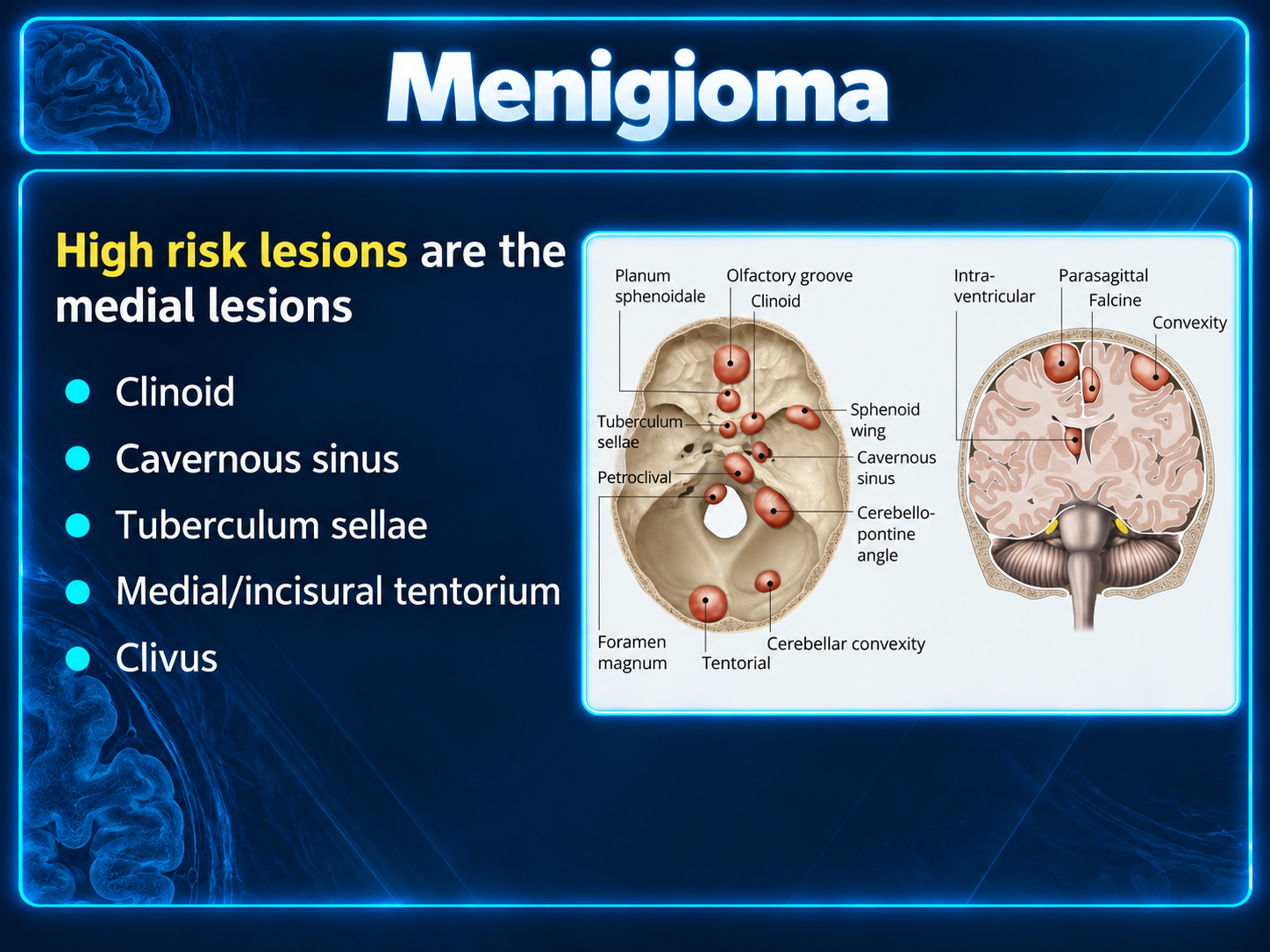
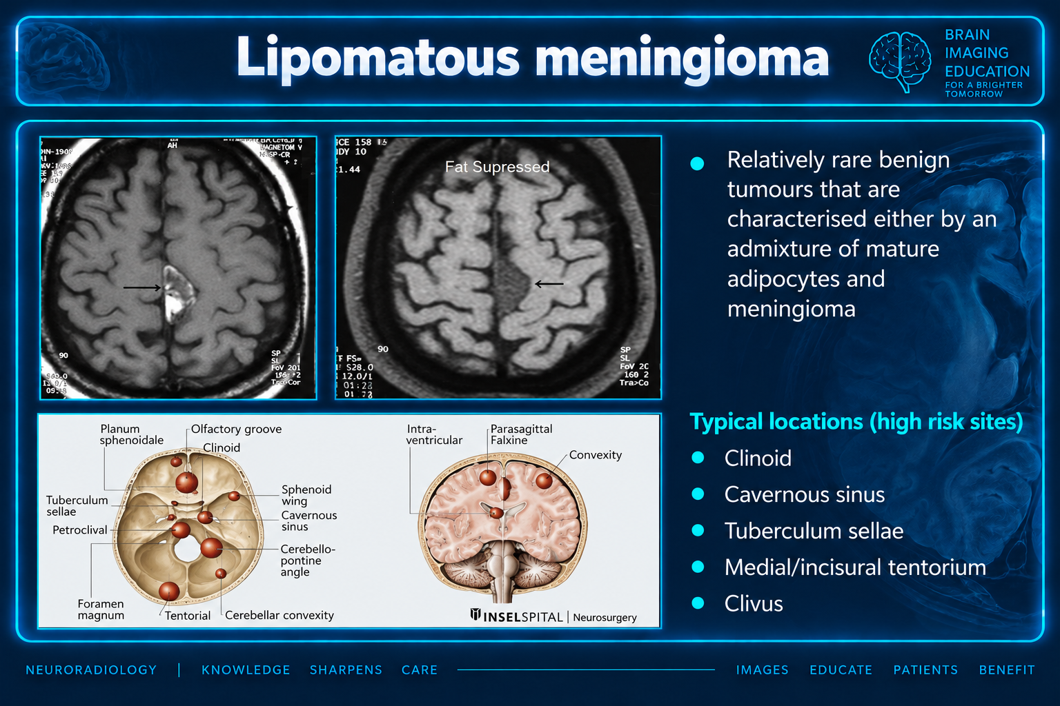
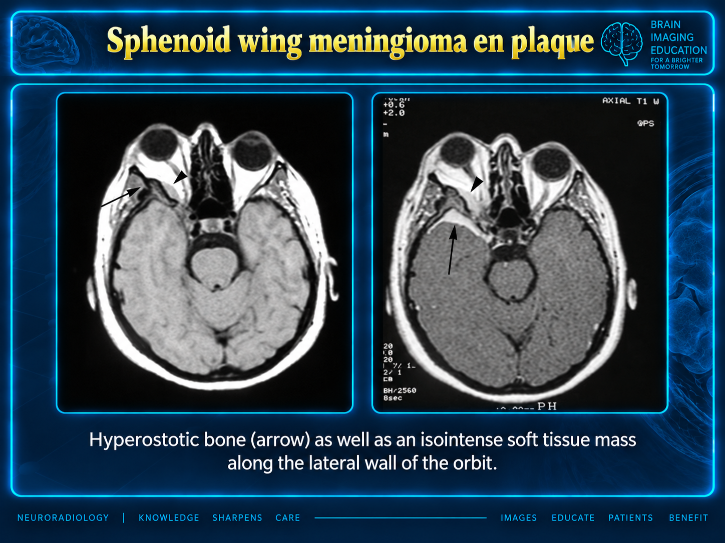
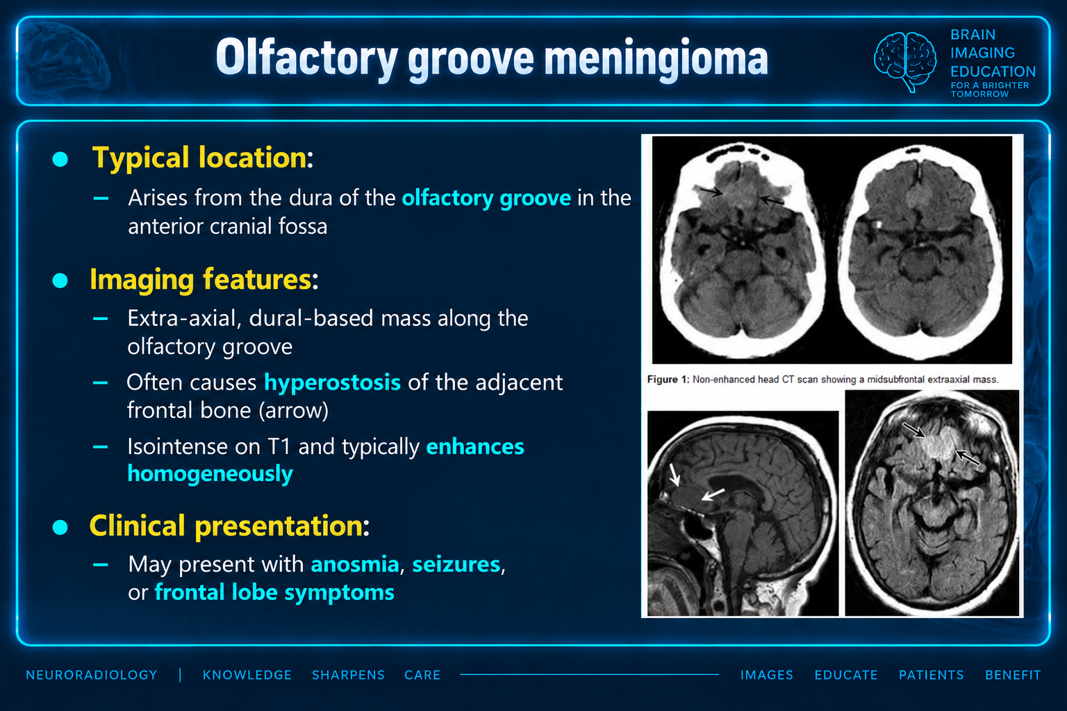
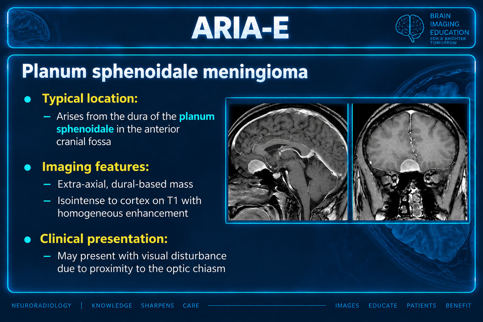
Extra-axial, well-circumscribed mass with a broad dural base, usually isointense to grey matter on T1 and T2, with vivid enhancement.
Contrast-enhanced MRI delineates tumor extent and its relationship to the brain. CT is useful for bony anatomy and calcified or ossified tumors.
A dural tail is not specific. Diffusion restriction is not universally useful for predicting grade. Benign secretory tumors can have disproportionately extensive edema.
The supplied discussion uses the WHO 5th Edition (2021). Histology and molecular features determine grade.
| Criterion | Grade 2 | Grade 3 |
|---|---|---|
| Mitotic figures | 4?19 in 10 consecutive high-power fields (HPF) | ?20 in 10 consecutive HPF |
| Other listed criteria | Overt brain invasion; chordoid or clear cell subtype; or three or more of the five listed histological features | Homozygous CDKN2A/B deletion; TERT promoter mutation; or sarcoma-, carcinoma-, or melanoma-like appearance |
| Histological feature set | Increased cellularity, prominent nucleoli, necrosis, sheet-like growth, small cells with a high nuclear-to-cytoplasmic ratio | See the full grading discussion below |
Papillary or rhabdoid histology alone is no longer sufficient to denote a higher grade in the supplied 2021 classification discussion.
When describing meningiomas, a variety of terms can be used to describe these common tumors more accurately.
Most commonly, they are either classified according to the histological subtype (e.g. rhabdoid or papillary, etc.), location (e.g. skull base, spinal, intraosseous, intraventricular, etc.), or by etiology (e.g. radiation-induced, etc.).
A broad division of meningiomas into primary intradural (which may or may not have a secondary extradural extension) and primary extradural is also used, although the latter is rare, accounting for only 1-2% of cases 25. Ectopic primary meningiomas include tumors residing in the head and neck, orbit, nose, paranasal sinuses, oropharynx, and even more remote sites (e.g. lung).
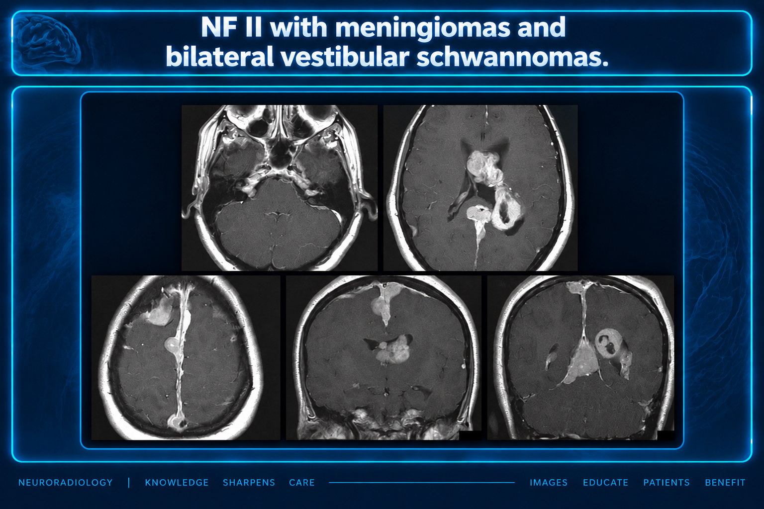
Meningiomas are more common in women, with a ratio of 2:1 intracranially and 4:1 in the spine. Atypical and malignant meningiomas are slightly more common in males. They are uncommon in patients before the age of 40 and should raise suspicion of neurofibromatosis type 2 when found in young patients.
Many small meningiomas are found incidentally and are entirely asymptomatic. Often, they cause concern as they are mistakenly deemed to be the cause of vague symptoms, most frequently headaches. Larger tumors or those with adjacent edema or abutting particularly sensitive structures can present with a variety of symptoms. Most common presentations include 8:
Occasionally, transosseous, or intraosseous involvement with prominent hyperostosis may result in local mass effect (e.g. proptosis).
Although dural venous sinus invasion and occlusion do occur, it usually occurs very gradually. Therefore, most cases of venous invasion are asymptomatic as collateral veins have had time to enlarge.
Meningiomas are thought to arise from meningocytes or arachnoid cap cells, which themselves arise from pluripotent mesenchymal progenitor cells, thereby accounting for the unusual location of primary extradural tumors 17,18.
Although the majority of tumors are sporadic, they are also seen in the setting of previous cranial irradiation and of course in patients with neurofibromatosis type 2 (Merlin gene on Chromosome 22). Additionally, meningiomas demonstrate estrogen and progesterone sensitivity and may grow during pregnancy.
In the 5th Edition (2021) WHO classification of CNS tumors a total of 15 subtypes of meningioma are recognized.
Unlike other tumors, the term "atypical" and "anaplastic"/"malignant" have been retained as histological subtypes with grade 2 and grade 3 tumors, respectively 31.
Otherwise, meningiomas are graded from grade 1 to 3 based on histological features (e.g. mitotic index), some histological subtypes (e.g. chordoid and clear cell meningiomas), and molecular features (see below) 7,11,21,31.
An important change in the 5th Edition (2021) WHO classification of CNS tumors is that the identification of some histological subtypes (e.g. papillary meningiomas and rhabdoid meningiomas) is no longer sufficient to denote a higher grade. However, they often have other histological features that will confer a higher grade 31.
Brain invasion as a stand-alone feature remains controversial. In prior editions of the WHO classification (e.g. 2016), if a meningioma (regardless of histology) demonstrated any brain invasion, it was designated as grade 2, as it was believed to indicate a poorer prognosis and a higher likelihood of recurrence 7. In many instances, growth is actually along perivascular spaces rather than truly into the brain parenchyma. The 5th Edition has backed away from this dogmatic recommendation recognizing the difficulty in assessing this in some instances 31,32. Nonetheless, overt brain invasion remains sufficient to denote a grade 2 tumor.
The cut surface reflects the various histologies encountered, ranging from very soft to extremely firm in fibrous or calcified tumors. They are usually light tan in color, although again this will depend on histological subtypes.
Increasingly, molecular markers are being incorporated into the diagnosis and grading of meningioma subtypes 31.
Meningiomas are best imaged with contrast-enhanced MRI, as this most accurately delineates the tumor, the presence of intra- and trans-osseous extension, and its relationship to the underlying brain. CT, however, is useful if bony anatomy is required (e.g. at the base of the skull), when patients cannot have an MRI, and especially when the meningioma is entirely ossified/calcified (see burnt-out meningioma).
Note that, in addition to histological variants, many of which have less typical imaging appearances, a number of 'special examples' of meningiomas are best discussed separately. These include:
The remainder of this section focuses on more typical imaging appearances of run-of-the-mill meningiomas.
As is the case with most other intracranial pathologies, MRI is the investigation of choice for the diagnosis and characterization of meningiomas. When appearance and location are typical, the diagnosis can be made with a very high degree of certainty. In some instances, however, the appearances are atypical and careful interpretation is needed to make a correct preoperative diagnosis.
Meningiomas typically appear as extra-axial masses with a broad dural base. They are usually homogeneous and well-circumscribed, although many variants are encountered. It seems that the signal intensity of meningiomas on T2-weighted images correlates with the histological subtypes 27.
DWI/ADC: grade 2 and 3 tumors may show greater than expected restricted diffusion, although this is not universally useful in prospectively predicting histological grade 14,15
this may aid in preoperative planning for meningioma resection by allowing planning of a safer access route that would result in less residual functional iatrogenic deficits 28
CSF cleft sign, which is not specific for meningioma, but helps establish the mass to be extra-axial; loss of this can be seen in grade II and grade III, which may suggest brain parenchyma invasion
More than half of the meningiomas demonstrate a variable amount of vasogenic edema in adjacent brain parenchyma 22. Correlations between age, gender, tumor size, rapid growth, location (convexity and parasagittal > elsewhere), histologic type, and invasion in malignant meningiomas have been suggested in the literature but not yet confirmed. Although in general, the presence of severe adjacent edema is considered more compatible with aggressive meningiomas, in some histologically benign types such as secretory type, edema can be disproportionately larger than the small tumor size.
The underlying mechanism is most likely multifactorial however it has been shown that there is a strong association between the presence and severity of the peritumoral vasogenic edema (i.e. edema index) and expression of the vascular endothelial growth factor (VEGF) or expression of CEA and CK 16,23.
Catheter angiography is rarely of diagnostic use but rather is performed for preoperative embolization to reduce intraoperative blood loss and alleviate resection of a tumor. This is especially useful for skull base tumors, or those thought to be particularly vascular (e.g. microcystic variants or those with very large vessels). Particles are favored typically 7-9 days prior to surgery although they are not free of complications, particularly one study showed a high prevalence of complications associated with particles smaller than 150 μm, so risks and benefits should be thoroughly assessed 24.
Meningiomas can have a dual blood supply. The majority of tumors are predominantly supplied by meningeal vessels; these are responsible for the sunburst or spoke-wheel pattern observed on MRI/DSA. Some tumors also have a significant pial supply to the periphery of a tumor.
A well-known angiographic sign of meningiomas is the mother-in-law sign, in which the tumor contrast blush "comes early, stays late, and is very dense".
Treatment is usually with surgical excision. If only incomplete resection is possible (especially at the base of the skull) or if surgery is deemed high-risk, then other treatments may be used as well or instead:
No systemic chemotherapeutic or other therapy has been proven efficacious, although some mTOR inhibitors and antiangiogenic treatments show promise 34.
The Simpson grade correlates the degree of surgical resection completeness with symptomatic recurrence rate, which also varies with grade and length of follow-up 8,20. Metastatic disease is rare but has been reported 8.
The 2023 NCCN guidelines for surgically treated Grade 1 and 2 meningiomas recommend a follow-up brain MRI at 3, 6, and 12 months, then every 6-12 months for 5 years, and subsequently every 1-3 years. For WHO Grade 3 meningiomas, recommendations include a brain MRI every 2-4 months for the first 3 years, followed by every 3-6 months thereafter 36.
The term "meningioma" was first introduced by Harvey Cushing (1869-1939), a renowned American neurosurgeon, in 1922 9,22.
The differential diagnosis generally includes other dural masses as well as some location-specific entities.
Source limitations: This reference preserves the supplied article?s WHO 2021 classification and NCCN 2023 follow-up statements; it does not establish their current guideline status. Numbered citations are retained as supplied, but no corresponding bibliography was provided. The ?ARIA-E? heading on figure 6 is preserved from the uploaded slide; the user caption and slide body identify a planum sphenoidale meningioma. The lipomatous slide lists general meningioma high-risk sites without establishing subtype-specific location frequencies.