At a Glance
Why it mattersThe AChA is a small ICA branch supplying a compact territory rich in motor, sensory, and visual pathways.
Origin
- Arises from the posterior wall of the supraclinoid internal carotid artery.
- Usually 2–5 mm distal to the posterior communicating artery.
- Usually 2–5 mm proximal to the ICA termination.
- Approximately 1 mm in diameter.
Key relationships
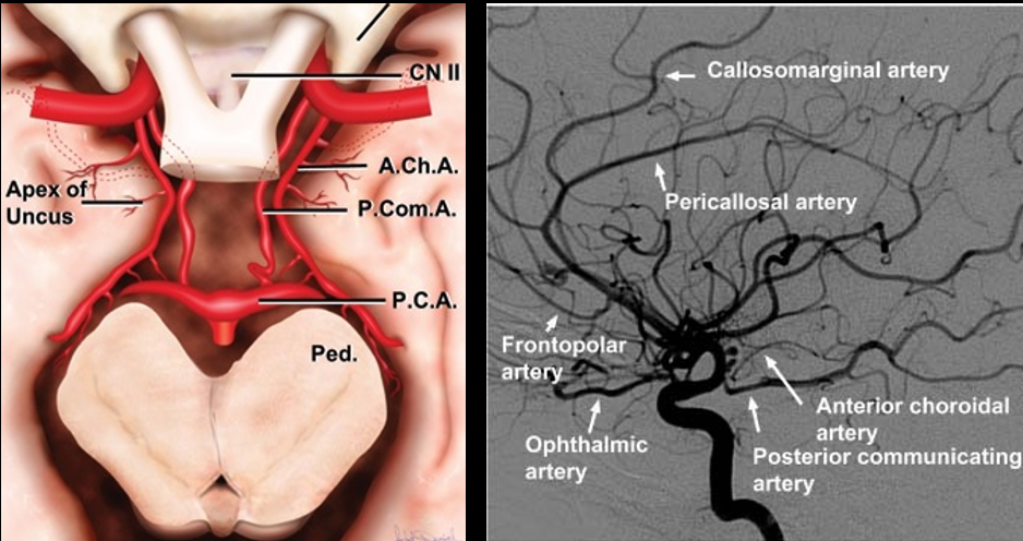
The artery follows the optic tract around the cerebral peduncle toward the lateral geniculate body, then enters the choroidal fissure at the plexal point.
Course
Perioptic course
From its ICA origin, the AChA begins lateral to the optic tract, curves to its inferomedial surface, then turns laterally to follow the tract.
Posterolateral course
- Passes above the uncus.
- Curves around the lateral cerebral peduncle.
- Approaches the lateral geniculate body.
- Traverses the carotid, crural, and ambient cisterns.
Plexal point
The vessel enters the choroidal fissure at the plexal point. On lateral angiography this is the center of a checkmark-like descent followed by sharp posterior ascent.
Segments
Cisternal segment
- Origin to the choroidal fissure.
- Approximately 2.5 cm long; reported range 1.5–3.5 cm.
- Gives penetrating branches to deep brain structures.
Intraventricular segment
- Begins after entry through the choroidal fissure.
- Supplies choroid plexus in the anterior temporal horn.
Pathway shorthandSupraclinoid ICA → cisternal AChA → plexal point → choroidal fissure → temporal-horn choroid plexus
Deep Vascular Territory
Motor and sensory pathways
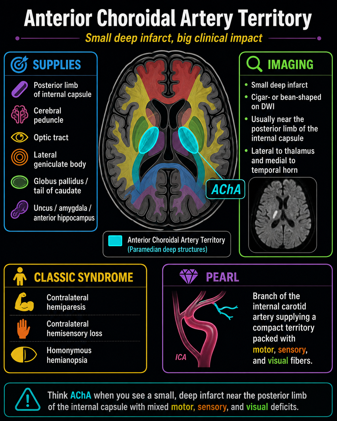
- Posterior limb and retrolenticular internal capsule.
- Lateral cerebral peduncle.
- Globus pallidus internus and tail of caudate.
Visual pathway
- Optic tract.
- Lateral geniculate body and lateral thalamus.
- Retrolenticular optic radiations.
Mesial temporal structures
- Uncus and amygdala.
- Head of hippocampus.
Anterior territory overlaps variably with lenticulostriate arterial supply.
Variants
Origin variants
- Intracerebral carotid bifurcation.
- Middle cerebral artery.
- Posterior communicating artery.
Duplication
A duplicated AChA is reported in approximately 5% of cases.
Hyperplastic AChA
A hyperplastic AChA may retain an expanded embryologic territory and supply regions normally served by the posterior cerebral artery.
Interpretive cautionTerritorial distribution is variable; an infarct pattern may extend beyond the classic compact deep territory.
Imaging & Clinical Pearl
Angiography / MRA
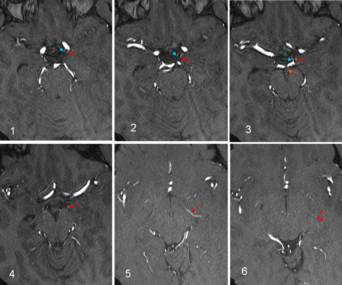
- Identify the small posterior ICA origin between PCOM and carotid termination.
- Trace the vessel alongside the optic tract to the plexal point.
- Distinguish it from PCOM and small perforators.
Infarct pattern
AChA infarcts are typically small and deep, often involving the posterior limb of the internal capsule, lateral to the thalamus and medial to the temporal horn.
Classic syndrome
- Contralateral hemiparesis.
- Contralateral hemisensory loss.
- Contralateral homonymous hemianopia.
The full triad is not required because collateral supply and territorial variation are common.