At a Glance
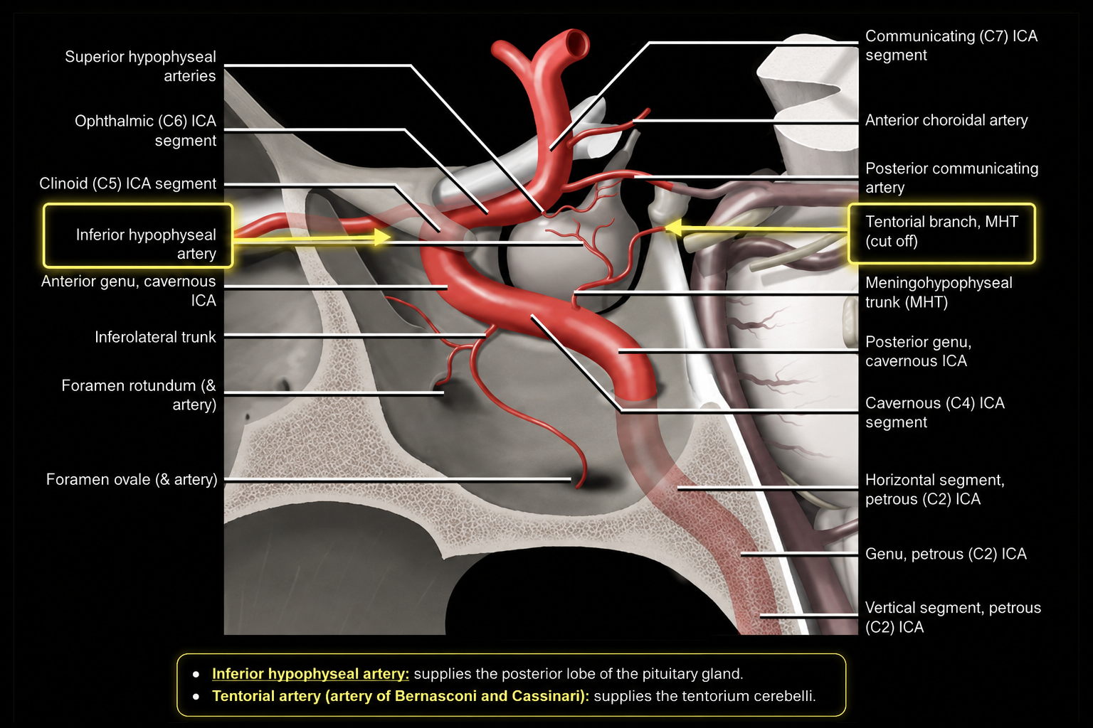
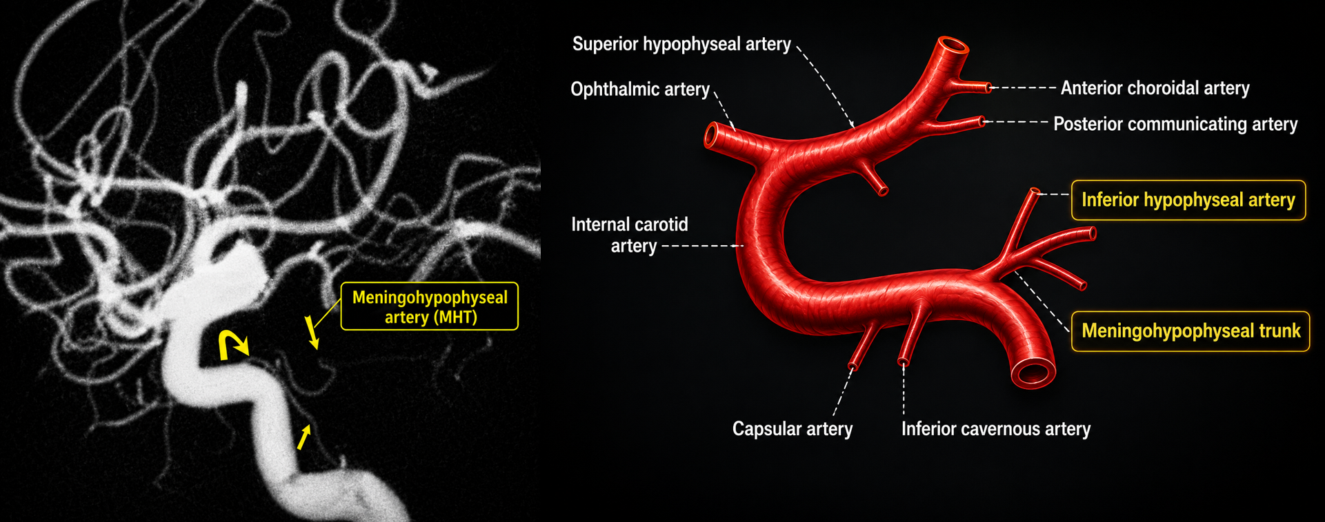
DefinitionThe meningohypophyseal trunk (MHT), or posterior trunk, is a small but important branch of the C4 cavernous internal carotid artery.
Visibility
- Almost always identifiable at anatomic dissection.
- Seen on only a minority of routine cerebral angiograms because of its small caliber.
- More conspicuous with selective injection or pathologic enlargement.
Primary territory
Posterior pituitary gland, tentorium cerebelli, clival dura, and adjacent skull-base meninges.
Origin & Relationships
Cavernous ICA
The MHT typically arises from the posterior aspect of the posterior genu or horizontal cavernous ICA, directed posteriorly toward the dorsum sellae and tentorium.
Different from the ILT
The inferolateral trunk usually arises more laterally from the horizontal cavernous ICA and supplies the cavernous sinus, cranial nerves, and foraminal territories.
Angiographic profile
- Tiny posteriorly directed trunk.
- Best appreciated on selective ICA angiography.
- May share origins or branch patterns that vary considerably.
Hypophyseal Branch
Inferior hypophyseal artery
- Supplies the posterior pituitary and inferior sellar region.
- Contributes to the inferior hypophyseal arterial circle.
- Often forms an anastomotic network with the contralateral artery.
DistinctionInferior hypophyseal arteries arise from the cavernous ICA/MHT system; superior hypophyseal arteries usually arise from the ophthalmic or communicating ICA segments.
Imaging relevance
Enlargement may accompany sellar, parasellar, cavernous sinus, or dural vascular pathology.
Tentorial Branches
Marginal tentorial artery
The Bernasconi–Cassinari artery courses upward along the medial free edge of the tentorium cerebelli.
Lateral tentorial artery
Courses downward along the lateral attached edge of the tentorium.
Radiology relevance
- Tentorial branches may enlarge as feeders to tentorial or cavernous sinus dural arteriovenous fistulas.
- They may supply tentorial and skull-base meningiomas.
- Direction and dural course help distinguish the marginal and lateral branches.
Clival & Meningeal Branches
Dorsal meningeal artery
- Supplies dura over the clivus and dorsum sellae.
- Courses near Dorello canal and the abducens nerve.
- Not synonymous with the posterior meningeal artery.
Clival branches
Small branches descend or course posteriorly to supply the meninges covering the clivus and may anastomose with ascending pharyngeal and contralateral cavernous ICA branches.
Safety implicationPotential cranial-nerve and ICA anastomoses demand careful angiographic mapping before embolization.
Angiography & Intervention
When it becomes visible
- Cavernous sinus or tentorial DAVF.
- Skull-base or tentorial meningioma.
- Hypervascular sellar and parasellar lesions.
Before embolization
- Define the precise MHT origin and all distal branches.
- Search for dangerous anastomoses to ophthalmic, ascending pharyngeal, middle meningeal, and contralateral ICA territories.
- Respect proximity to cranial nerves in the cavernous sinus and Dorello canal.
Selective catheterization can be technically demanding because the trunk is small and arises from the cavernous ICA.