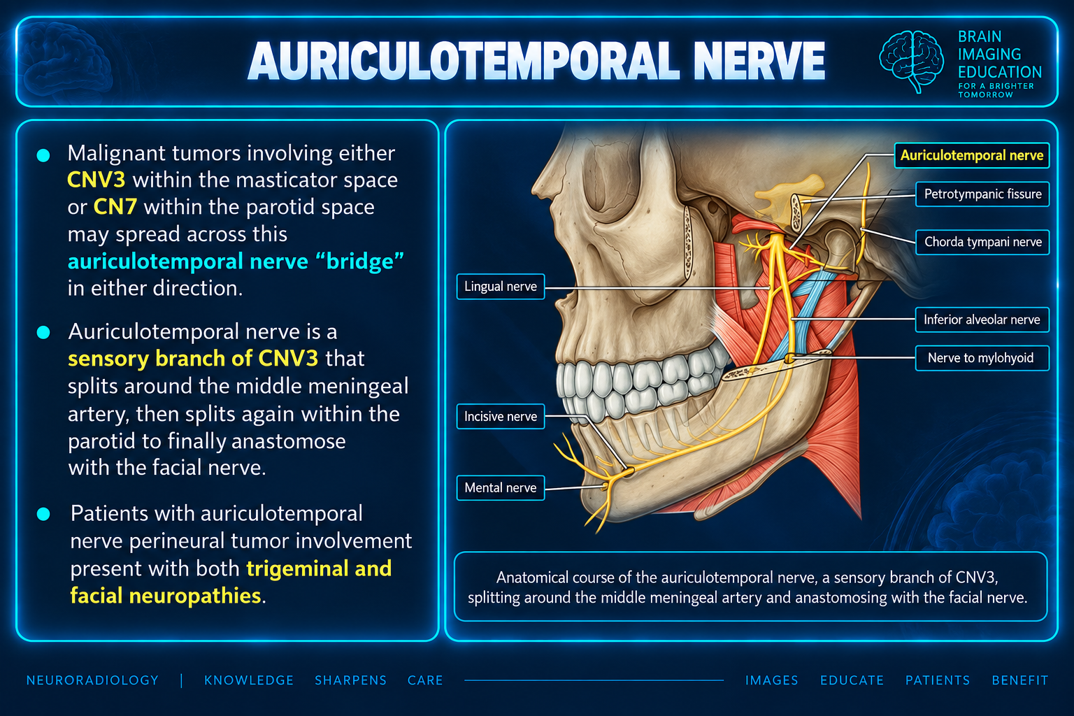
The auriculotemporal nerve is a sensory branch of the posterior division of the mandibular division (V3) of the trigeminal nerve. Communicating rami within the parotid gland connect it with the facial nerve.
Gross anatomy
Origin and course
- Arises posteriorly from the posterior division of the mandibular nerve.
- Two separate roots encircle the middle meningeal artery as the artery ascends to enter the foramen spinosum.
- The roots rejoin and pass medial to the neck of the mandible, deep to the sphenomandibular ligament.
- The nerve ascends between the temporomandibular joint and external auditory meatus, giving off small articular branches to the joint.
- At the upper border of the parotid gland, it supplies the gland sheath and terminates as auricular and temporal cutaneous branches 1–3.
Parotid fibers and connections
The supplied text describes sympathetic fibers received from the middle meningeal artery and secretomotor parasympathetic fibers (derived from the otic ganglion) from the lesser petrosal nerve; both supply the parotid gland 1–3.
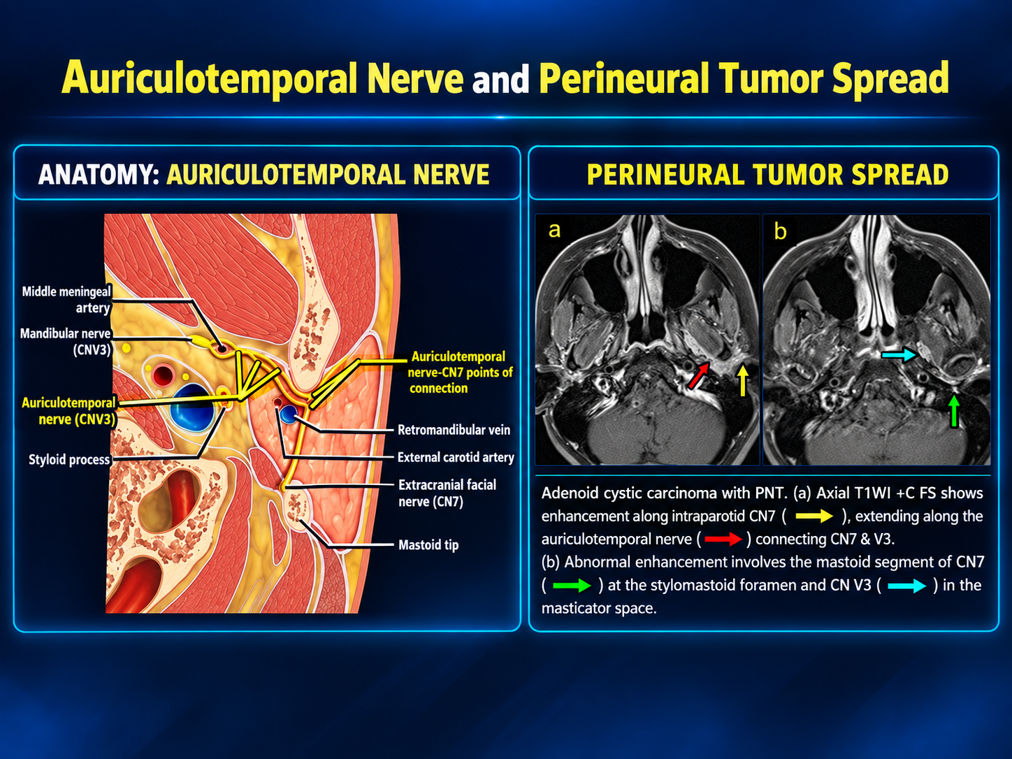
Communicating rami link the auriculotemporal and facial nerves within the parotid gland 4.
Radiographic features
The auriculotemporal nerve is usually not seen on CT or MRI.
Expected location
Its expected course lies parallel to the posterior border of the mandible, just superior to the terminal bifurcation of the external carotid artery into the maxillary and superficial temporal arteries 4.
Related pathology
Perineural spread of head and neck malignancies, including cutaneous squamous cell carcinoma and salivary gland adenoid cystic carcinoma, can occur along the auriculotemporal nerve.
This route permits potential spread from the mandibular division (V3) of the trigeminal nerve to the facial nerve, or vice versa.
Teaching graphic context
Figure 1 describes the auriculotemporal nerve as a “bridge” between CNV3 in the masticator space and CN7 in the parotid space. It states that patients with perineural tumor involvement present with both trigeminal and facial neuropathies.
Source limitations
The supplied prose includes reference markers 1–3 and 4, but no bibliography was provided. These markers are retained without invented citations.
The first graphic’s statement that patients present with both trigeminal and facial neuropathies is preserved as a teaching-slide claim; the supplied prose does not establish its frequency or whether it applies to every patient. The original graphics, labels, arrows, and embedded captions are preserved unchanged.