Buccal Space
The buccal space, also known as the buccinator space, is one of the seven suprahyoid deep compartments of the head and neck.
Gross anatomy
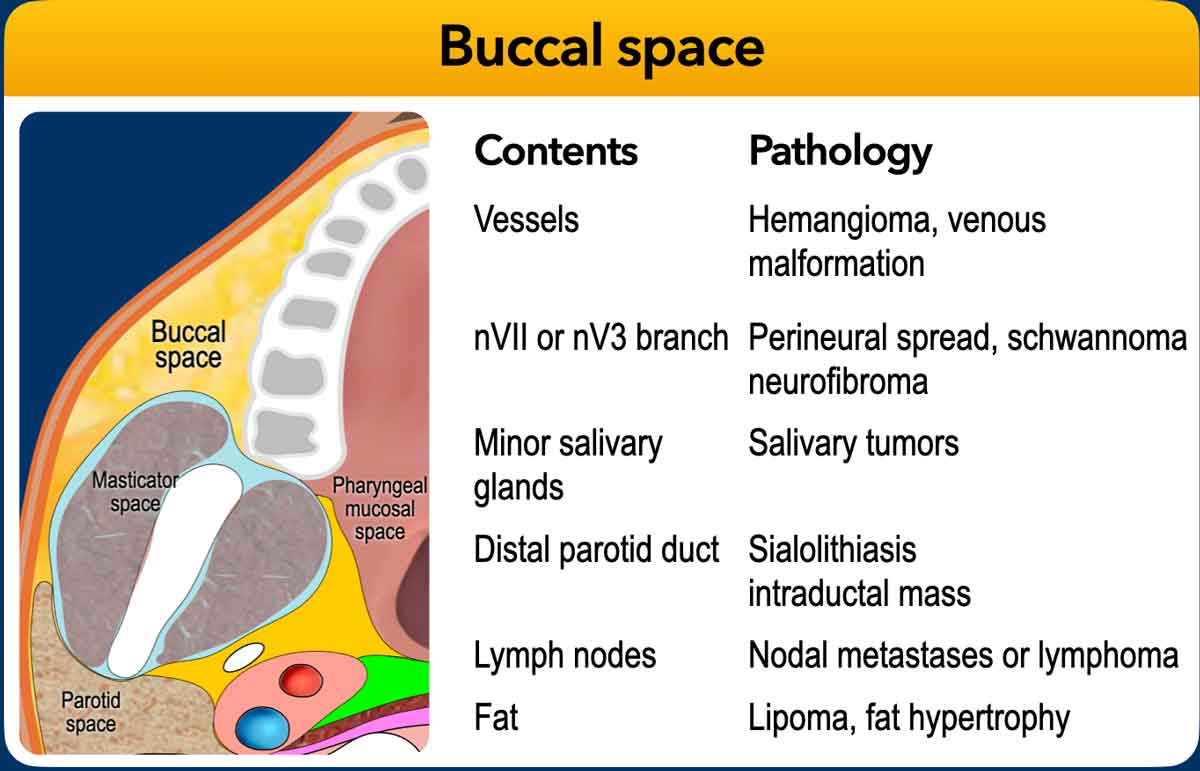
The buccal spaces are paired fat-containing spaces on each side of the face, forming the cheeks.
Each space is enveloped by the superficial (investing) layer of the deep cervical fascia.
The supplied text describes it as a small potential space with limited contents, located between the buccinator and platysma muscles.
Contents
- Fat: cheek padding
- Parotid duct
- Accessory parotid gland in 20% of people; may cause facial asymmetry and is readily seen on MRI
- Facial and buccal arteries and corresponding veins
- Facial nerve (CN VII): buccal branch
- Trigeminal nerve (CN V): buccal nerve of the mandibular division
Related pathology
- Parotid duct calculi
- Odontogenic infection
- Minor salivary gland tumors
- Vascular lesions, such as hemangiomas
These entities are listed in the supplied text. The first reference image includes additional content–pathology associations.
Boundaries and Relations
| Aspect | Boundary or relation |
|---|
| Anterior | Orbicularis oris muscle and the angle of the mouth |
|---|
| Posterior | Masseter muscle, mandible, medial pterygoid and lateral pterygoid muscles |
|---|
| Superior | Zygomatic process of the maxilla and zygomaticus muscles |
|---|
| Inferior | Depressor anguli oris muscle and the deep fascia attaching to the mandible |
|---|
| Medial (deep) | Buccinator muscle |
|---|
| Lateral (superficial) | Platysma muscle and subcutaneous tissues with the skin |
|---|
Communications
Buccal space infection can spread to or from the teeth.
There is no real boundary between the buccal space and the submandibular space inferiorly. There is also potential communication with the pterygomandibular region, infratemporal space, and parapharyngeal space posteriorly.
Source Scope
The supplied text describes a fascial envelope and an inferior boundary, while also stating that there is no real boundary with the submandibular space inferiorly. Both descriptions are retained; the source does not explain their reconciliation.
The teaching slide additionally lists minor salivary glands and lymph nodes, with pathology associations beyond the prose summary. These remain visible in the original figure. No detailed diagnostic criteria or management guidance were supplied.
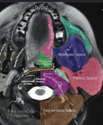
The second figure is a small annotated source image; enlargement preserves its original resolution, so some labels may remain difficult to read.