Extent
A roughly cylindrical space extending from the skull base to the aortic arch.
The carotid space, the suprahyoid portion of which is also known as the poststyloid parapharyngeal space, is a deep compartment of the head and neck bound by the carotid sheath.
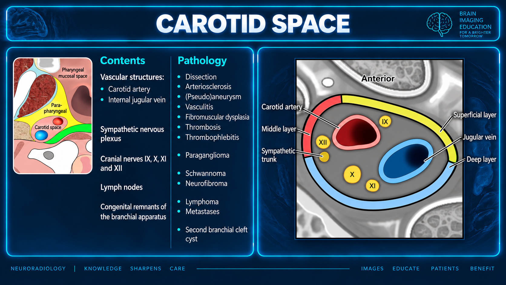
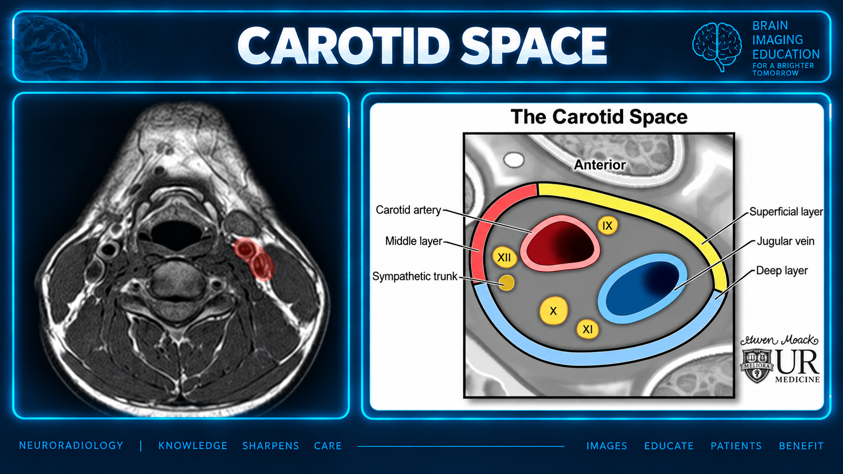
A roughly cylindrical space extending from the skull base to the aortic arch.
Circumscribed by the carotid sheath, with contributions from all three layers of the deep cervical fascia.
A mass originating in the carotid space displaces parapharyngeal space fat anteriorly.
The "carotid space" terminology was introduced by some radiologists to facilitate differential diagnosis and is used in this article [1]. However, in much of the surgical and anatomical literature, the carotid space within the suprahyoid neck is considered part of the parapharyngeal space [2,3]. Under this nomenclature, the parapharyngeal space is divided into prestyloid and poststyloid (retrostyloid) compartments by the tensor-vascular-styloid fascia [9]. The poststyloid parapharyngeal space consists of or includes the carotid space or sheath around the cervical internal carotid artery.
The carotid space is a roughly cylindrical space that extends from the skull base through to the aortic arch. Functionally, it is often considered in terms of its contents within the suprahyoid neck, and the infrahyoid neck [11]. It may further be divided into nasopharyngeal, oropharyngeal, cervical, and mediastinal segments [11].
It is circumscribed by the carotid sheath, which has contributions from all three layers of the deep cervical fascia [4]. Above the carotid bifurcation, the contribution of the middle layer of cervical fascia can be inconsistent 9, and the sheath is interrupted [10].
The carotid artery bifurcation occurs near the level of the hyoid bone [1].
| Region | Vessels | Nerves |
|---|---|---|
| Suprahyoid neck [11] | Internal carotid artery; internal jugular vein | In the nasopharyngeal segment: glossopharyngeal nerve (CN IX), anterior to vessels; vagus nerve (CN X), posterior to vessels in the posterior notch; accessory nerve (CN XI); hypoglossal nerve (CN XII). The vagus is the only nerve present from the oropharyngeal segment of the carotid sheath inferiorly. |
| Infrahyoid neck [11] | Common carotid artery; internal jugular vein | Vagus nerve (CN X); ansa cervicalis, embedded in the anterior wall of the sheath. |
Throughout: deep cervical lymph node chain (levels II, III, and IV).
| Direction | Suprahyoid carotid space | Infrahyoid carotid space |
|---|---|---|
| Anteriorly | Masticator space; parapharyngeal space | Anterior cervical space |
| Laterally | Parotid space | Posterior cervical space |
| Posteriorly | Perivertebral space | Perivertebral space |
| Medially | Retropharyngeal space | Retropharyngeal and visceral space |
The suprahyoid portion of the carotid space is synonymous with the poststyloid compartment of the parapharyngeal space.
Superior margin: lower border of the jugular foramen. Inferior margin: aortic arch.
The carotid sheath is made from the various regional fascia, including contributions from all three layers of the deep cervical fascia [2]:
| Margin | Fascial contribution |
|---|---|
| Lateral | Fascia of the sternocleidomastoid muscle (superficial layer of the deep cervical fascia) |
| Anterior | Stylopharyngeal aponeurosis or tensor-vascular-styloid fascia (middle layer of the deep cervical fascia) |
| Medial | Cloison sagittale (either part of the deep or middle layer of the deep cervical fascia) |
| Posterior | Prevertebral fascia and/or alar fascia (deep layer of the deep cervical fascia) |
Ultrasound is good for identifying vascular structures in the lateral neck. The origin of the internal and external carotid arteries is the main point of sonographic interest as the carotid space superior to this cannot be visualized due to the mastoid process.
The internal carotid is usually posterolateral to the external carotid however in 5-10% of the population the internal carotid lies medial or anterior or the two vessels may lie within the same frontal plane [5].
The external carotid can be identified from the internal carotid as:
it branches within the neck; the first immediate branch is the superior thyroid artery
Doppler tracing of vessel flow within the internal carotid is typical of low peripheral resistance (high diastolic peak), whereas the external carotid has high-resistance flow (lower diastolic peak, systolic peak higher than that of the internal carotid) [5]
often modality of choice when evaluating for pathology within the carotid space because CT is able to identify any benign or malignant tumors within the carotid space and allows for better visualization of any associated bone involvement in comparison to MRI
best identifies vascular lesions on CT angiography [6]
useful for identifying between the different types of benign tumors involving the carotid space
provides better soft tissue contrast in comparison to CT and can be useful at elucidating any signs of intramural metastatic nodal invasion of the carotid artery which is a relative contraindication for surgical tumor resection [6]
A mass originating from the carotid space will cause anterior displacement of the parapharyngeal space fat. Lesions include [7]:
Cellulitis/abscess.
Figure 1 also lists arteriosclerosis, (pseudo)aneurysm, vasculitis, thrombosis, thrombophlebitis and second branchial cleft cyst. Its contents list includes the sympathetic nervous plexus and congenital remnants of the branchial apparatus.
The supplied prose contains numbered citation markers, retained in brackets, but no bibliography or reference links. The cited sources cannot be identified from the supplied material.
The slides are reproduced unchanged. No separate captions were supplied; captions describe visible content. Figure 2 retains the visible Steven Moack / UR Medicine attribution. The slide lists a sympathetic nervous plexus/trunk among contents, while the prose gives segment-specific nerve contents without explaining this relationship. Both descriptions are retained without resolving the difference.