General Salivary Gland Information
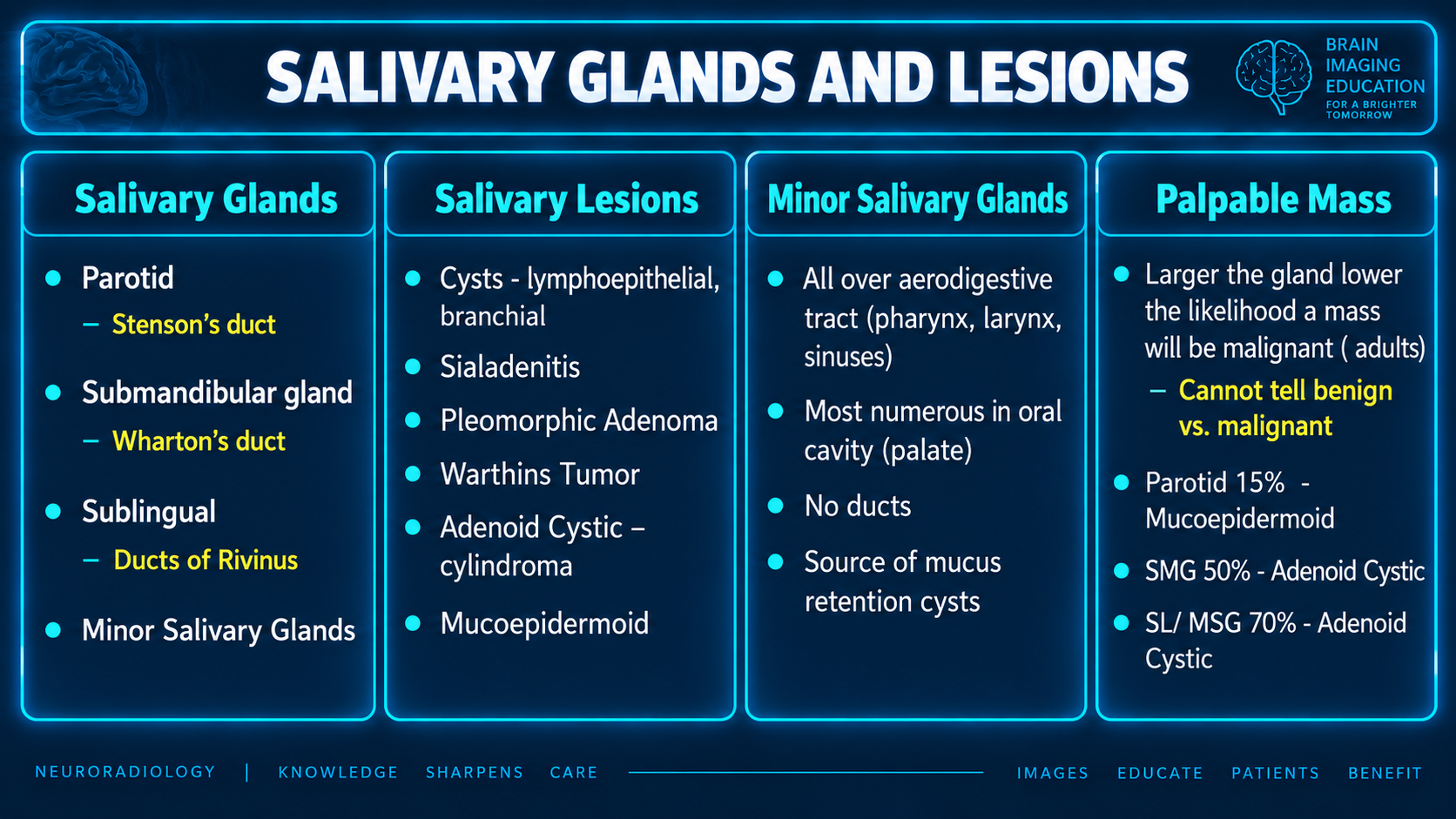
The organizing rule: the smaller the gland, the more likely a mass is malignant: parotid ~20–25%, submandibular ~40–50%, sublingual and minor glands 50–90%. Imaging narrows the differential but cannot exclude low-grade malignancy, so most solid masses still get tissue.
Reference Images
Source limitations
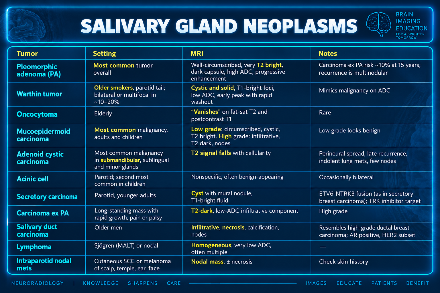
The first supplied graphic gives malignancy proportions of 15% for parotid, 50% for submandibular, and 70% for sublingual/minor gland masses. The supplied prose gives approximately 20–25%, 40–50%, and 50–90%, respectively. Both are preserved; their population and reference basis are not supplied. The graphic calls pleomorphic adenoma well-circumscribed, while the prose describes it as bosselated. No bibliography or staging edition was supplied; the staging labels and imaging thresholds below are reproduced from the source.
Both sources describe minor glands as having no ducts (the prose says “None”); this wording is retained as supplied, without an explanatory reference. Image captions identify the visible slide titles; no separate captions were supplied.
Anatomy
| Gland | Location and relations | Duct | Imaging relevance |
|---|---|---|---|
| Parotid | Parotid space. The CN VII plane separates superficial and deep "lobes"; the retromandibular vein is the surrogate (nerve just lateral). Deep lobe extends through the stylomandibular tunnel toward the prestyloid parapharyngeal space | Stensen: over masseter, pierces buccinator, opens opposite the 2nd maxillary molar | Only gland with intraglandular nodes (Warthin, lymphoma, skin cancer mets). Accessory tissue along the duct in ~20%. Fatty replacement with age |
| Submandibular | Wraps the posterior free edge of mylohyoid: superficial part in the submandibular space, deep part in the posterior sublingual space | Wharton: runs anteriorly to the sublingual caruncle; lingual nerve loops beneath it | No intraglandular nodes. Viscous, calcium-rich saliva and an uphill duct, hence most stones |
| Sublingual | Sublingual space, above mylohyoid, lateral to genioglossus | Multiple ducts of Rivinus ± Bartholin duct | Source of ranulas; masses are usually malignant |
| Minor (~500–1,000) | Submucosal throughout the upper aerodigestive tract, densest in the palate | None | Submucosal palate mass; staged by site of origin |
Secretomotor supply to the parotid runs CN IX → Jacobson nerve → lesser petrosal → otic ganglion → auriculotemporal nerve. The submandibular and sublingual glands are supplied via chorda tympani → lingual nerve → submandibular ganglion. The auriculotemporal nerve is also the CN VII–V3 bridge for perineural spread.
Imaging toolkit
- US: first line for superficial lesions and FNA/core guidance; blind to the deep lobe and skull base.
- CT: noncontrast is most sensitive for stones; contrast for abscess and bone.
- MRI: characterization and staging. Non-fat-sat precontrast T1 is the key sequence (lesion against fatty gland, foraminal fat pads), plus T2, DWI/ADC, DCE, and fat-sat postcontrast for perineural spread.
- MR sialography: noninvasive assessment of strictures and sialectasis; conventional sialography is now rarely needed.
- Nuclear: Warthin and oncocytoma retain Tc-99m pertechnetate. FDG does not separate benign from malignant (Warthin and pleomorphic adenoma can be avid). PSMA ligands show intense physiologic uptake.
Neoplasms
| Tumor | Setting | MRI | Notes |
|---|---|---|---|
| Pleomorphic adenoma (PA) | Most common tumor overall | Bosselated, very T2 bright, dark capsule, high ADC, progressive enhancement | Carcinoma ex PA risk ~10% at 15 years; recurrence is multinodular |
| Warthin | Older smokers, parotid tail; bilateral or multifocal in ~10–20% | Cystic and solid, T1-bright foci, low ADC, early peak with rapid washout | Mimics malignancy on ADC |
| Oncocytoma | Elderly | "Vanishes" on fat-sat T2 and postcontrast T1 | Rare |
| Mucoepidermoid | Most common malignancy, adults and children | Low grade: circumscribed, cystic, T2 bright. High grade: infiltrative, T2 dark, nodes | Low grade looks benign |
| Adenoid cystic | Most common malignancy in submandibular, sublingual and minor glands | T2 signal falls with cellularity | Perineural spread, late recurrence, indolent lung mets, few nodes |
| Acinic cell | Parotid; second most common in children | Nonspecific, often benign-appearing | Occasionally bilateral |
| Secretory carcinoma | Parotid, younger adults | Cyst with mural nodule, T1-bright fluid | ETV6-NTRK3 fusion (as in secretory breast carcinoma); TRK inhibitor target |
| Carcinoma ex PA | Long-standing mass with rapid growth, pain or palsy | T2-dark, low-ADC infiltrative component | High grade |
| Salivary duct carcinoma | Older men | Infiltrative, necrosis, calcification, nodes | Resembles high-grade ductal breast carcinoma; AR positive, HER2 subset |
| Lymphoma | Sjögren (MALT) or nodal | Homogeneous, very low ADC, often multiple | — |
| Intraparotid nodal mets | Cutaneous SCC or melanoma of scalp, temple, ear, face | Nodal mass, ± necrosis | Check skin history |
For DCE and ADC (Yabuuchi criteria):
- PA: time to peak over 120 s with high ADC (roughly above 1.4 × 10⁻³ mm²/s).
- Warthin: early peak with washout of 30% or more and low ADC.
- Malignancy: early peak with washout under 30% and intermediate ADC.
This inverts breast kinetics, where washout is the suspicious curve.
Perineural spread and what to report
- Routes:
- Parotid: along CN VII to the stylomastoid foramen, or via the auriculotemporal nerve to V3, foramen ovale and Meckel cave.
- Palate: via the palatine nerves to the pterygopalatine fossa, then V2 or the vidian nerve.
- Submandibular and sublingual: via the lingual nerve (V3) and CN XII.
- Signs: effaced fat at the stylomastoid foramen or pterygopalatine fossa on T1, nerve thickening and enhancement, foraminal widening, denervation of masticator muscles. Skip lesions occur.
- Report:
- Lobe, and relation to the retromandibular vein and stylomastoid foramen.
- Extraparenchymal extension (T3).
- Skin, mandible, ear canal or facial nerve involvement (T4a).
- Skull base, pterygoid plates or carotid encasement (T4b).
- Intraparotid, level IB and level II–III nodes.
- Multiplicity and bilaterality.
- Deep lobe vs parapharyngeal origin: deep lobe tumors widen the stylomandibular tunnel and have no fat plane with the gland.
Non-neoplastic disease
- Sialolithiasis: 80–90% submandibular. CT detects nearly all stones; radiographs miss many parotid stones. Number, size and position (distal duct, hilum, intraglandular) determine sialendoscopic, transoral or open management.
- Acute sialadenitis: bacterial (S. aureus; elderly, dehydrated, postoperative) with an enlarged enhancing gland ± abscess. Viral (mumps) is usually bilateral parotid.
- Juvenile recurrent parotitis: recurrent swelling with punctate sialectasis, resolving around puberty.
- Sjögren: enlarged glands, then innumerable small T2-bright cysts with fat ("salt and pepper"), then atrophy. Lifetime lymphoma risk is roughly 5–10%, so a dominant solid mass or new adenopathy warrants biopsy.
- IgG4-related disease: bilateral lacrimal and submandibular more than parotid enlargement (Mikulicz), or a firm unilateral submandibular mass (Küttner). Look for orbital, infraorbital nerve and pancreatic disease.
- Sarcoid: bilateral parotid enlargement or nodules; Heerfordt syndrome adds uveitis and facial palsy.
- HIV: bilateral lymphoepithelial cysts with adenoidal hypertrophy and reactive nodes.
- Sialadenosis: painless bilateral parotid enlargement with diabetes, alcohol, malnutrition or bulimia.
- Iatrogenic: radiation (swelling, then atrophy), I-131 sialadenitis, iodinated contrast sialadenitis ("iodide mumps"), xerostomia after PSMA radioligand therapy.
Cystic lesions, pediatrics and pitfalls
- Ranula: simple (sublingual space) or plunging (pseudocyst in the submandibular space with a tail into the sublingual space). The differential is lymphatic malformation, dermoid or epidermoid, and second branchial cleft cyst.
- First branchial cleft cyst (Work type II): in or beside the parotid with a tract toward the external auditory canal; its relation to CN VII matters surgically.
- Pediatric:
- Infantile hemangioma is the most common parotid mass in infants.
- In older children, PA is the most common benign epithelial tumor and mucoepidermoid the most common malignant one.
- Solid nonvascular masses carry a higher malignancy rate than in adults.
- Pitfalls:
- A resected, absent or irradiated submandibular gland makes the contralateral one look like a mass or asymmetric on FDG PET.
- Mylohyoid boutonnière with herniated sublingual tissue.
- Stafne defect.
- Accessory parotid tissue.
- Masseter hypertrophy mimicking parotid enlargement.
- Normal intraparotid nodes.
- Recurrent PA nodules scattered through a parotidectomy bed.