Localize completely
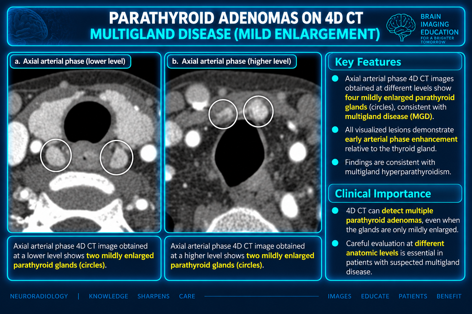
Continue the search after finding an apparent adenoma. Multiple candidates, small lesions or a nonlocalizing study raise concern for multigland disease.
Continue the search after finding an apparent adenoma. Multiple candidates, small lesions or a nonlocalizing study raise concern for multigland disease.
Describe relationships to the cricoid, tracheoesophageal groove, thyroid poles or isthmus, and suprasternal notch. Height alone does not determine gland origin.
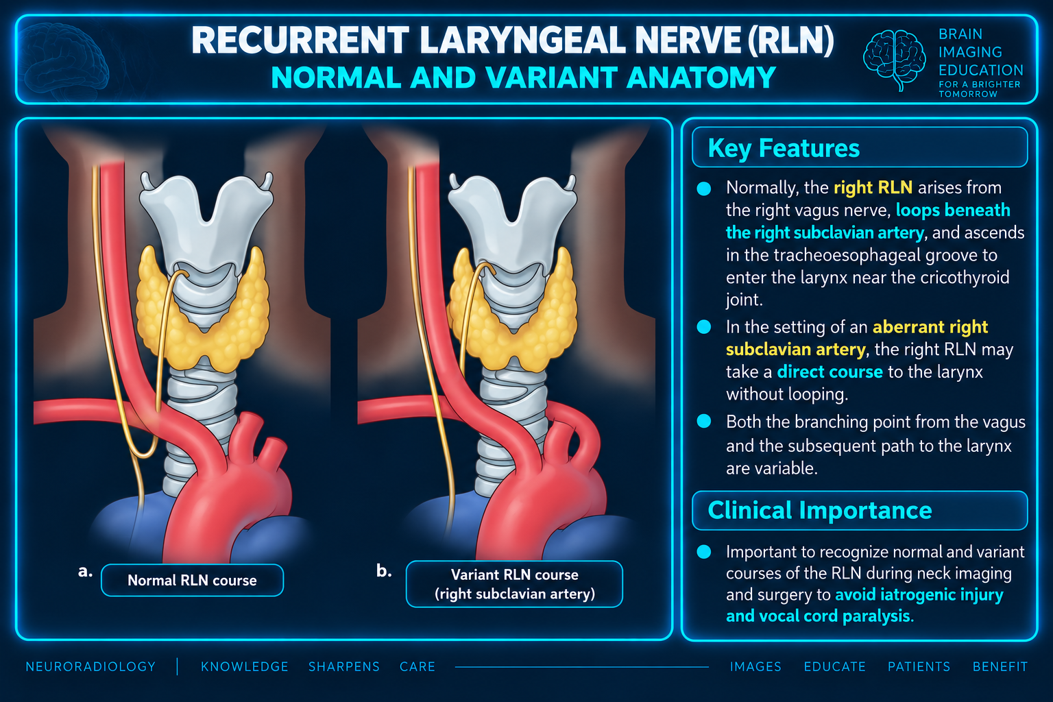
Explicitly identify arterial variants associated with a nonrecurrent laryngeal nerve and state confidence for each candidate lesion.
PHPT is the most common cause of hypercalcemia and most often results from a single parathyroid adenoma (85%–90%). Less common causes are multiglandular hyperplasia (~6%), double adenomas (~4%), and carcinoma (<1%). Definitive treatment is surgical removal of abnormal parathyroid tissue.
Preoperative imaging helps select the surgical approach:
| Surgical strategy | Role of imaging | Key implications |
|---|---|---|
| Minimally invasive parathyroidectomy (MIP) | Requires confident, precise localization of a single abnormal gland | Unilateral, smaller operation; shorter operative time and stay, lower cost, and better cosmesis; in experienced hands, cure rates are equivalent to BNE |
| Bilateral neck exploration (BNE) | Used for multigland disease, nonlocalizing examinations, or uncertain disease distribution | All glands are sought/examined; long-term cure rate exceeds 95% in experienced hands |
If imaging does not localize a gland, the patient still remains a surgical candidate, but BNE is more likely required. Localization is particularly important in persistent or recurrent PHPT, where guidelines regard preoperative localization as mandatory because reoperation carries higher risk.
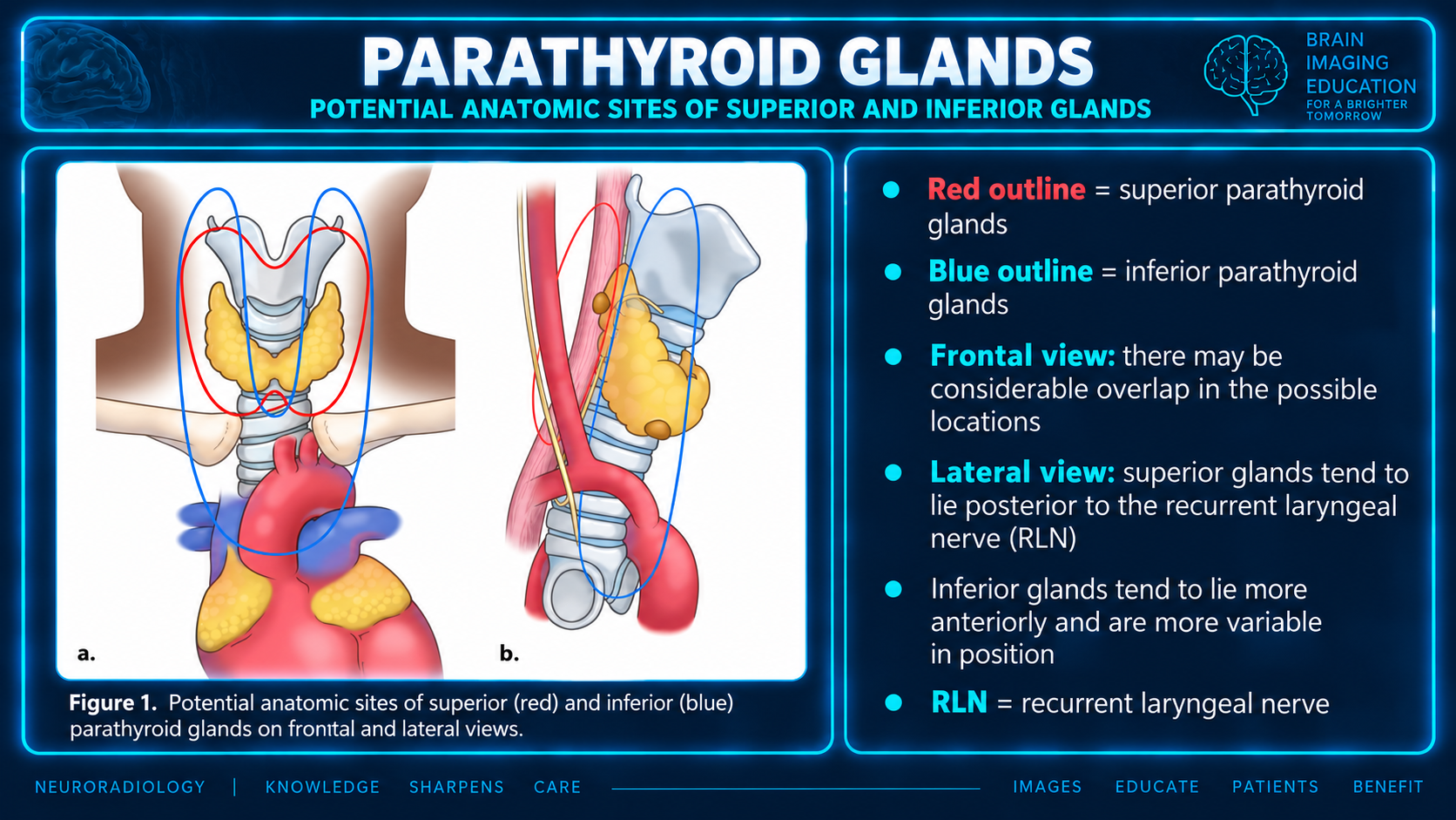
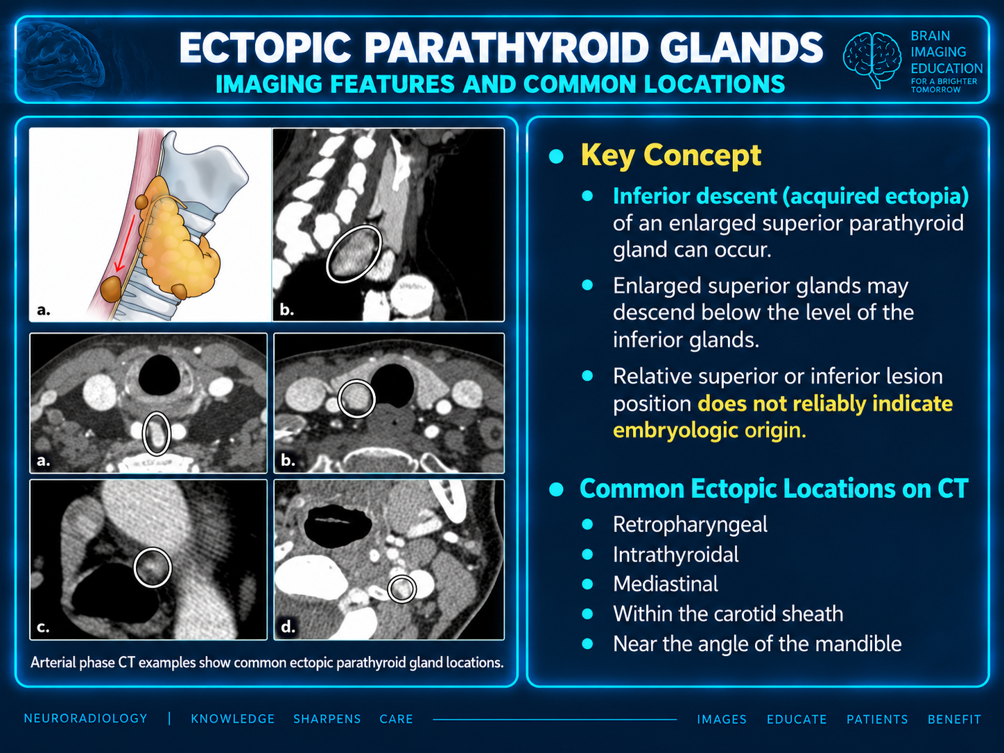
Most individuals have four glands—two superior and two inferior—although fewer than four occur in 3%–6% and supernumerary glands in 5%–13%. The key embryologic point is that a gland’s cranio-caudal position does not reliably establish whether it is a superior or inferior gland.
Up to 16% of glands are ectopic, more often inferior glands. Important ectopic locations include:
The authors recommend reporting lesion location relative to the cricoid cartilage and the tracheoesophageal groove, rather than primarily by cervical vertebral level. These landmarks are visible or usable in the operative field and are especially valuable in reoperative necks. Also useful are the upper/lower poles and isthmus of the thyroid and the suprasternal notch.
A critical additional responsibility is identifying arterial anatomy associated with a nonrecurrent laryngeal nerve:
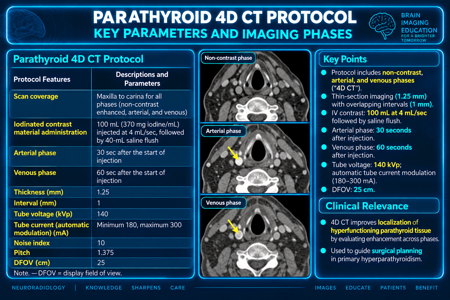
The article uses a three-phase protocol:
The authors favor three phases because the true noncontrast acquisition helps distinguish parathyroid lesions from thyroid tissue. Dual-energy CT with virtual noncontrast imaging could reduce dose by eliminating true noncontrast imaging, but it also removes intrinsic thyroid iodine contrast and is therefore an imperfect substitute.
Practical quality measures include shoulder-lowering positioning and, when feasible, right-arm contrast injection to decrease streak/scatter artifact in the central neck and superior mediastinum.
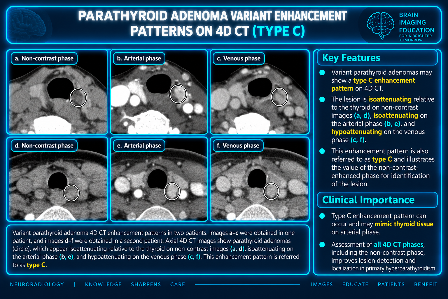
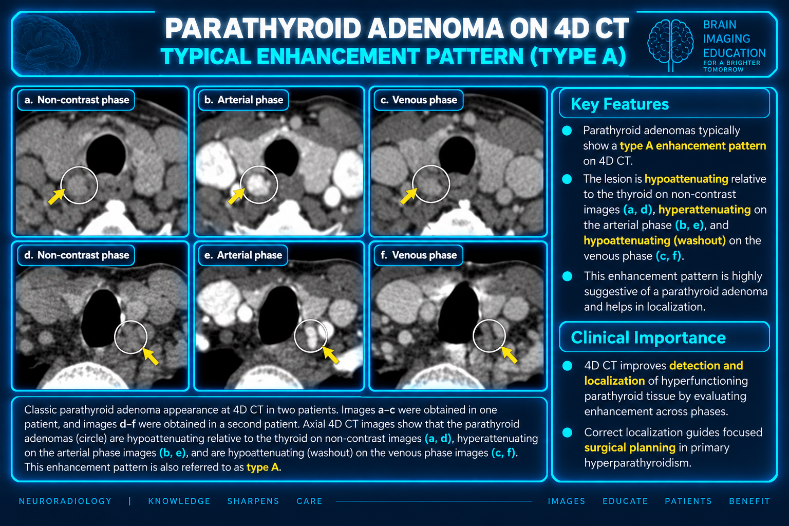
A parathyroid adenoma is usually lower in attenuation than the normal iodine-rich thyroid on noncontrast CT. The classic postcontrast behavior is arterial hyperenhancement relative to thyroid followed by venous washout, but this classic “type A” pattern occurs in only about 20% of adenomas.
Other enhancement patterns are common:
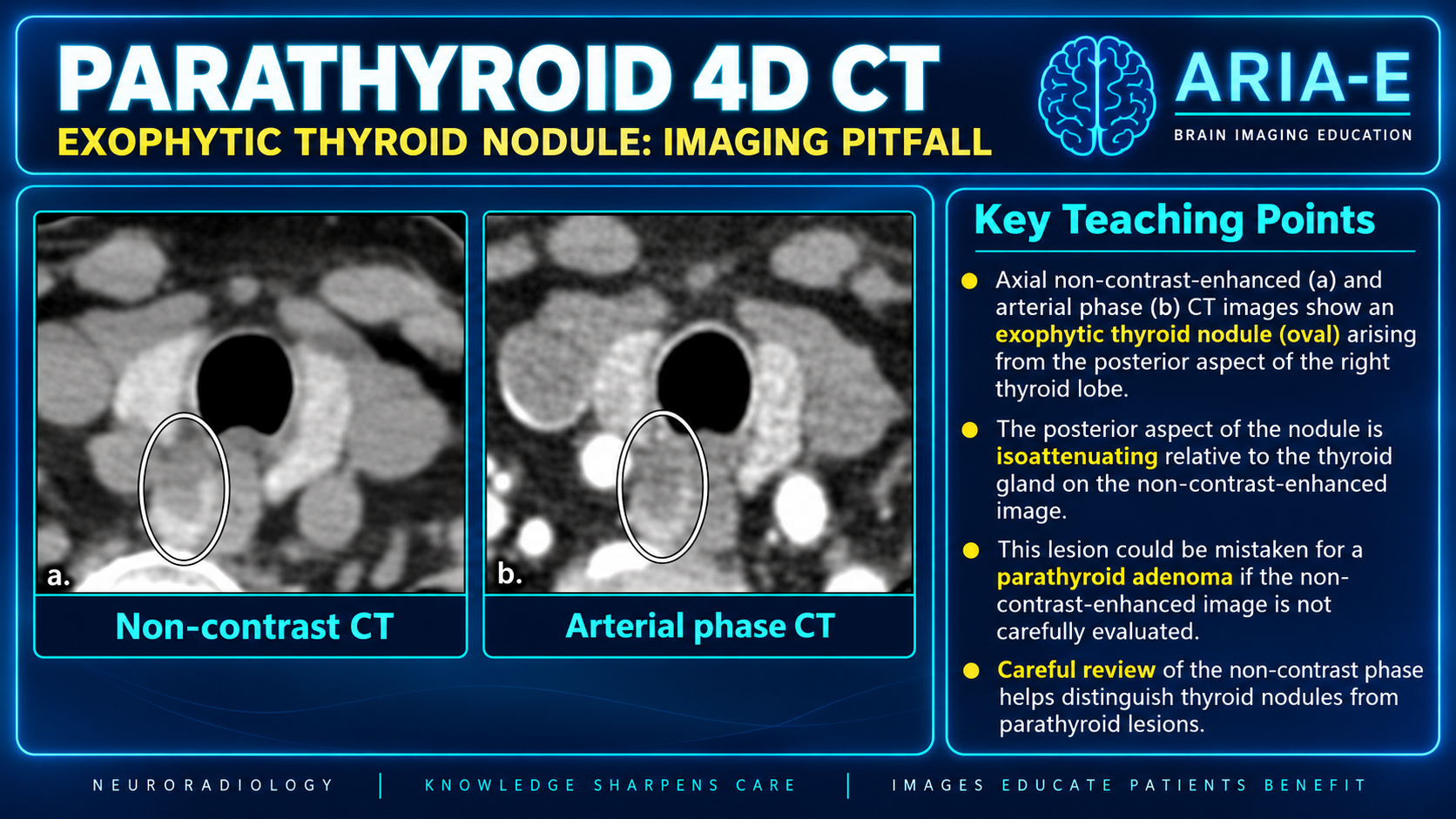
Therefore, the noncontrast images are important. In Hashimoto thyroiditis or chronic hypothyroidism, low thyroid iodine content can make thyroid tissue hypoattenuating, reducing the usefulness of the usual noncontrast distinction.
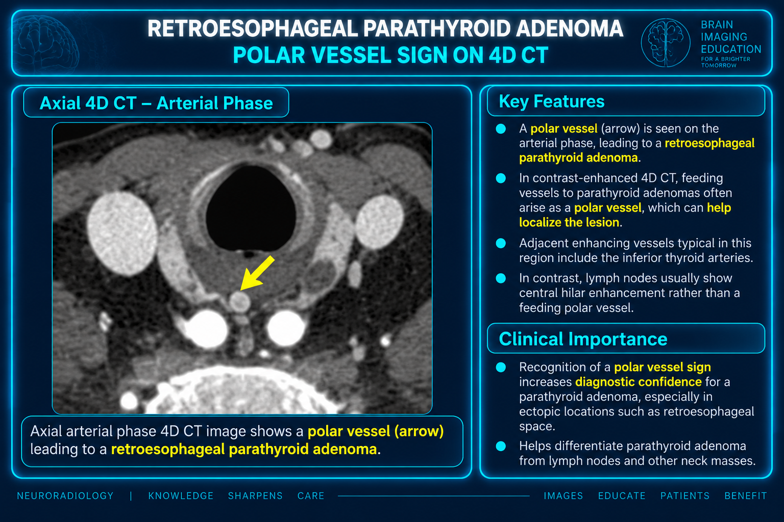
A polar vessel entering or exiting a lesion supports parathyroid origin; it differs from the hilar vascular pattern of a lymph node.
| Pattern (supplied prose) | Arterial vs thyroid | Venous vs thyroid |
|---|---|---|
| Type A | Hyperattenuating | Hypoattenuating (washout) |
| Type B | Isoattenuating | Hypoattenuating |
| Type C | Isoattenuating | Isoattenuating |
The authors advise being suspicious of any structure that cannot confidently be classified as thyroid tissue, lymph node, or vessel, while also correlating with ultrasound and nuclear medicine studies when available. Correlation can improve confidence, especially in difficult or reoperative cases.
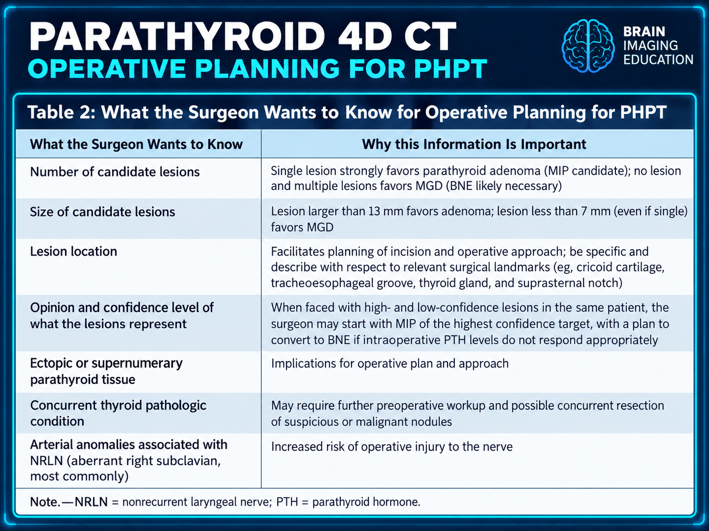
Multiple candidate parathyroid lesions favor multiglandular disease (MGD), but a key counterintuitive point is that no visible lesion also favors MGD, with reported 88% specificity. MGD lesions are often small.
Features that should raise concern for MGD include:
These distinctions matter directly because likely MGD shifts surgical planning toward BNE rather than a focused MIP.
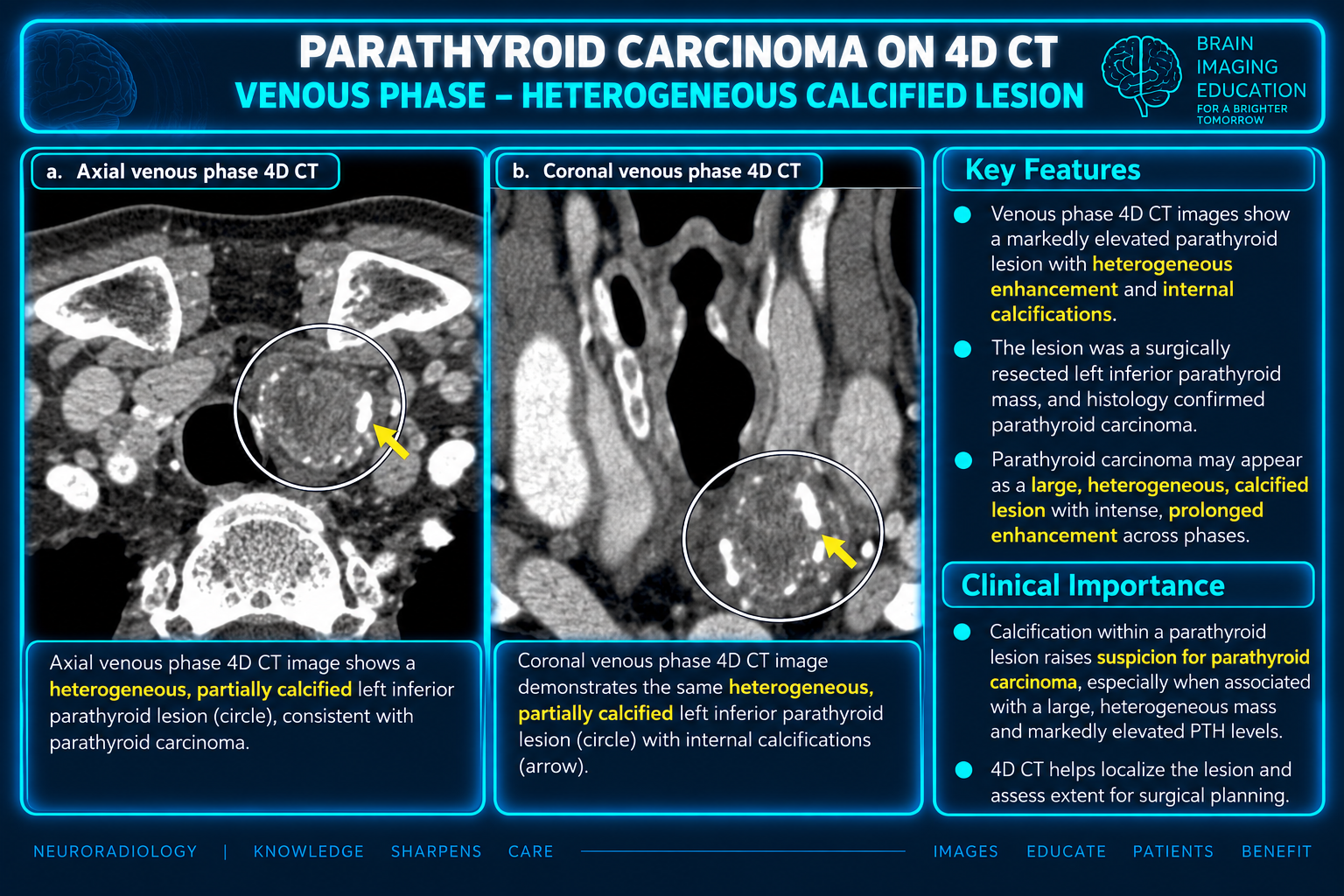
Parathyroid carcinoma is rare and difficult to diagnose preoperatively. Large lesions with calcification, very high PTH levels, and younger patient age should increase suspicion.
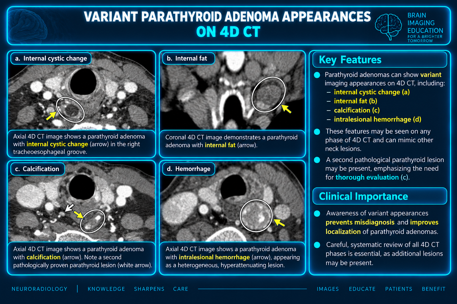
The authors stress completing a systematic search even after finding an apparently obvious adenoma, because additional abnormal or supernumerary glands may coexist.
A clinically useful report should provide a surgical roadmap, not merely state “probable adenoma.” For every candidate lesion, report:
A single lesion strongly supports adenoma and potential MIP. Multiple lesions or no identified lesion favors MGD and makes BNE more likely.
Size helps stratify single-gland versus multigland disease: >13 mm favors adenoma; <7 mm favors MGD.
Specify side, relation to the cricoid cartilage, tracheoesophageal groove/RLN plane, thyroid poles or isthmus, and suprasternal notch. Note ectopic locations.
State whether each candidate is high-confidence parathyroid adenoma, lower-confidence possible adenoma, lymph node, exophytic thyroid nodule, etc. A surgeon may begin with a high-confidence target and convert to BNE if intraoperative PTH fails to fall appropriately.
This may alter incision, approach, and extent of exploration.
Identify thyroid nodules that may require preoperative ultrasound/FNA or simultaneous surgery.
Explicitly report aberrant right subclavian artery or the relevant left-sided arch/subclavian configuration.
Parathyroid 4D CT is a high-value preoperative localization study when interpreted as a detailed surgical map. The radiologist should use embryology, RLN-based anatomy, systematic ectopic-gland search patterns, multiphasic enhancement assessment, and awareness of mimics to identify every credible candidate lesion. The greatest practical value comes from a concise structured report that states lesion number, size, exact surgically meaningful location, diagnostic confidence, ectopic/supernumerary tissue, thyroid disease, and arterial variants that increase RLN risk. Regular correlation with operative findings is the authors’ recommended method for improving future localization accuracy.
Parathyroid 4D CT reference PDF · Parathyroid reference PDF
The supplied article-derived percentages, size thresholds and acquisition parameters are preserved as source statements. The listed protocol is an example from the supplied material.