Localize first
Determine whether the epicenter is muscle, mandible, nerve, or an adjacent primary.
The key is localization: determine whether the lesion arises from the muscles, mandible, or V3, versus secondarily invades from an adjacent space. Infection is more common than tumor; secondary extension from an adjacent head-and-neck malignancy is the most common malignant involvement. Always assess the V3 pathway for perineural spread. pmc.ncbi.nlm.nih
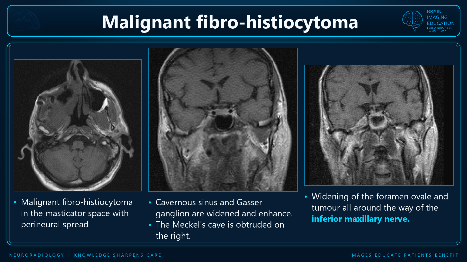
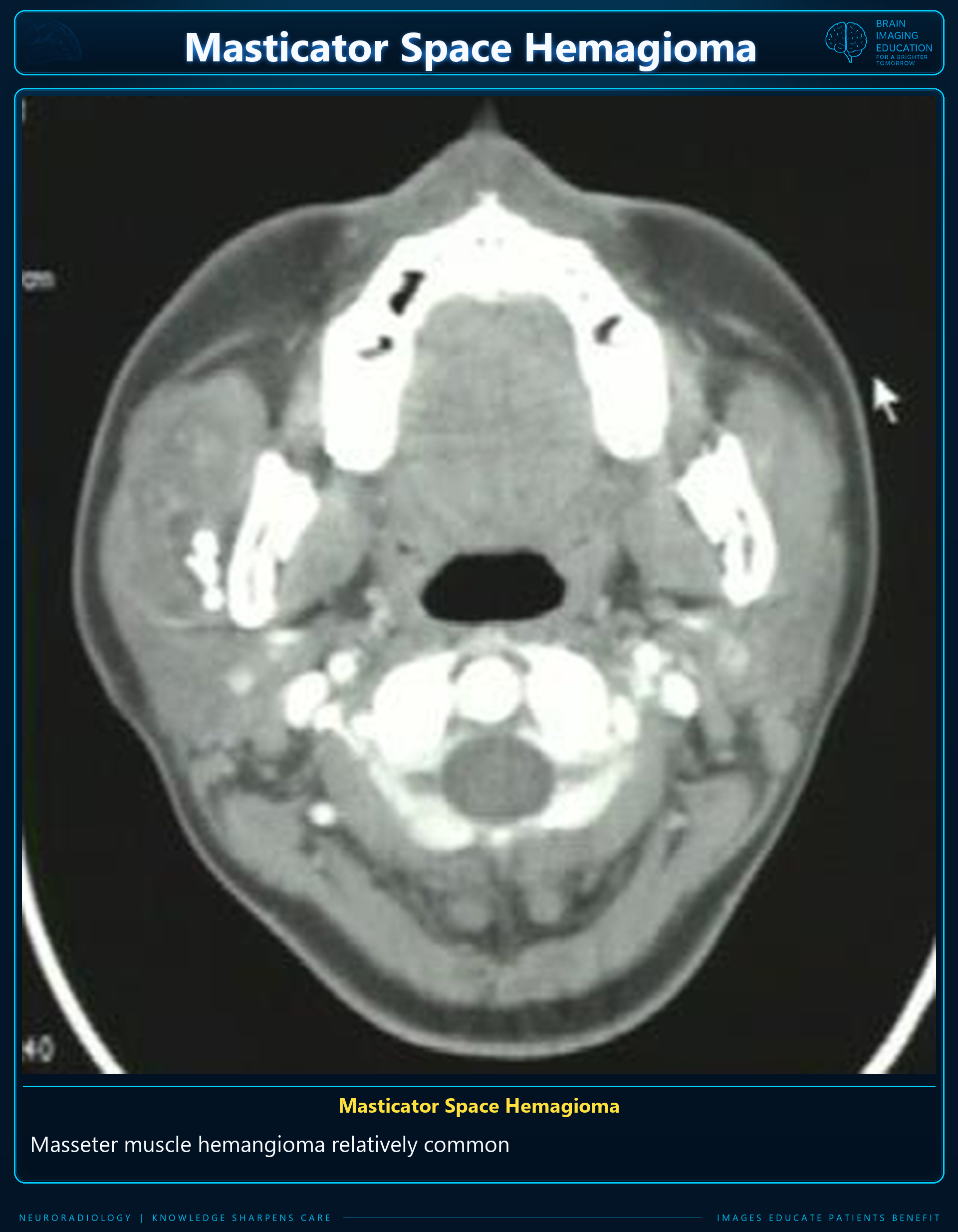
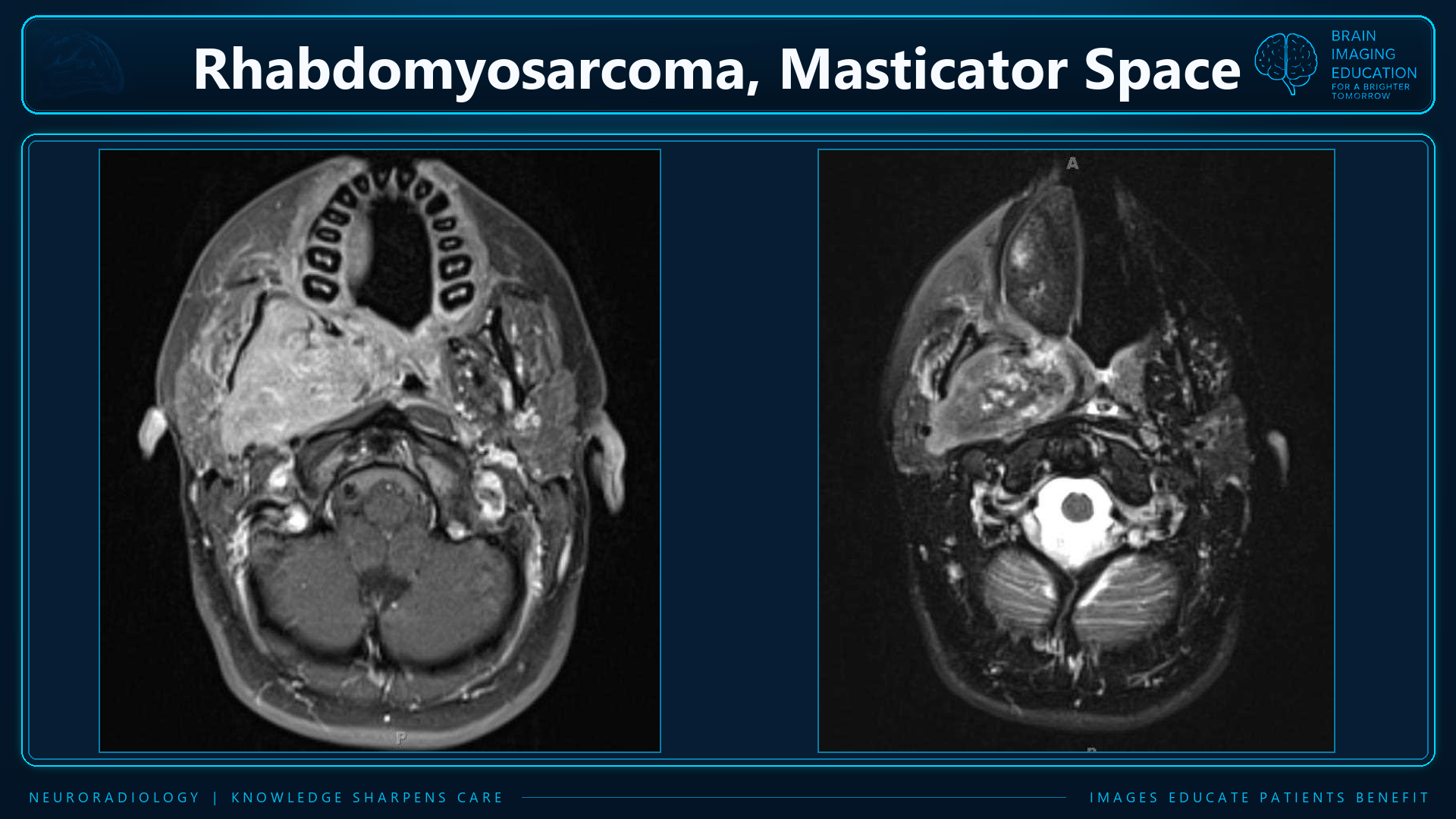
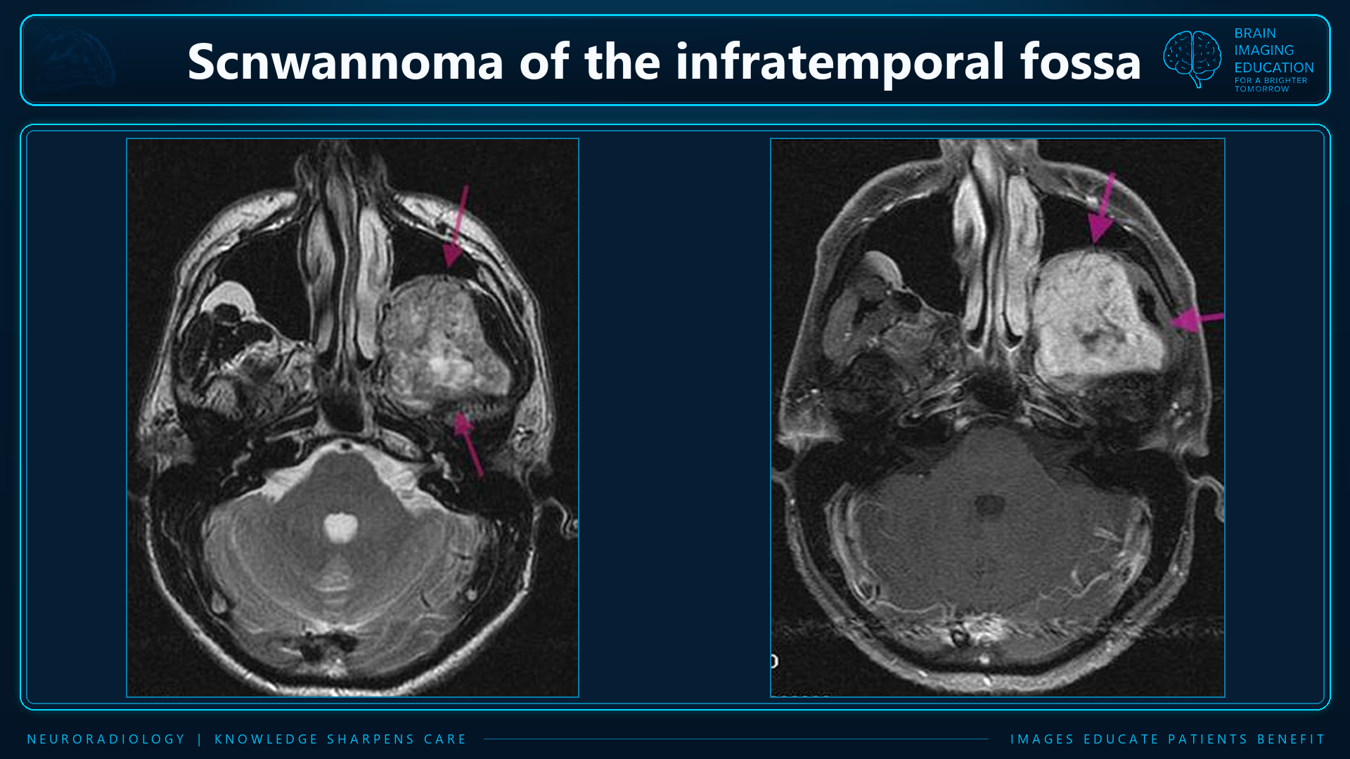
Determine whether the epicenter is muscle, mandible, nerve, or an adjacent primary.
Infection is more common than tumor. Secondary extension is the most common malignant involvement.
Assess the mandibular canal, foramen ovale, Meckel cave and masticatory muscle denervation.
| Category | Principal lesions | Useful imaging clues |
|---|---|---|
| Infection | Odontogenic cellulitis/myositis, phlegmon, abscess; associated mandibular osteomyelitis | Dental source; enlarged, heterogeneously enhancing muscles; surrounding inflammatory change. Abscess: rim-enhancing fluid collection ± gas; may cross fascial boundaries. pmc.ncbi.nlm.nih |
| Neural tumors | V3 schwannoma, neurofibroma; malignant peripheral nerve sheath tumor | Fusiform enhancing mass along V3, often near foramen ovale; associated denervation. Neurofibroma may show a T2 target sign; plexiform morphology suggests NF1. Rapid growth, necrosis, and destructive foraminal change raise concern for malignancy. pmc.ncbi.nlm.nih |
| Vascular malformations | Venous, lymphatic, arteriovenous malformations | Venous: markedly T2-bright channels, enhancement, phleboliths. Lymphatic: multiloculated cystic lesion ± fluid–fluid levels. AVM: serpiginous vessels and prominent flow voids. Often transspatial. pmc.ncbi.nlm.nih |
| Benign mandibular lesions | Dentigerous cyst, odontogenic keratocyst, ameloblastoma, fibrous dysplasia, ossifying fibroma | Tooth relationship and matrix are key. Dentigerous cyst surrounds an unerupted crown; ameloblastoma is expansile, often multilocular with enhancing solid components; fibrous dysplasia has ground-glass matrix. pmc.ncbi.nlm.nih |
| Malignant mandibular lesions | Osteosarcoma, chondrosarcoma, Ewing sarcoma, metastasis, myeloma/plasmacytoma | Destructive bone lesion ± extraosseous mass. Osteosarcoma: osteoid matrix and aggressive periosteal reaction. Chondrosarcoma may occur near the TMJ with mineralized matrix. pmc.ncbi.nlm.nih |
| Primary soft-tissue malignancy | Sarcoma, including pediatric rhabdomyosarcoma; lymphoma | Usually nonspecific enhancing masses. Sarcomas may be necrotic or destructive; lymphoma may show relatively little bone destruction. Smooth margins and intact fascia do not establish benignity. pmc.ncbi.nlm.nih |
| Secondary malignancy | Oral cavity/retromolar trigone SCC, oropharyngeal or nasopharyngeal carcinoma, parotid malignancy | Identify an adjacent primary and continuity of invasion, loss of fat planes, muscle infiltration, mandibular destruction, or perineural spread. pmc.ncbi.nlm.nih |
Trace the inferior alveolar or auriculotemporal nerve → V3 → foramen ovale → Meckel cave/skull base. Look for:
| Modality | Main strengths |
|---|---|
| Contrast-enhanced CT with bone windows | Dental source, cortical erosion, gas, abscess, phleboliths, and mineralized tumor matrix. Particularly useful for suspected odontogenic infection. pmc.ncbi.nlm.nih |
| Contrast-enhanced MRI | Soft-tissue characterization, muscle invasion, mandibular marrow involvement, skull-base extension, and perineural spread. pmc.ncbi.nlm.nih |
The figures are reproduced unchanged, in supplied order. Figure 1 retains the slide’s diagnostic label “malignant fibro-histiocytoma” and wording “inferior maxillary nerve” and “Meckel’s cave is obtruded.” The supplied summary uses V3/mandibular nerve terminology. These source wordings have not been reconciled or reinterpreted.
Figure 2 is labeled “hemangioma” in the source, while the summary discusses vascular malformations separately. The supplied material does not establish a reclassification of this case. Unmistakable spelling errors in figure titles are corrected in webpage captions only; the original graphics remain unchanged. No patient histories or pathology confirmation details were supplied.
Reference supplied with the summary: Masticator-space review (PubMed Central).