Masticator Space

The masticator space is a deep compartment of the head and neck that contains the muscles of mastication.

Reference Images

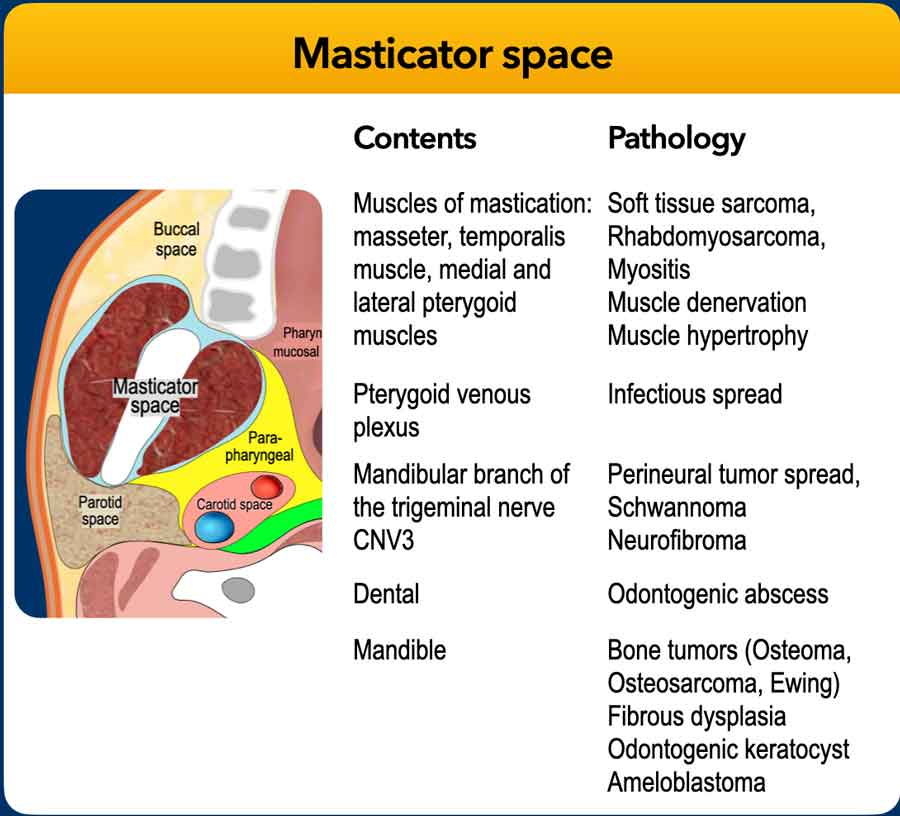
Teaching slide with a masticator space diagram and a contents–pathology table.
Masticator space — contents and pathologySelect to enlarge. Original annotations preserved.
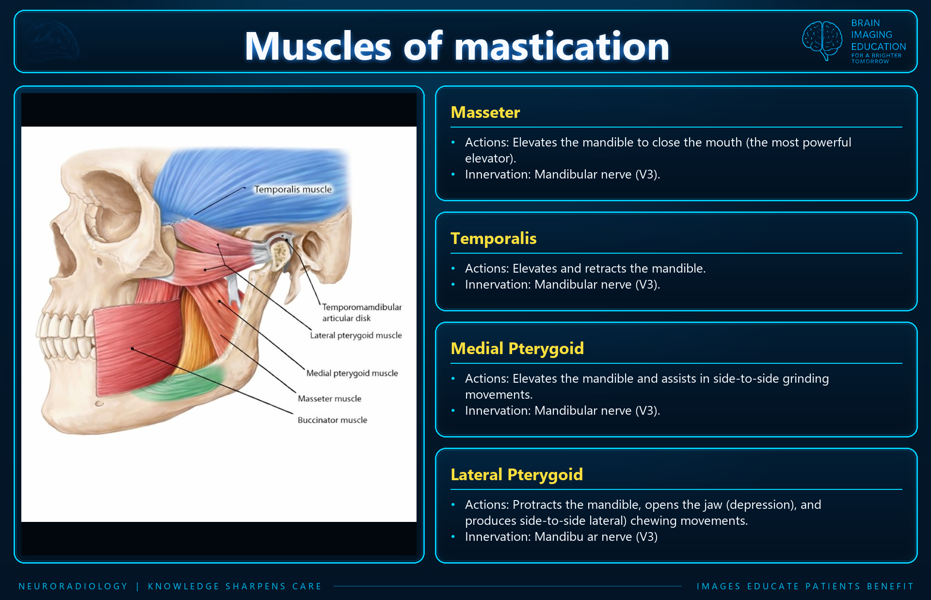
Muscles of mastication teaching slide showing the masseter, temporalis, medial pterygoid and lateral pterygoid muscles with their actions and innervation.
Muscles of masticationSelect to enlarge. Original annotations preserved.
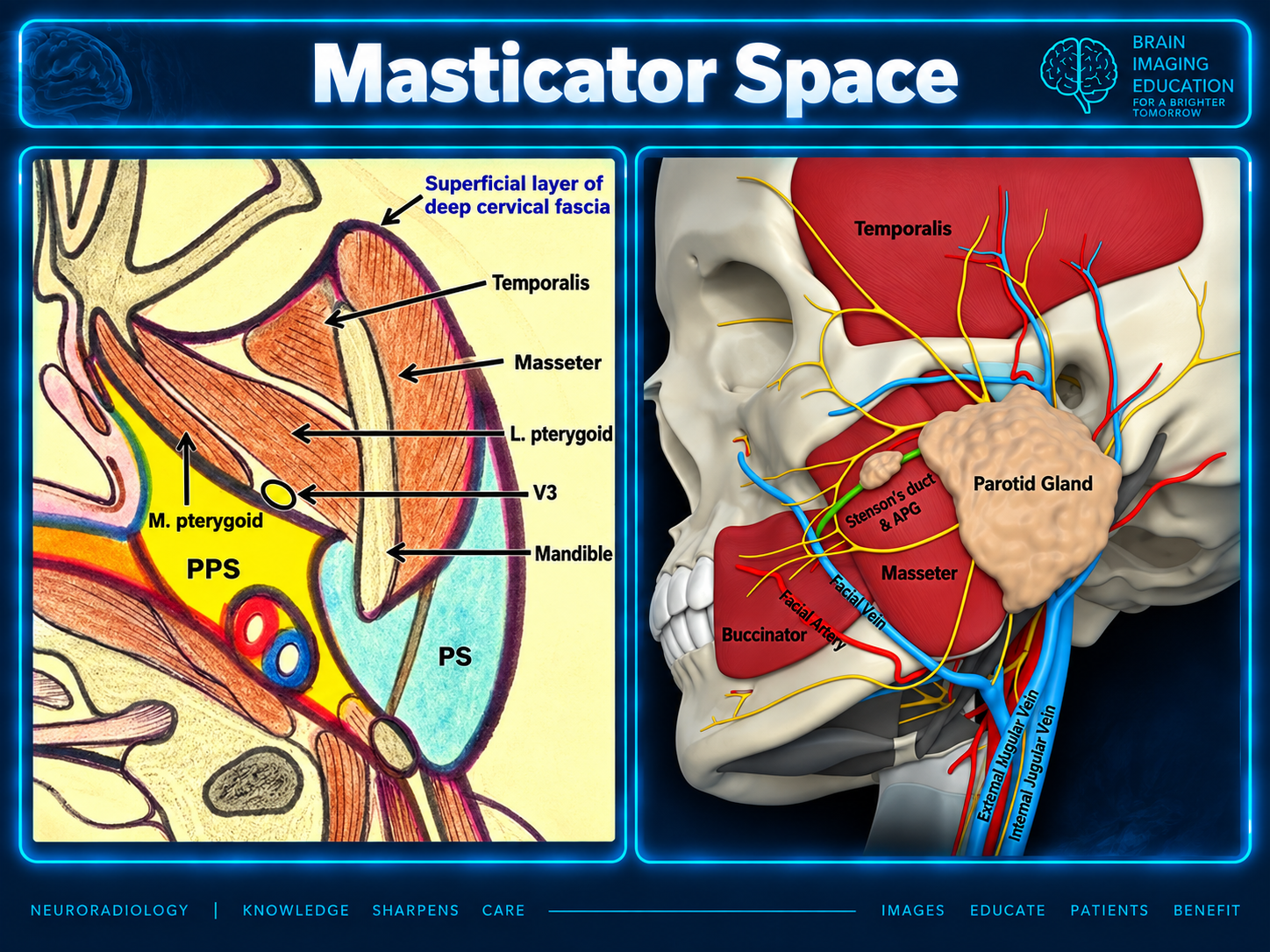
Anatomic teaching graphic labeling fascia, temporalis, masseter, pterygoid muscles, V3 and mandible, beside a lateral skull illustration.
Masticator anatomy and adjacent structuresSelect to enlarge. Original annotations preserved.
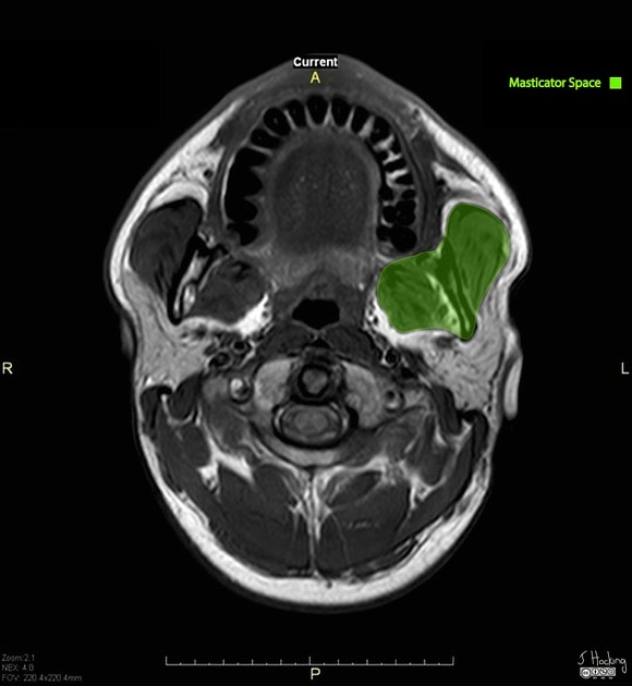
Axial MRI with the left masticator space highlighted in green and original orientation markers preserved.
Axial MRI — masticator space highlighted in greenSelect to enlarge. Original annotations preserved.
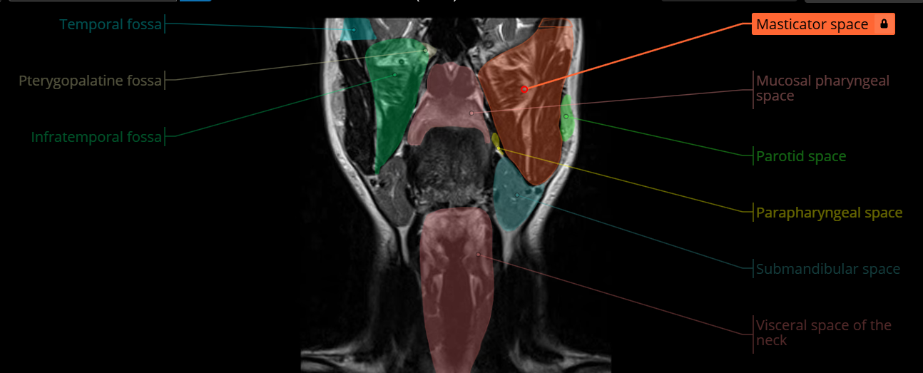
Annotated coronal MRI with an orange masticator space overlay and labels for adjacent head and neck spaces.
Coronal MRI — masticator space and neighboring spacesSelect to enlarge. Original annotations preserved.
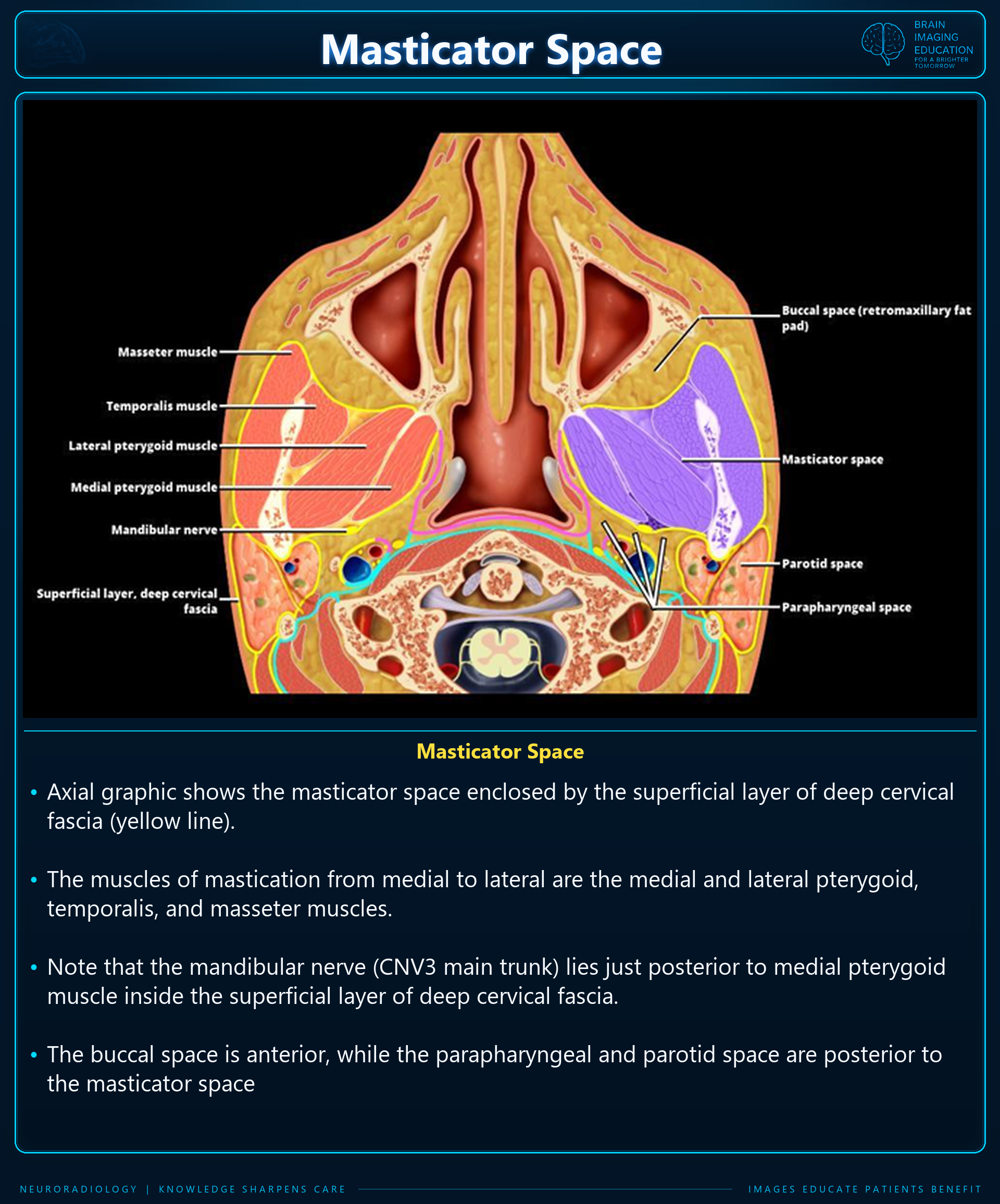
Axial anatomic illustration of the masticator space, muscles of mastication, mandibular nerve, fascia and neighboring spaces, with explanatory text.
Masticator space — axial anatomySelect to enlarge. Original annotations preserved.
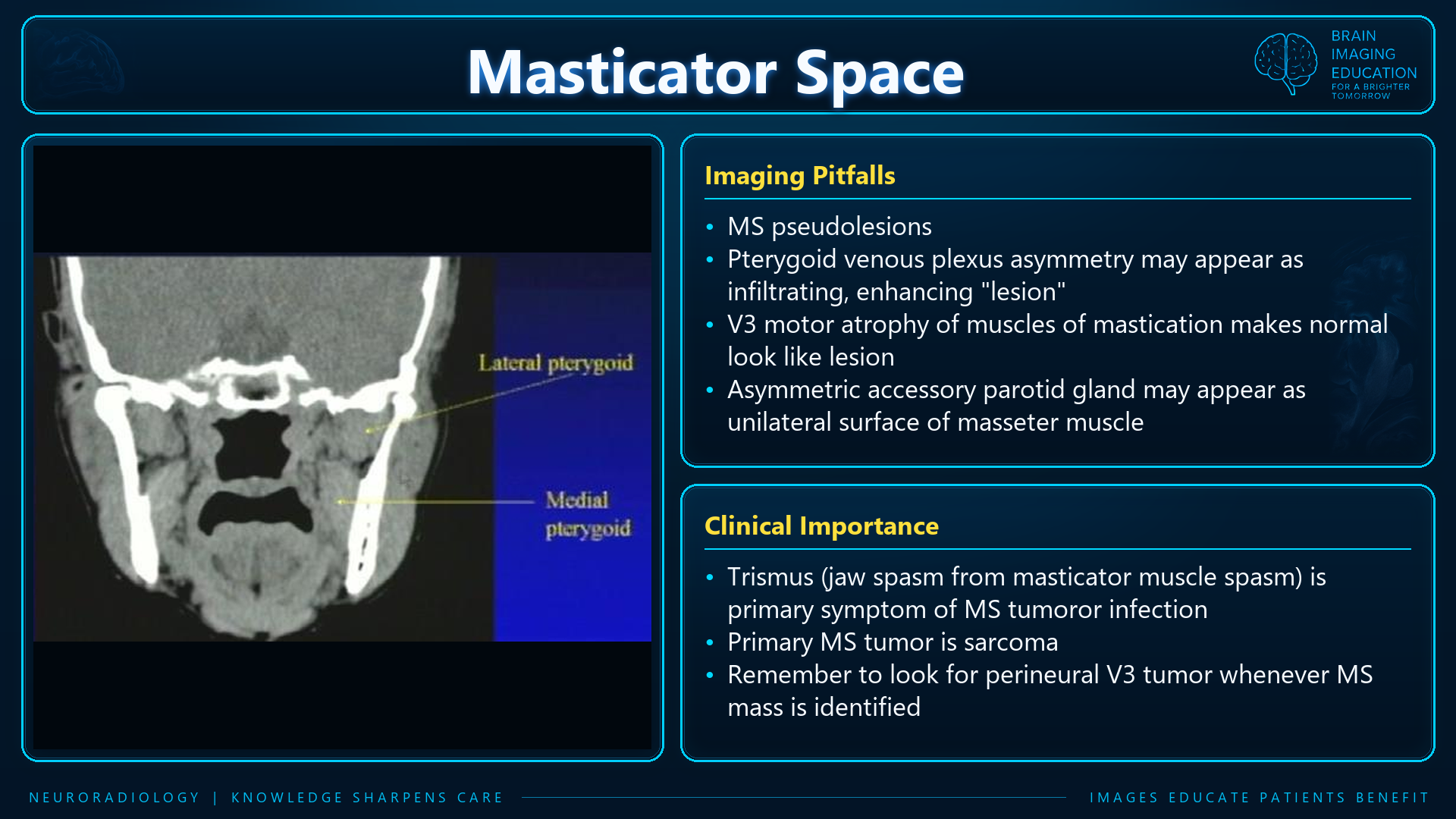
Coronal CT showing the pterygoid muscles alongside teaching notes on imaging pitfalls and clinical importance.
Masticator space — imaging pitfalls and clinical importanceSelect to enlarge. Original annotations preserved.
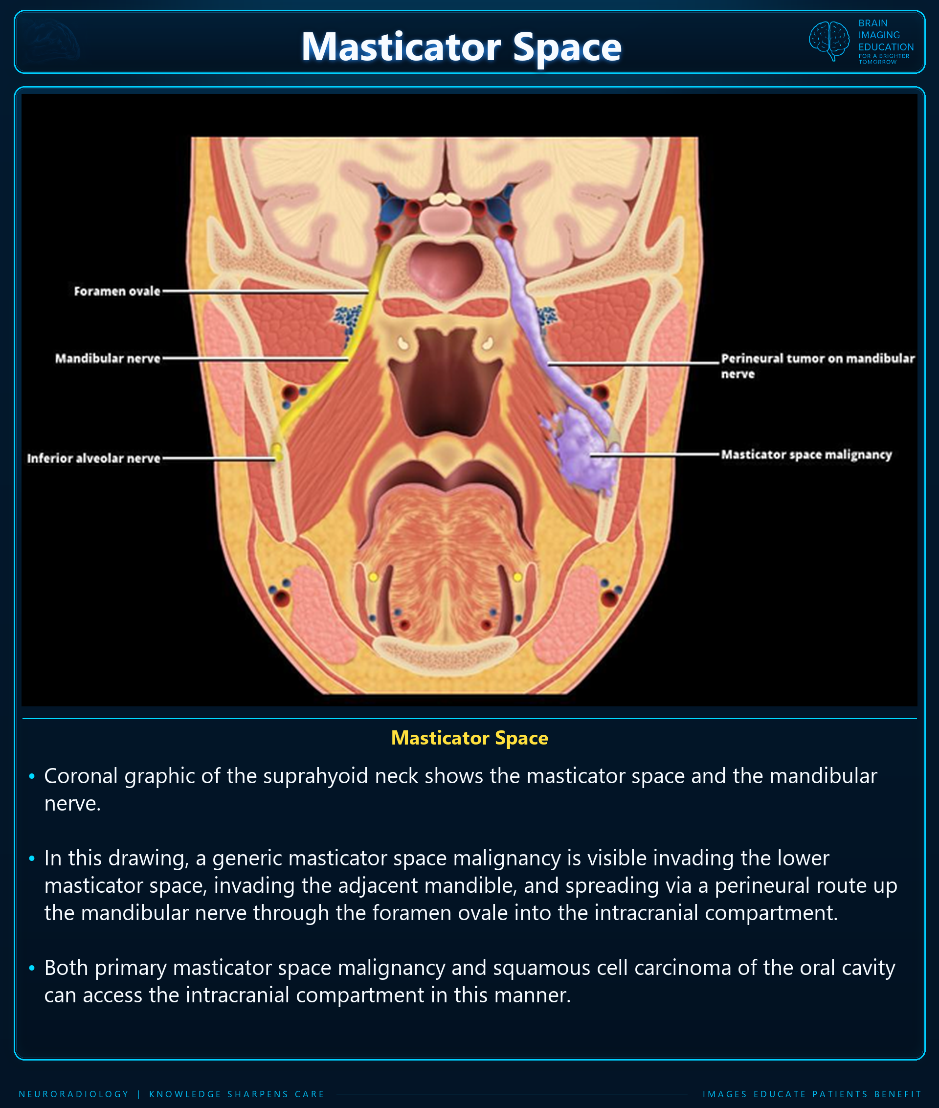
Coronal illustration showing masticator space malignancy spreading along the mandibular nerve through the foramen ovale, with explanatory text.
Masticator space — perineural tumor spreadSelect to enlarge. Original annotations preserved.

Gross anatomy

The masticator spaces are paired suprahyoid cervical spaces on each side of the face. Each is enveloped by the superficial (investing) layer of the deep cervical fascia.

This fascial layer splits into two at the lower border of the mandible:

  • Inner layer: runs deep to the medial pterygoid muscle and attaches to the skull base medial to foramen ovale.
  • Outer layer: covers the masseter and temporalis muscles and attaches superiorly to the parietal calvaria.

Contents

  • Muscles of mastication: masseter, temporalis, medial and lateral pterygoid muscles (named in the supplied figures)
  • Ramus and body of the mandible
  • Mandibular division of the trigeminal nerve; enters via the foramen ovale
  • Inferior alveolar nerve
  • Inferior alveolar artery and vein
  • Pterygoid venous plexus

Subcompartments

Fibroadipose tissue in the potential spaces between muscles and bones can be described by these subcompartments:

  • Submasseteric space
  • Pterygomandibular space
  • Temporal space
    • Superficial temporal space
    • Deep temporal space
  • Infratemporal space

Boundaries and Relations

AspectRelated space
AnteriorBuccal space
PosterolateralParotid space
MedialParapharyngeal space
InferomedialSubmandibular space

Communications

Masticator space malignancy or infection can spread perineurally via the foramen ovale and along the course of the mandibular division of the trigeminal nerve into the middle cranial fossa.

Radiographic Appearance — Ultrasound

Ultrasound has limited use when imaging the masticator space.

Structures that can be visualized

  • Masseter muscles
  • Zygomatic arch
  • Outer cortex of the mandibular ramus
  • Suprazygomatic segment of the temporal muscle

Structures with limited visualization

  • Pterygoid muscles
  • Pterygoid venous plexus
  • Mandibular branch of the trigeminal nerve

Related Pathology

Conditions listed in the supplied text

  • Odontogenic abscess
  • Osteomyelitis
  • Direct spread of squamous cell carcinoma
  • Lymphoma
  • Minor salivary gland tumors
  • Muscle sarcoma
  • Bone sarcoma
  • Osteoradionecrosis
  • Schwannoma
  • Neurofibroma
  • Benign masseteric hypertrophy
  • Accessory parotid tissue

Content–pathology associations in Figure 1

ContentAssociation shown
MusclesSoft tissue sarcoma, rhabdomyosarcoma, myositis, muscle denervation, muscle hypertrophy
Pterygoid venous plexusInfectious spread
Mandibular nerve (V3)Perineural tumor spread, schwannoma, neurofibroma
DentalOdontogenic abscess
MandibleBone tumors (osteoma, osteosarcoma, Ewing), fibrous dysplasia, odontogenic keratocyst, ameloblastoma

Source Notes

The supplied prose labels the mandibular division of the trigeminal nerve as “Vc”; the supplied diagrams label it V3 (CN V3). This notation discrepancy is retained here explicitly.

The text includes reference markers 1, 3 and 9 without a corresponding bibliography. Their sources cannot be identified from the supplied material. No detailed CT/MRI diagnostic criteria or management guidance were supplied.

Figures are reproduced unchanged. Some labels in the original coronal screenshot are low contrast or partly clipped; enlargement preserves the original resolution.

Secondary Links