Localize first
Identify the epicenter and displacement of PPS fat before relying on signal characteristics.
The key is localization: determine whether the lesion arises in the prestyloid parapharyngeal space (PPS), the carotid space, or an adjacent compartment. Displacement of PPS fat and the carotid–jugular vessels often narrows the differential more than signal characteristics alone. radiopaedia
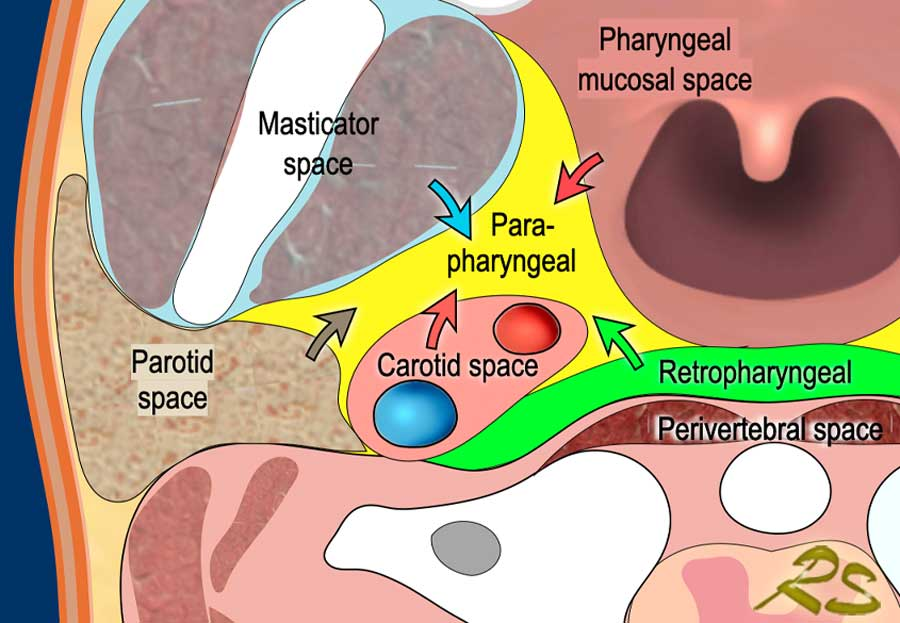
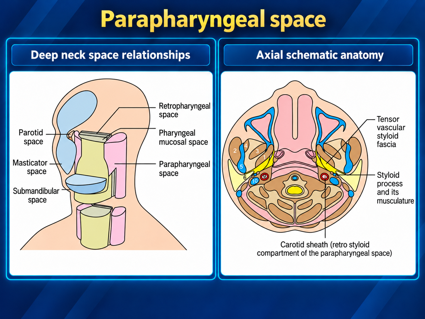
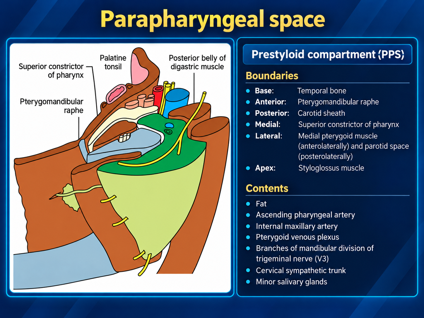
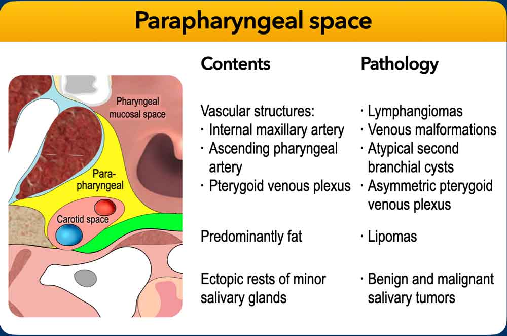
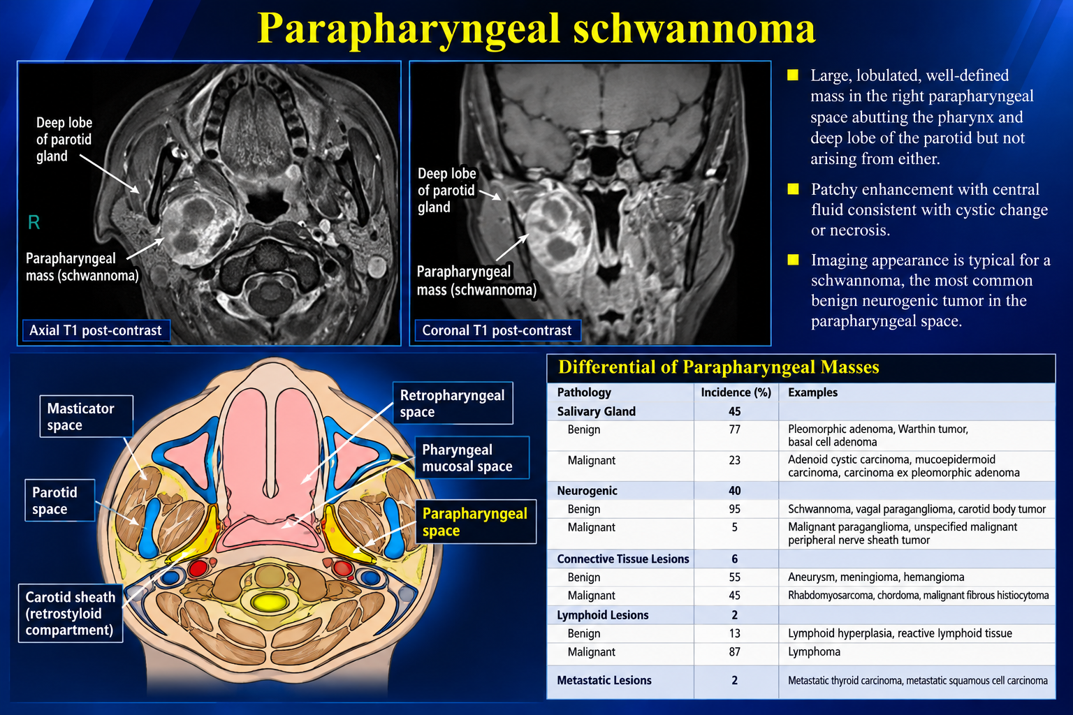
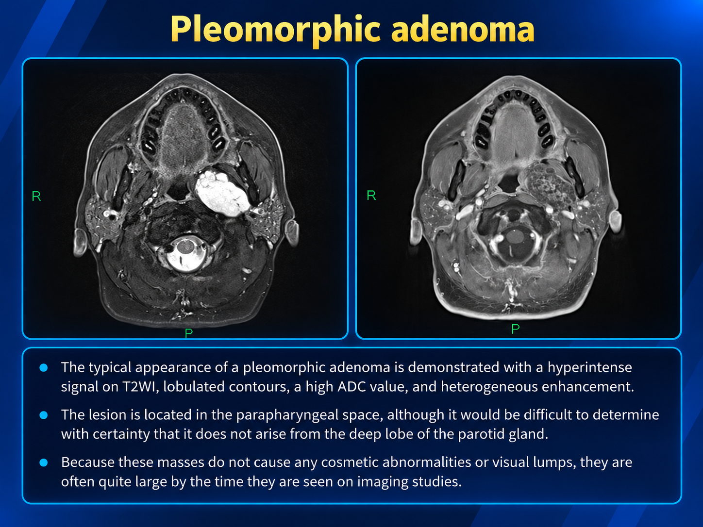
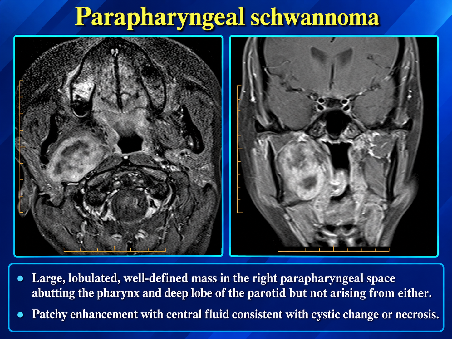
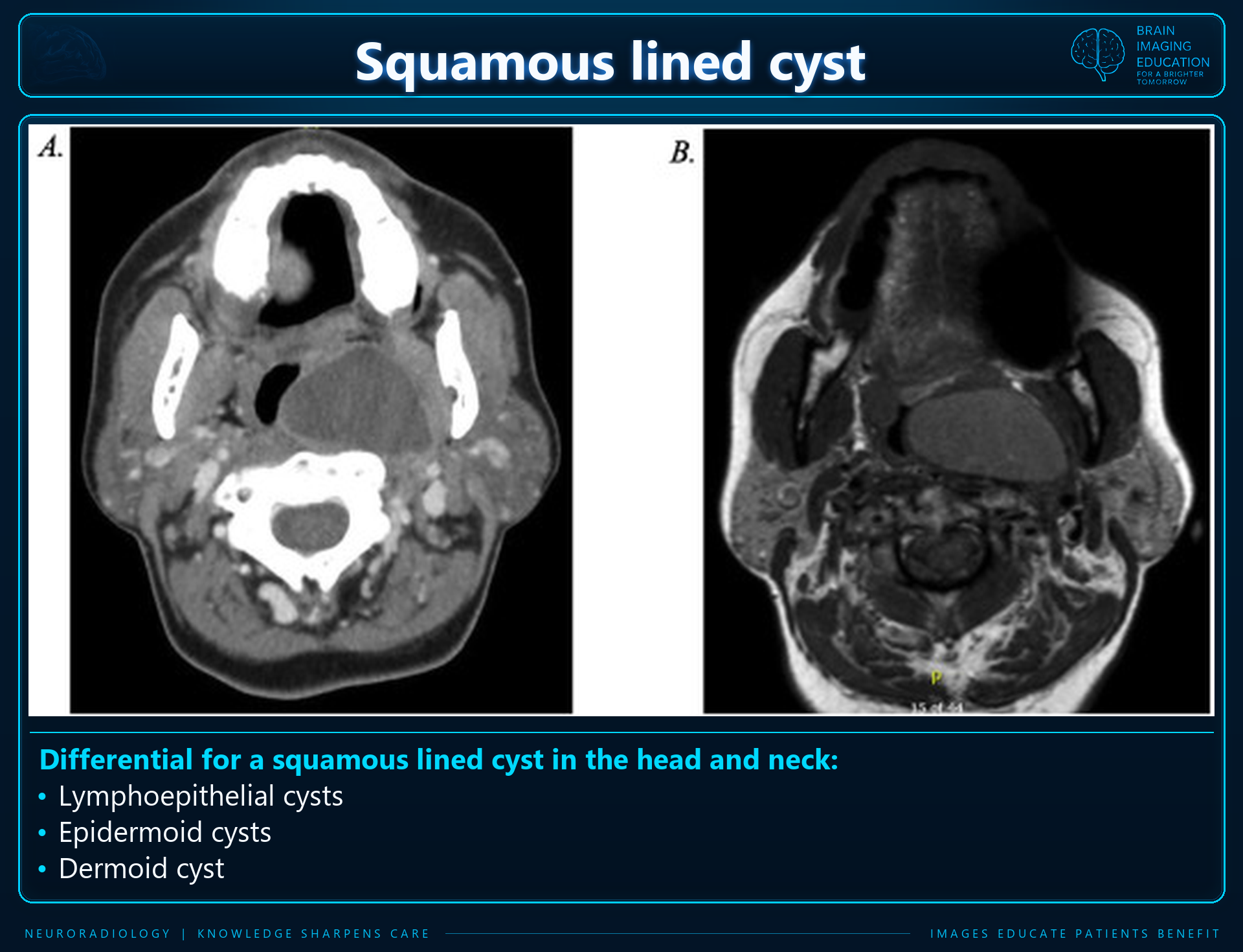
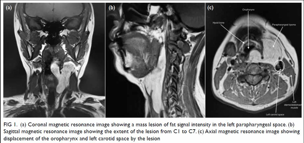
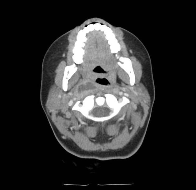
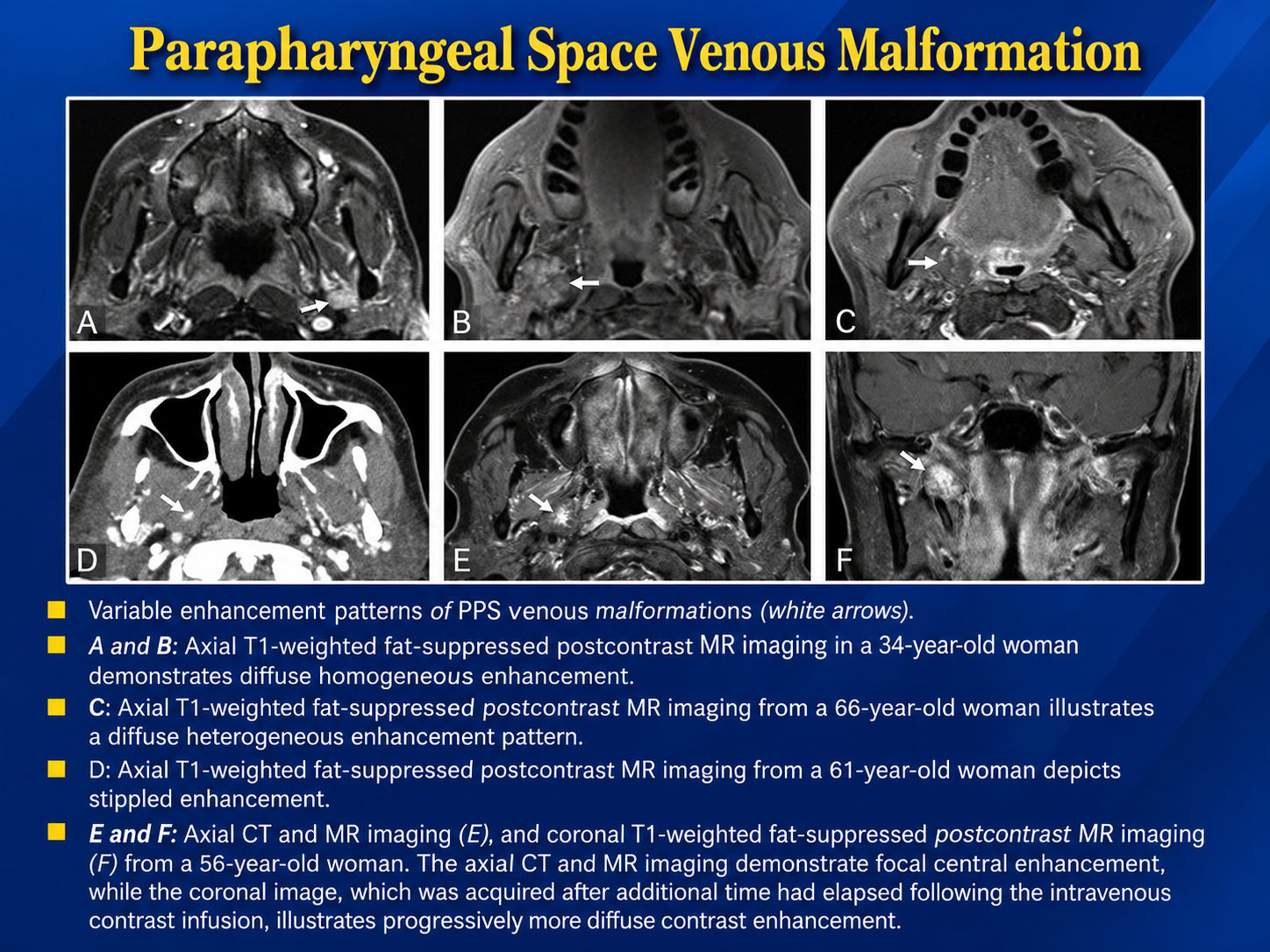
The supplied teaching graphics are preserved unchanged. Some use the traditional poststyloid PPS terminology; the summary below uses PPS for the prestyloid compartment and carotid space for the poststyloid compartment. Figure 3 lists the cervical sympathetic trunk under prestyloid contents, which conflicts with the compartment distinction in the supplied prose. Treat that slide list as source material rather than a resolved compartment assignment.
Figure 5 includes incidence percentages without a supplied study population or citation; they are not generalized in this summary. Figures 5 and 7 may show the same schwannoma example and are retained in the requested order. Figure 11 contains inconsistent modality labels in its embedded text (panel D visibly shows CT); the original text is retained. The supplied single CT abscess image has no clinical history, diffusion imaging or additional planes, so the full imaging criteria below cannot be confirmed from that image alone.
Identify the epicenter and displacement of PPS fat before relying on signal characteristics.
Separate prestyloid salivary lesions from carotid-space neurogenic and vascular lesions.
Exclude a vascular lesion before biopsy; suspected paraganglioma should not undergo routine biopsy.
The PPS is an inverted pyramid extending from the skull base to the hyoid, predominantly containing fat and minor/ectopic salivary tissue. Traditional descriptions divide it into prestyloid and poststyloid compartments; modern radiologic terminology generally calls the prestyloid compartment the PPS and the poststyloid compartment the carotid space. This distinction separates predominantly salivary lesions from neurogenic and vascular lesions. radiopaedia
| Space of origin | Typical displacement of PPS fat |
|---|---|
| Parotid space/deep parotid lobe | Anteromedial |
| Masticator space | Posteromedial |
| Pharyngeal mucosal space | Posterolateral |
| Carotid space | Anterior |
| Retropharyngeal space | Anterolateral |
| True prestyloid PPS lesion | Expands/replaces PPS fat; displaces the carotid space posteriorly and pharynx medially |
These are typical patterns; large or infiltrative masses may obscure the original epicenter. radiopaedia
| Lesion | Key imaging findings |
|---|---|
| Pleomorphic adenoma | Most common salivary tumor in this compartment. Well-defined, often lobulated and markedly T2 hyperintense, with variable enhancement. Determine whether it arises from the deep parotid lobe or minor/ectopic salivary tissue. |
| Malignant salivary tumor | Consider with infiltrative margins, adjacent invasion, perineural spread or suspicious nodes; imaging cannot reliably determine histology. |
| Lipoma | Fat attenuation/signal, with suppression on fat-saturated sequences. |
| Abscess/phlegmon | Fat stranding and inflammatory change; abscess shows a fluid center, peripheral enhancement and often restricted diffusion, sometimes gas. Frequently extends from tonsillar or odontogenic infection. |
| Developmental cyst / lymphatic or venous malformation | Cystic or transspatial lesion; atypical second branchial cleft cyst is a consideration. |
| Secondary tumor extension | Particularly nasopharyngeal/oropharyngeal carcinoma or lesions arising in the parotid or masticator spaces. Identify the actual epicenter rather than labeling these primary PPS tumors. |
Salivary tumors—especially pleomorphic adenoma—lead the primary prestyloid differential, but lesions extending from adjacent spaces are an important and common explanation for PPS involvement. radiopaedia
Look for continuity with the deep parotid lobe and the intervening fat planes. A mass arising from the parotid pushes PPS fat medially/anteromedially, whereas a primary PPS mass is centered within that fat. This distinction matters for operative planning. radiologykey
| Lesion | Vessel displacement | Other distinguishing findings |
|---|---|---|
| Vagal schwannoma | Separates ICA and IJV: ICA usually anteromedial, IJV posterolateral | Well-circumscribed, T2 hyperintense, variable/heterogeneous enhancement and possible cystic degeneration; generally lacks prominent intratumoral flow voids |
| Sympathetic-chain schwannoma | Displaces ICA and IJV together, usually anterolaterally | Similar tissue characteristics to vagal schwannoma; vessel separation is usually absent |
| Vagal paraganglioma | Displaces carotid arteries together anteromedially, separating them from the IJV | Marked early enhancement and prominent flow voids; “salt-and-pepper” MRI appearance |
| Carotid-body paraganglioma | Splays ICA and ECA at the carotid bifurcation—the lyre sign | Hypervascular; epicenter at the bifurcation rather than the upper PPS |
| Aneurysm/pseudoaneurysm | Direct continuity with an artery rather than a simple extrinsic displacement pattern | Vascular lumen and possible mural thrombus; characterize with angiographic imaging |
Vessel displacement is a guide, not an absolute rule: schwannomas can occasionally produce atypical patterns, including carotid-bifurcation splaying. Combine the epicenter, vessel relationships and vascularity. pmc.ncbi.nlm.nih
In the report, document: