Definition used here
The radiological convention calls the prestyloid compartment the parapharyngeal space and the poststyloid compartment the carotid space. This convention facilitates differential diagnosis. [4–6]
The parapharyngeal space (PPS), also called the prestyloid parapharyngeal space, is a deep head and neck compartment around which most other suprahyoid fascial spaces are arranged. It consists largely of fat and neurovascular structures, with the retromandibular part of the deep lobe of the parotid gland included in some definitions.
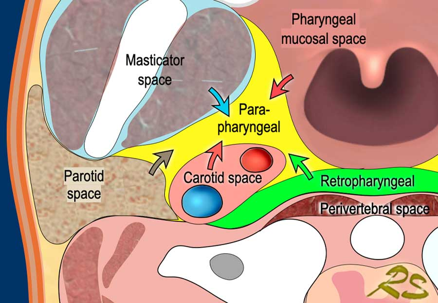
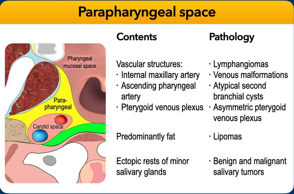
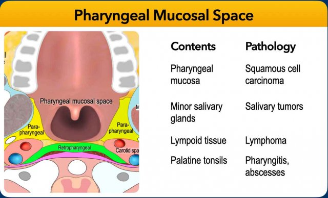
The radiological convention calls the prestyloid compartment the parapharyngeal space and the poststyloid compartment the carotid space. This convention facilitates differential diagnosis. [4–6]
An inverted pyramid with its base at the skull base and its inferior apex pointing toward the greater cornu of the hyoid bone. [2]
On axial CT/MRI it appears triangular, with density or signal consistent with fat.
Displacement of PPS fat helps localize lesions in adjacent deep neck spaces.
A primary PPS lesion displaces the carotid space posteriorly and the pharynx medially.
Two naming conventions coexist. The convention familiar to most head and neck surgeons divides the parapharyngeal space into prestyloid and poststyloid (retrostyloid) compartments. [1–3,10] This page uses the radiological convention described above.
Other names include the lateral pharyngeal space and pharyngomaxillary space; less common terms are pterygomaxillary, pterygopharyngeal, peripharyngeal and pharyngomasticatory space. [1] Although pterygomandibular space has rarely been used for this location, that term is best reserved for the subcompartment of the masticator space.
Lymph nodes and muscle are excluded from the radiological definition. The supplied mnemonic for the contents is FATPIG; an expansion was not supplied.
The complex fascial margins occupy the space between the muscles of mastication and the muscles of deglutition. [1–6]
| Aspect | Boundary |
|---|---|
| Superior | Base of skull. |
| Inferior | Greater cornu of the hyoid bone; some authors describe a functional end higher, at the styloglossus muscle at the level of the angle of the mandible. [1] |
| Medial | Middle layer of the deep cervical fascia covering the superior pharyngeal constrictor and levator and tensor veli palatini muscles. |
| Lateral | Superficial layer of the deep cervical fascia extending between the styloid process and mandibular ramus, covering the parotid and lateral pterygoid muscle. Some authors describe the fascia as creating the stylomandibular tunnel through which part of the deep lobe of the parotid protrudes into the space. [1] |
| Anterior | Pterygomandibular raphe and superficial layer of the deep cervical fascia covering the medial pterygoid muscle. |
| Posterior | An extension of tensor veli palatini fascia, termed the tensor-vascular-styloid fascia [1,10–12]; alternatively, an extension of fascia of the stylopharyngeus, styloglossus and levator veli palatini muscles [13,14]. |
| Relationship | Adjacent space or structure |
|---|---|
| Posteromedial to | Masticator space, particularly the medial pterygoid muscle. |
| Anteromedial to | Parotid space. |
| Posterolateral to | Pharyngeal mucosal space. |
| Anterolateral to | Prevertebral space, retropharyngeal space and danger space. |
| Anterior to | Carotid (poststyloid parapharyngeal) space. |
| Overlaps with | Infratemporal fossa. [5] |
The space is triangular in the axial plane and has fat density/signal. The displacement patterns below aid localization of lesions in adjacent deep head and neck spaces.
| Lesion origin | Direction of fat displacement |
|---|---|
| Masticator space | Posteromedially |
| Parotid space | Anteromedially |
| Pharyngeal mucosal space | Posterolaterally |
| Retropharyngeal, danger or prevertebral space | Anterolaterally |
| Carotid (poststyloid parapharyngeal) space | Anteriorly |
Primary PPS involvement: the carotid space is displaced posteriorly and the pharynx medially.
Lesions involving the prestyloid PPS include: [7]
Tumor source markers: [8,9].
The teaching slide also lists venous malformations, asymmetric pterygoid venous plexus, and benign and malignant salivary tumors. The third graphic compares the adjacent pharyngeal mucosal space; its pathology list applies to that space.
The supplied prose contains numbered citation markers, preserved here in brackets, but no bibliography or reference links. The references therefore cannot be identified from the supplied material. Alternative definitions of the inferior limit, posterior fascia and parotid inclusion are retained as supplied.
The three teaching graphics are reproduced unchanged. No separate figure captions or attribution details were supplied; captions describe their visible annotations.