Perineural tumor spread
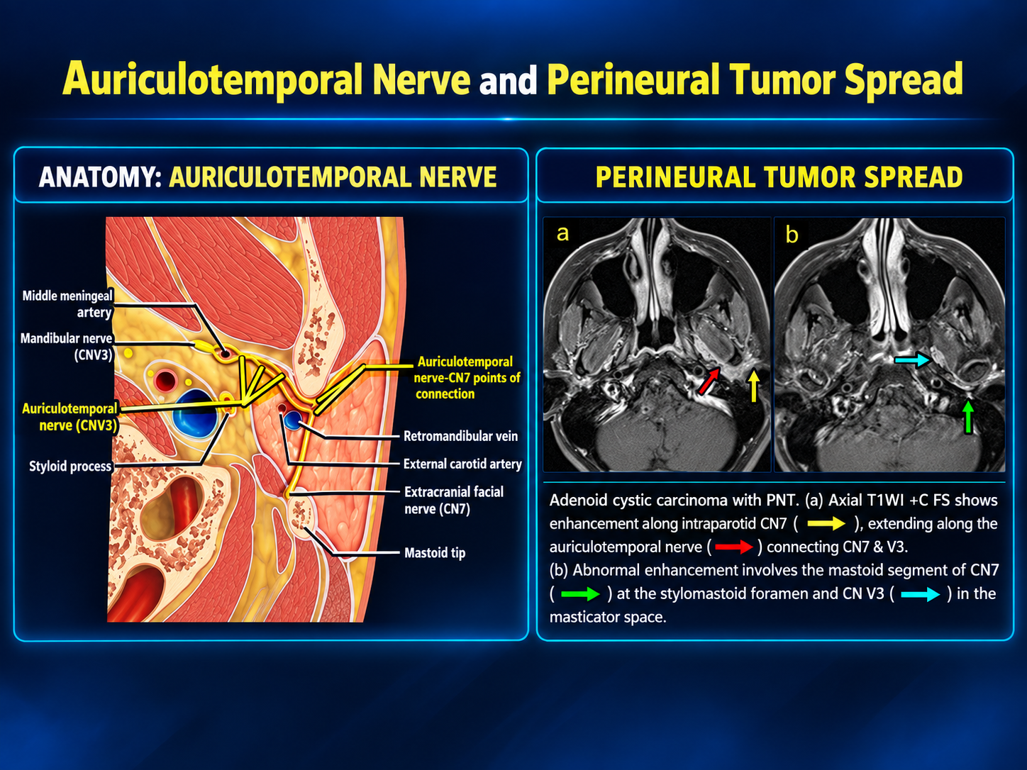
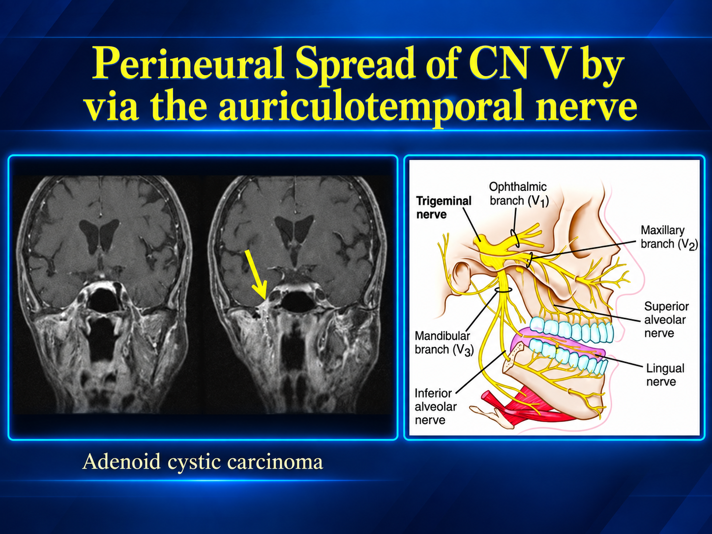
Perineural tumor spread (PNS) from a parotid malignancy is macroscopic tumor extension along a named nerve, most often the facial nerve (CN VII) or the auriculotemporal nerve connecting the parotid plexus with V3. It is an important adverse prognostic and staging feature that alters the surgical field and radiation-treatment volume.[pmc.ncbi.nlm.nih][jnm.snmjournals][pmc.ncbi.nlm.nih]
Facial nerve pathway
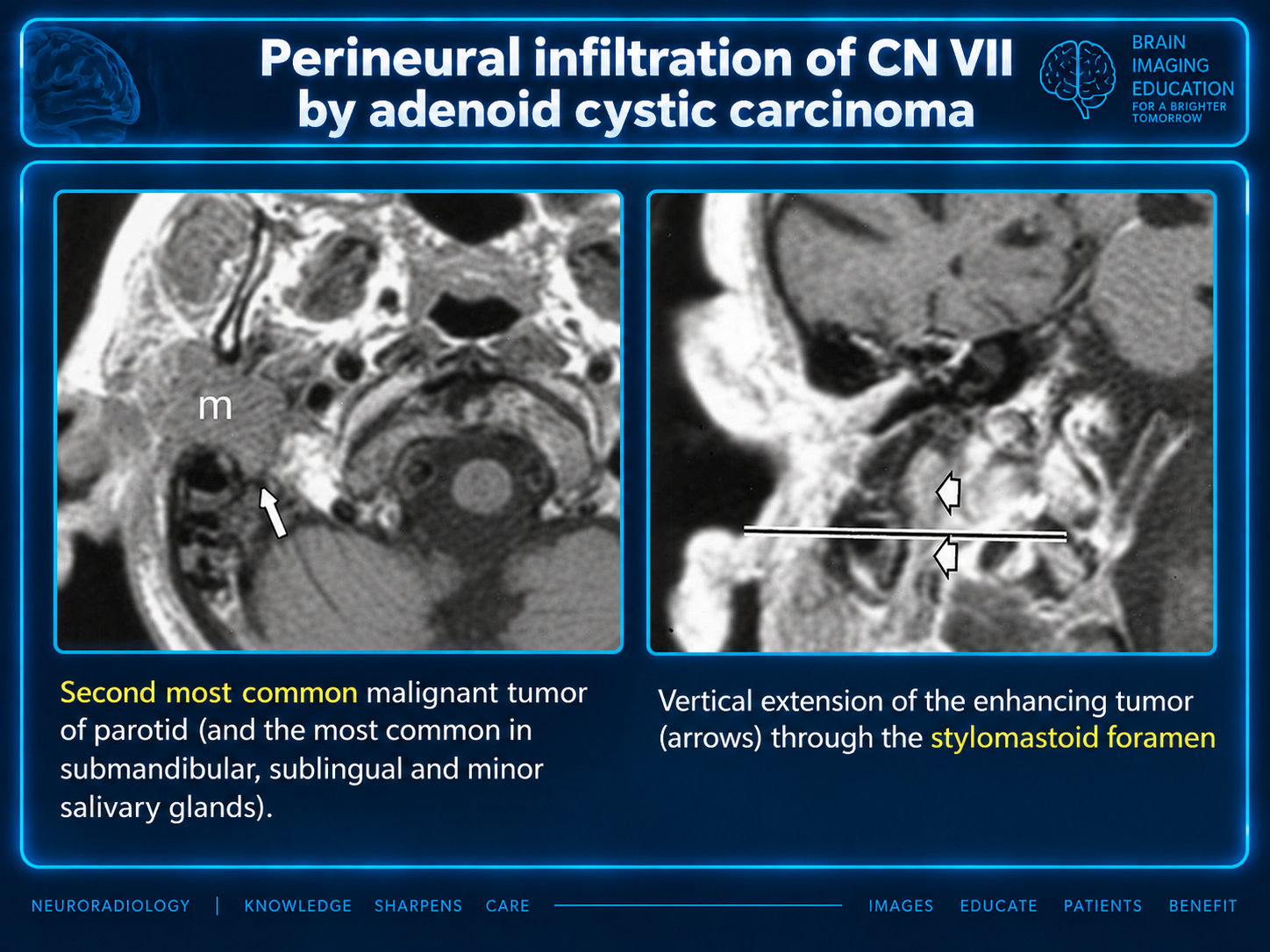
Tumor may extend proximally from intraparotid facial-nerve branches into the main trunk, then through the stylomastoid foramen → mastoid facial canal → tympanic/labyrinthine segments → geniculate ganglion → internal auditory canal. Normal facial movement does not exclude microscopic or radiographic nerve involvement.[pmc.ncbi.nlm.nih][pmc.ncbi.nlm.nih][pmc.ncbi.nlm.nih]
Auriculotemporal pathway
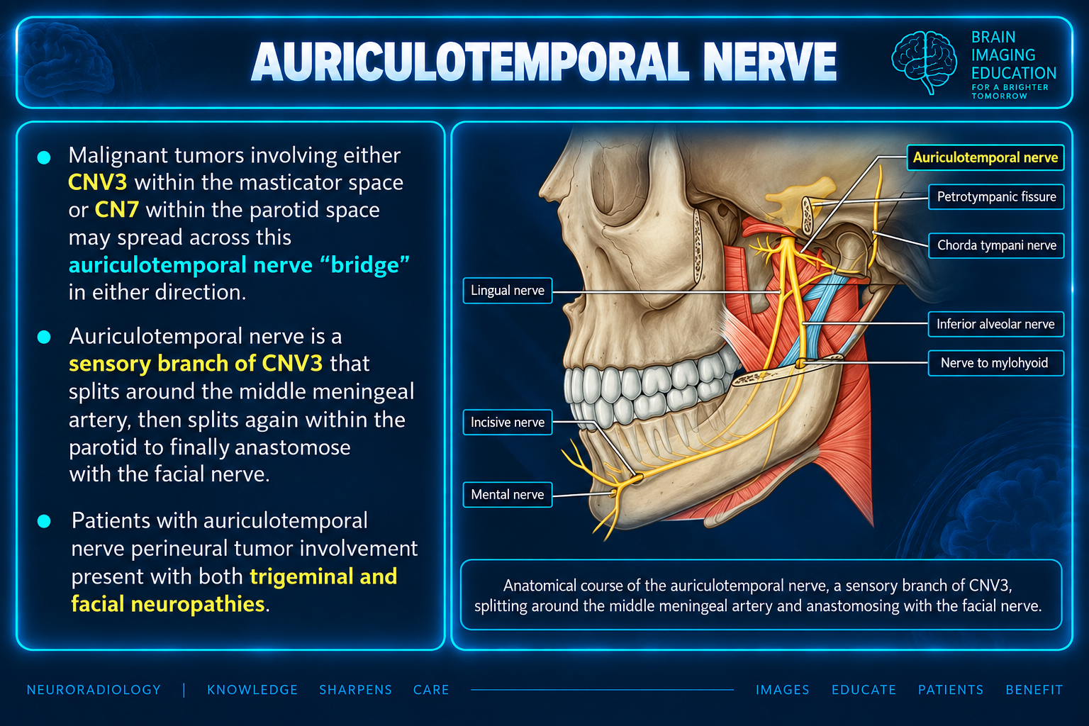
The auriculotemporal nerve provides a connection between CN VII and V3 within the parotid/retromandibular region. Tumor can track around the posterior mandibular ramus into the masticator space, then proximally along V3 through the foramen ovale → Meckel cave → cavernous sinus.[ajronline][pmc.ncbi.nlm.nih][pmc.ncbi.nlm.nih]
Combined facial weakness and trigeminal sensory symptoms should particularly raise concern for spread through the auriculotemporal connection.[pmc.ncbi.nlm.nih][onlinelibrary.wiley]
Associated tumors
PNS may occur with any parotid malignancy but is particularly associated with:
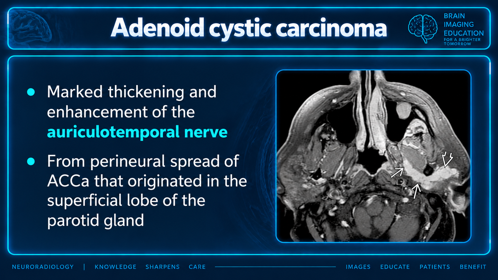
- Adenoid cystic carcinoma, including relatively small primary tumors.
- Salivary duct carcinoma.
- High-grade mucoepidermoid carcinoma.
- Carcinoma ex pleomorphic adenoma.
- Metastatic cutaneous squamous cell carcinoma involving intraparotid nodes or the gland.[jnm.snmjournals][pmc.ncbi.nlm.nih][pmc.ncbi.nlm.nih][pmc.ncbi.nlm.nih]
Imaging findings
MRI
Contrast-enhanced MRI with thin-section, fat-suppressed T1 imaging is the preferred examination. Important findings include:[pmc.ncbi.nlm.nih][pmc.ncbi.nlm.nih]
- Asymmetric nerve enlargement and enhancement.
- Linear or curvilinear enhancement extending away from the tumor.
- Replacement of fat at the stylomastoid foramen or foramen ovale.
- Enlargement or enhancement of the facial canal, foramen ovale, Meckel cave, or cavernous sinus.
- Abnormal enhancement extending to the geniculate ganglion or internal auditory canal.
- V3-related denervation edema or later fatty atrophy of the muscles of mastication.
- Facial-nerve denervation changes in the muscles of facial expression.
- Apparent discontinuous or “skip” involvement, requiring evaluation of the entire nerve to the skull base.[insightsimaging.springeropen][ajnr][geiselmed.dartmouth][pmc.ncbi.nlm.nih][pmc.ncbi.nlm.nih]
CT and PET/CT
CT is less sensitive for early neural involvement but may demonstrate foraminal widening, facial-canal expansion, loss of foraminal fat, or skull-base erosion. PET/CT may show linear FDG uptake along the nerve but is complementary to MRI.[jnm.snmjournals][jnm.snmjournals][ajronline]
Evaluate the entire nerve
Apparent discontinuous or “skip” involvement requires evaluation of the entire nerve to the skull base.
Reporting checklist
A radiology report should identify:
- Involved nerve and side.
- Most distal and proximal extent.
- Facial-nerve involvement relative to the stylomastoid foramen, mastoid segment, geniculate ganglion, and IAC.
- Auriculotemporal/V3 involvement through the masticator space, foramen ovale, Meckel cave, and cavernous sinus.
- Denervation changes.
- Skull-base, intracranial, nodal, and adjacent-space involvement.
- Whether the proximal nerve margin appears radiographically free of disease.
Sample impressions
Facial nerve spread
Findings concerning for perineural tumor spread along the right facial nerve, extending from the parotid mass through the stylomastoid foramen into the mastoid facial canal. No extension to the geniculate ganglion or internal auditory canal.
Auriculotemporal spread
Curvilinear enhancing soft tissue extends from the parotid mass around the posterior mandibular ramus along the expected course of the auriculotemporal nerve, with contiguous involvement of V3 through the foramen ovale. Findings are consistent with perineural tumor spread. No extension into Meckel cave or the cavernous sinus.
These are supplied reporting examples, not laterality or extent assigned to the reference images.
Source limitations
The supplied prose contains bracketed source labels (for example, [pmc.ncbi.nlm.nih] and [ajronline]) without article titles, complete URLs, or a bibliography. They are retained beside the corresponding text; individual references cannot be resolved from the supplied material.
The original teaching graphics and their captions are preserved. Figure 1 states that affected patients present with both trigeminal and facial neuropathies; the supplied prose treats the combined symptoms as a reason for concern and also states that normal facial movement does not exclude involvement. The graphic’s statement should not be interpreted as a requirement in every patient.
Figure 4 includes a relative-frequency claim for adenoid cystic carcinoma that is not further sourced in the supplied prose. No frequency estimates or formal staging thresholds were supplied.