Two compartments
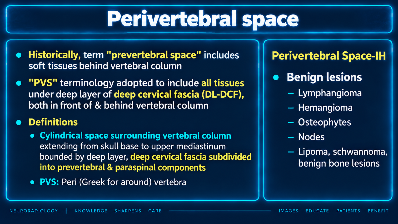
Anterior prevertebral and posterior paraspinal compartments are enclosed by the prevertebral layer of deep cervical fascia.
Localize the lesion before narrowing the differential: the supplied entities include processes within the perivertebral space, secondary extension from the spine or adjacent tissues, and nonneoplastic mass mimics.
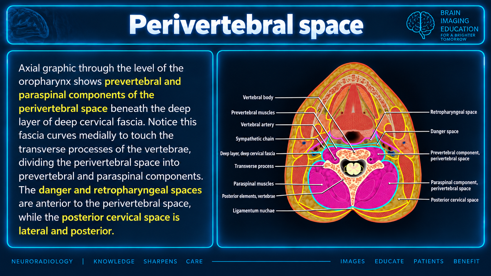
Anterior prevertebral and posterior paraspinal compartments are enclosed by the prevertebral layer of deep cervical fascia.
Vertebral-origin lesions tend to displace the prevertebral muscles and retropharyngeal fat anteriorly; retropharyngeal lesions tend to compress the muscles posteriorly against the spine.
Retropharyngeal nodes are outside the perivertebral space proper. “Pseudotumors” and “benign bone lesions” are descriptive categories, not specific diagnoses.
The perivertebral space includes the anterior prevertebral compartment and posterior paraspinal compartment, enclosed by the prevertebral layer of deep cervical fascia. Its contents include the cervical spine, associated muscles, vertebral vessels, and proximal nerve roots. It lies posterior to the retropharyngeal and danger spaces. Thus, some entities in your list arise within the perivertebral space, whereas others invade it or mimic a mass there. radiopaedia
A useful localization clue: vertebral-origin lesions tend to displace the prevertebral muscles and retropharyngeal fat anteriorly; retropharyngeal lesions tend to compress the prevertebral muscles posteriorly against the spine. radiopaedia
| Entity | Relationship to the perivertebral space | Key imaging findings / distinguishing points |
|---|---|---|
| Retropharyngeal abscess | Primarily an adjacent-space process, not a primary perivertebral lesion. Can coexist with or extend from cervical spinal infection. | Retropharyngeal fluid collection with peripheral enhancement, surrounding inflammation, and often restricted diffusion of purulent contents. Evaluate the vertebrae, discs, prevertebral muscles, and epidural space for a spinal source or associated extension. pmc.ncbi.nlm.nih |
| Osseous metastases | Vertebral metastases may extend into the prevertebral or paraspinal soft tissues and epidural space. | Marrow replacement with low T1 signal and enhancement; cortical destruction and an associated soft-tissue mass may occur. Compared with vertebral lymphoma, metastases more often produce destructive cortical change and an altered vertebral contour. radiopaedia |
| Chordoma | Malignant notochordal tumor arising in the clivus or vertebral body, with secondary perivertebral extension. | Destructive, usually midline osseous lesion with a lobulated soft-tissue mass, striking T2 hyperintensity, and heterogeneous/septal enhancement. CT may show intratumoral calcifications or residual bone fragments. Can extend across several vertebral levels and into the epidural space. radiopaedia |
| Osteomyelitis | Infection originating in the vertebrae, often with associated disc and perivertebral involvement. | Early MRI findings include low T1/high STIR marrow signal and enhancement. More advanced disease produces osseous destruction and prevertebral/paraspinal inflammatory change or abscess. Assess for epidural extension. radiopaedia |
| Schwannoma | Benign nerve-sheath tumor that may involve the perivertebral region along cervical nerve roots or the proximal brachial plexus. | A nerve-sheath tumor is a recognized lesion of the prevertebral/adjacent deep-neck spaces. Localization to a nerve or neural foramen helps distinguish it from a vertebral-origin tumor or retropharyngeal mass. pmc.ncbi.nlm.nih |
| Osteochondromas | Benign osseous outgrowths from the cervical spine projecting into adjacent perivertebral tissues or toward the canal/foramina. | Defining feature is continuity of both cortex and medullary cavity with the parent vertebra, with an overlying cartilage cap. CT best defines the bony origin and relationship to the canal and neural foramina. pubs.rsna |
| Direct invasion by squamous cell carcinoma | Secondary invasion from an adjacent mucosal primary rather than a primary perivertebral tumor. | Look for continuity with the primary mass, posterior spread through the retropharyngeal tissues, and infiltration/replacement of the prevertebral musculature. Distinguish direct invasion from retropharyngeal nodal metastases. pmc.ncbi.nlm.nih |
| Pseudotumors | A descriptive category of nonneoplastic abnormalities that mimic a mass, rather than a single diagnosis. | Potential mimics include edema, hematoma, inflammatory collections, and degenerative disc-related protrusions. The specific diagnosis depends on tissue characteristics, relation to the spine, and clinical history. The slide’s unqualified term “pseudotumors” does not establish one particular entity. pmc.ncbi.nlm.nih |
| Anterior herniated disc | Disc material projecting anteriorly from the intervertebral space into the prevertebral region; a mass mimic. | Demonstrating continuity with the disc and a disc-level epicenter is the key clue. Anterior disc bulges are recognized causes of prevertebral mass-like abnormalities. pmc.ncbi.nlm.nih |
| Vertebral osteophytes | Degenerative bony projections from vertebral margins that encroach on the prevertebral region. | A bone-density projection continuous with the vertebra, rather than a separate soft-tissue mass. Best categorized as a spine-origin mass mimic; anterior displacement of adjacent soft tissues supports this localization. radiopaedia |
| Calcific tendinitis of the longus colli | Sterile hydroxyapatite-associated inflammation of the longus colli tendon within the prevertebral compartment, producing reactive retropharyngeal edema/effusion. | Amorphous calcification typically anterior to C1–C2, with longus colli/prevertebral edema and a smooth retropharyngeal effusion. Unlike abscess, the effusion generally lacks rim enhancement and restricted diffusion. CT is best for detecting the calcification. pmc.ncbi.nlm.nih |
| Lymphoma | May involve vertebral marrow, perivertebral soft tissues, or adjacent nodal spaces. | A relatively bulky paraspinal soft-tissue component with less cortical destruction than expected can suggest lymphoma. The vertebral “wrap-around” pattern describes marrow and surrounding soft-tissue involvement with relative preservation of vertebral shape; it is suggestive, not diagnostic. radiopaedia |
Osteophytes, schwannoma, and osteochondroma are covered above. The remaining terms need some compartmental and terminology clarification.
| Slide term | Perivertebral-space relevance and imaging summary |
|---|---|
| Lymphangioma | More appropriately termed a lymphatic malformation. Can involve or traverse multiple deep-neck spaces, including the prevertebral region. Typically multiloculated and markedly T2 hyperintense, with enhancement mainly of the walls and septa; hemorrhage may produce fluid–fluid levels. pmc.ncbi.nlm.nih |
| Hemangioma | A vascular lesion may involve the prevertebral/adjacent neck tissues. True soft-tissue hemangiomas generally show high T2 signal and intense enhancement, sometimes with flow voids. “Hemangioma” should not automatically be used interchangeably with venous malformation, which is a different vascular-anomaly category. pmc.ncbi.nlm.nih |
| Nodes | Important localization correction: retropharyngeal lymph nodes lie anterior to the prevertebral muscles, in the suprahyoid retropharyngeal space—not within the perivertebral space proper. A nodal lesion should therefore prompt reconsideration of the assigned compartment. pmc.ncbi.nlm.nih |
| Lipoma | Rare benign fatty lesion of the deep-neck spaces, potentially involving the prevertebral region. Shows fat attenuation on CT and high T1 signal that suppresses on fat-saturated MRI. Thick enhancing septa or nodular nonfat components warrant consideration of a more complex lipomatous lesion. pmc.ncbi.nlm.nih |
| Benign bone lesions | An umbrella category rather than a specific diagnosis. These are localized to the vertebral component by their bony epicenter and may project into adjacent perivertebral tissues. Osteochondroma is a specific example; the supplied text does not identify which additional benign bone lesions the slide intended. radiopaedia |
The heading “Perivertebral Space-IH” can be retained as supplied, but the suffix “IH” remains undefined; it should not be expanded without the original source’s explanation.