Compartment
A cylinder of soft tissue enclosed by the prevertebral layer of the deep cervical fascia, extending from the skull base to the upper mediastinum.
The perivertebral space is a deep compartment of the head and neck comprising the prevertebral and paraspinal spaces, posterior to the retropharyngeal and danger spaces.
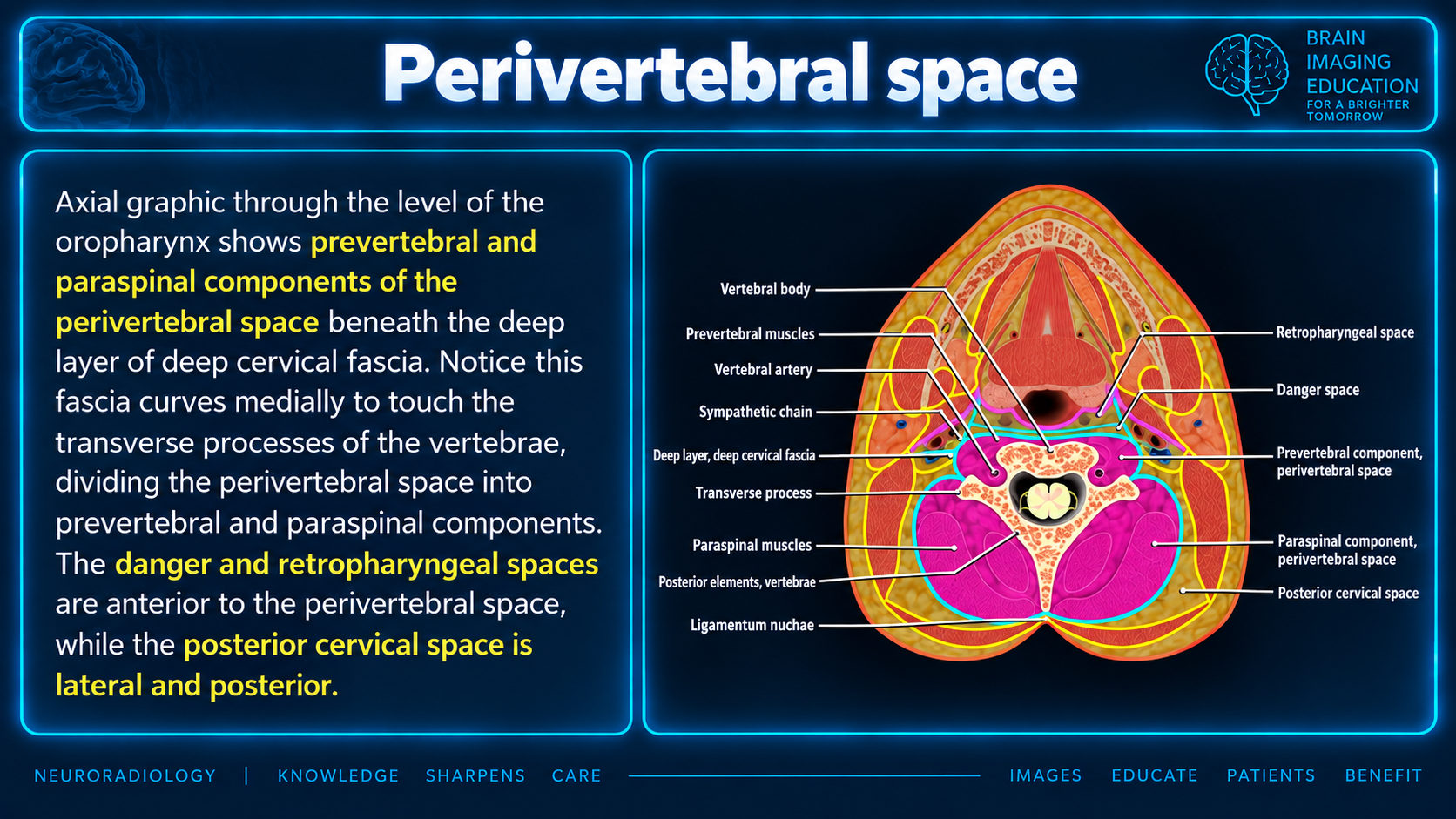
A cylinder of soft tissue enclosed by the prevertebral layer of the deep cervical fascia, extending from the skull base to the upper mediastinum.
A deep fascial slip to the transverse process separates the anterior prevertebral and posterior paraspinal portions.
CT depicts bone destruction. MRI is preferred for soft-tissue assessment and detection of early spinal infection. [1]
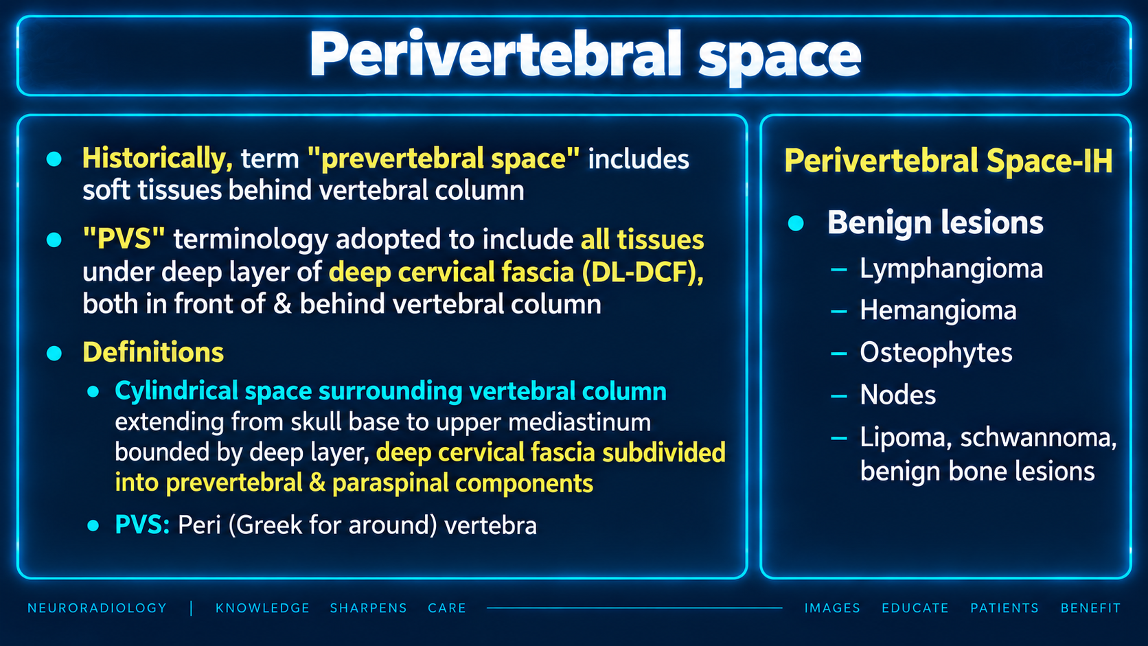
The supplied teaching slide describes the historical use of “prevertebral space” as including soft tissues behind the vertebral column. “Perivertebral space” (PVS; peri means around) includes tissues both in front of and behind the column beneath the deep layer of the deep cervical fascia.
| Boundary | Description |
|---|---|
| Anterior | Danger space and retropharyngeal space |
| Posterior | Fascia attaches to the spinous process and ligamentum nuchae |
| Lateral | Surrounded by the posterior cervical space |
| Superior | Base of skull |
| Inferior | Usually defined to the superior mediastinum; the supplied text also describes extension to the coccyx. |
The cervical definition above is the usual extent used in the supplied material; the broader description to the coccyx is retained as a source qualification.
On sagittal imaging, the prevertebral component is generally measured from the anterior vertebral-body border to the air within the pharynx or trachea. Thickness varies with body build and normal variation. No definitive cutoff perfectly separates patients with injury from those without injury; sensitivity and specificity depend on the threshold selected.
| Level | Measurement |
|---|---|
| C2/3 | <7 mm |
| C6/7 | <21 mm |
Values above these measurements produced a true positive rate of 53% and a false positive rate of 5% in that study. [7]
| Level | Thickness |
|---|---|
| C1 | 8.5 mm |
| C2 | 6 mm |
| C3 | 7 mm |
| C4/C5 | Variable because of variable cricoid/esophageal height |
| C6 | 18 mm |
| C7 | 18 mm |
Pediatric values differ and are more variable because of superior adenoidal tissue and smaller size.
| Age (years) | C2 | C6 |
|---|---|---|
| 0–2 | 7.6 mm | 9.0 mm |
| 3–6 | 8.4 mm | 9.8 mm |
| 7–10 | 6.8 mm | 12.1 mm |
| 11–15 | 6.8 mm | 14.5 mm |
Preferred for assessing soft tissues in this space and detecting early spinal infection. [1]
Acute marrow edema produces low T1-weighted and high T2-weighted signal, especially with fat-saturated techniques, spectroscopic inversion recovery (SPIR) or short tau inversion recovery (STIR) sequences. [4]
The described “wrap-around” sign is tumor in marrow and paraspinal soft tissue without alteration of the normal vertebral shape. This may help differentiate lymphoma from metastasis and myeloma, which tend to destroy the cortex and produce a bulging contour.
Source limitation: The T2 phrase is reproduced as supplied; the sequence attribution of contrast enhancement is ambiguous and has not been resolved.
Homogeneous hypointense/isointense lesions on T1-weighted images, hyperintense on T2-weighted images, with post-gadolinium high signal. Tumor develops within vertebral bodies, frequently invades the perivertebral/epidural spaces and may span several vertebral segments. [2]
Lymphangioma, hemangioma, osteophytes, nodes, lipoma, schwannoma and benign bone lesions. The slide’s “Perivertebral Space-IH” heading is retained in the original image; the suffix is not defined in the supplied material.
This page organizes the supplied text and two teaching slides. Bracketed numbers [1–8] preserve the source’s reference markers; no bibliography was supplied, so the cited studies cannot be identified from this material. The numerical values and study performance above are retained as source-specific findings rather than universal diagnostic cutoffs.