Contents
- Areolar fat (the main component)
- Retropharyngeal lymph nodes
- Small vessels
The retropharyngeal space (RPS) is a midline deep compartment of the head and neck, consisting largely of fatty areolar tissue and lymph nodes that drain the pharynx, nose and middle ear.
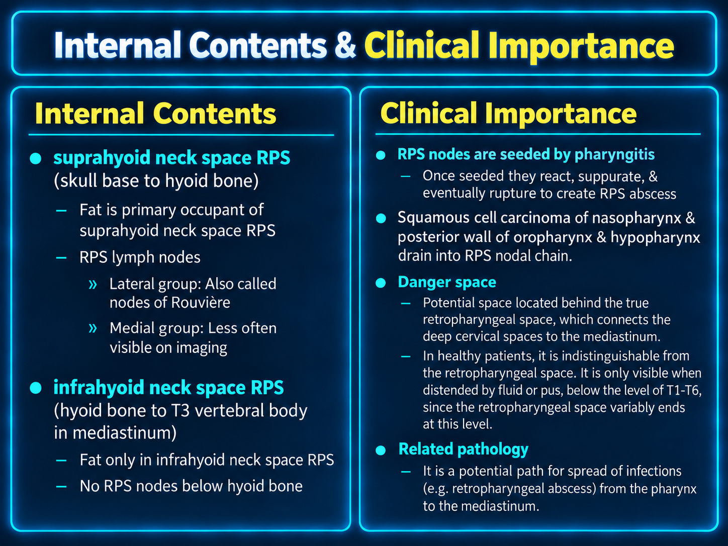
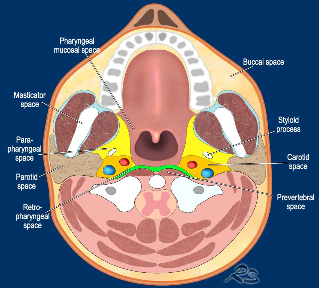
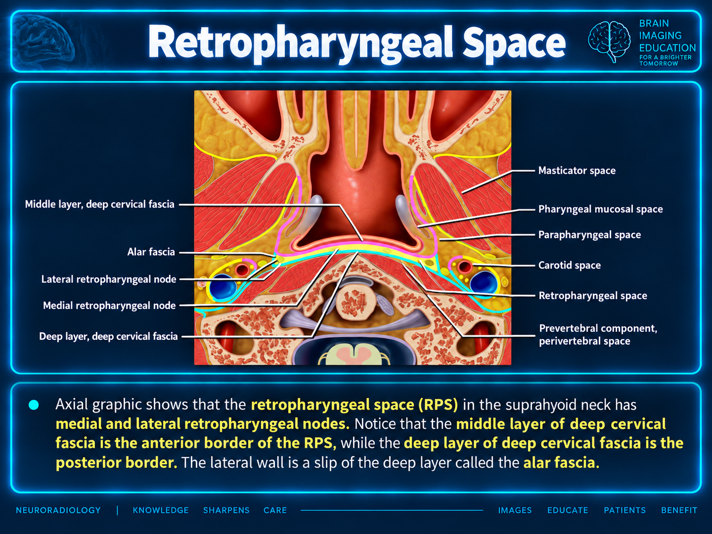
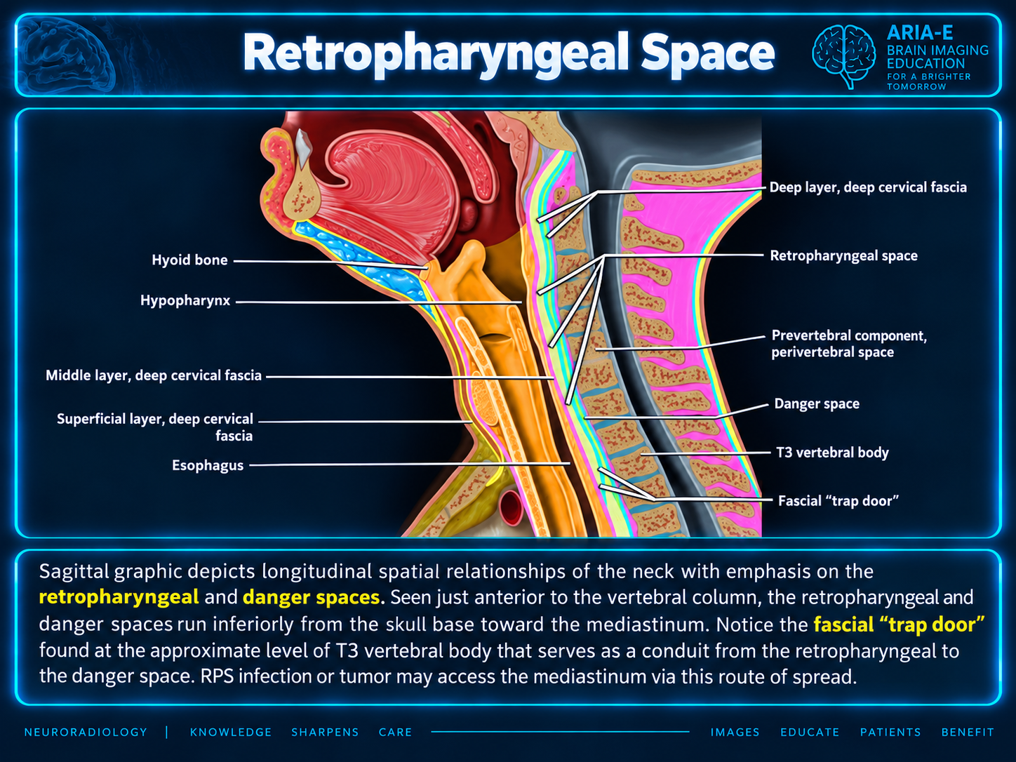
Nodes lie above the hyoid bone and drain the pharynx, nasal cavity, paranasal sinuses and middle ears. They are prominent in children and atrophy with age. [2]
The supplied slide identifies lateral nodes as nodes of Rouvière and notes that medial nodes are less often visible on imaging.
A small, roughly rectangular potential space on axial imaging: wider mediolaterally and thinner anteroposteriorly. Areolar tissue has fat density on CT and fat signal on MRI.
The very thin alar fascia makes separation from the danger space difficult. [3]
Here, retropharyngeal space is used loosely to include its inferior continuation below the pharynx, surrounding the esophagus and extending into the mediastinum. Some authors use retrovisceral space for its relationship to the visceral space. [6] In that convention, only the superior portion posterior to the pharynx is retropharyngeal; its inferior continuation is the retroesophageal space.
The thin alar fascia divides the interval between the visceral (buccopharyngeal) and prevertebral fascia into an anterior true retropharyngeal space and a posterior danger space. [4] Some authors include the true retropharyngeal space within the visceral space. [6]
The alar fascia is not discernible on cross-sectional imaging. [4] Many authors therefore use retropharyngeal space for the combined functional unit, treating danger space as a subcomponent or synonym. [7–9]
The space lies posterior to the pharynx and esophagus, extending from the skull base to the thoracocervical junction, where the alar fascia attaches to the buccopharyngeal fascia at a variable level between C6 and T6. [2]
| Margin | Boundary |
|---|---|
| Anterior | Middle layer of the deep cervical fascia [1] |
| Posterior | Alar fascia, separating the RPS from the danger space |
| Lateral | Deep layer of the deep cervical fascia [1] |
| Superior | Clivus |
| Inferior | Fusion of the alar fascia with the middle layer of the deep cervical fascia, typically around T4 [3] |
The supplied teaching slide describes pharyngitis seeding retropharyngeal nodes, which may react, suppurate and rupture to produce an RPS abscess. It also describes drainage of squamous cell carcinoma of the nasopharynx and posterior walls of the oropharynx and hypopharynx into the RPS nodal chain.
The danger space provides a potential route for infection to spread from the pharynx through the deep cervical spaces to the mediastinum. The supplied sagittal figure illustrates a fascial “trap door” near T3 through which infection or tumor may enter this route.
The supplied prose states that obliteration of retropharyngeal fat predicts involvement of the prevertebral fascia, indicating locally advanced disease. [5]
The supplied prose and teaching figures use differing fascial conventions. The prose defines the posterior boundary of the true RPS as the alar fascia and the lateral boundaries as the deep cervical fascia. The axial slide instead labels the deep layer as the posterior border and the alar fascia as the lateral wall. The original figure and caption are preserved; these descriptions should not be read as identical definitions.
Inferior extent also varies across the material: the prose gives C6–T6 and separately a typical fusion near T4; the contents slide uses T3 and elsewhere T1–T6; the sagittal slide depicts a T3 “trap door.” These are preserved source descriptions rather than a single fixed termination level.
The contents slide summarizes the infrahyoid RPS as fat only, with no nodes below the hyoid, whereas the prose lists small vessels among overall contents. The original wording is retained in the graphic.
Bracketed citation numbers are retained from the supplied prose. No corresponding bibliography or reference links were supplied, so those citations cannot be identified from this package.