Sublingual spaces pathology
Localize the epicenter first: sublingual gland, mucosal floor of mouth, Wharton duct, or another structure. Then map the relationship to the mylohyoid and adjacent submandibular space.
Reference Images
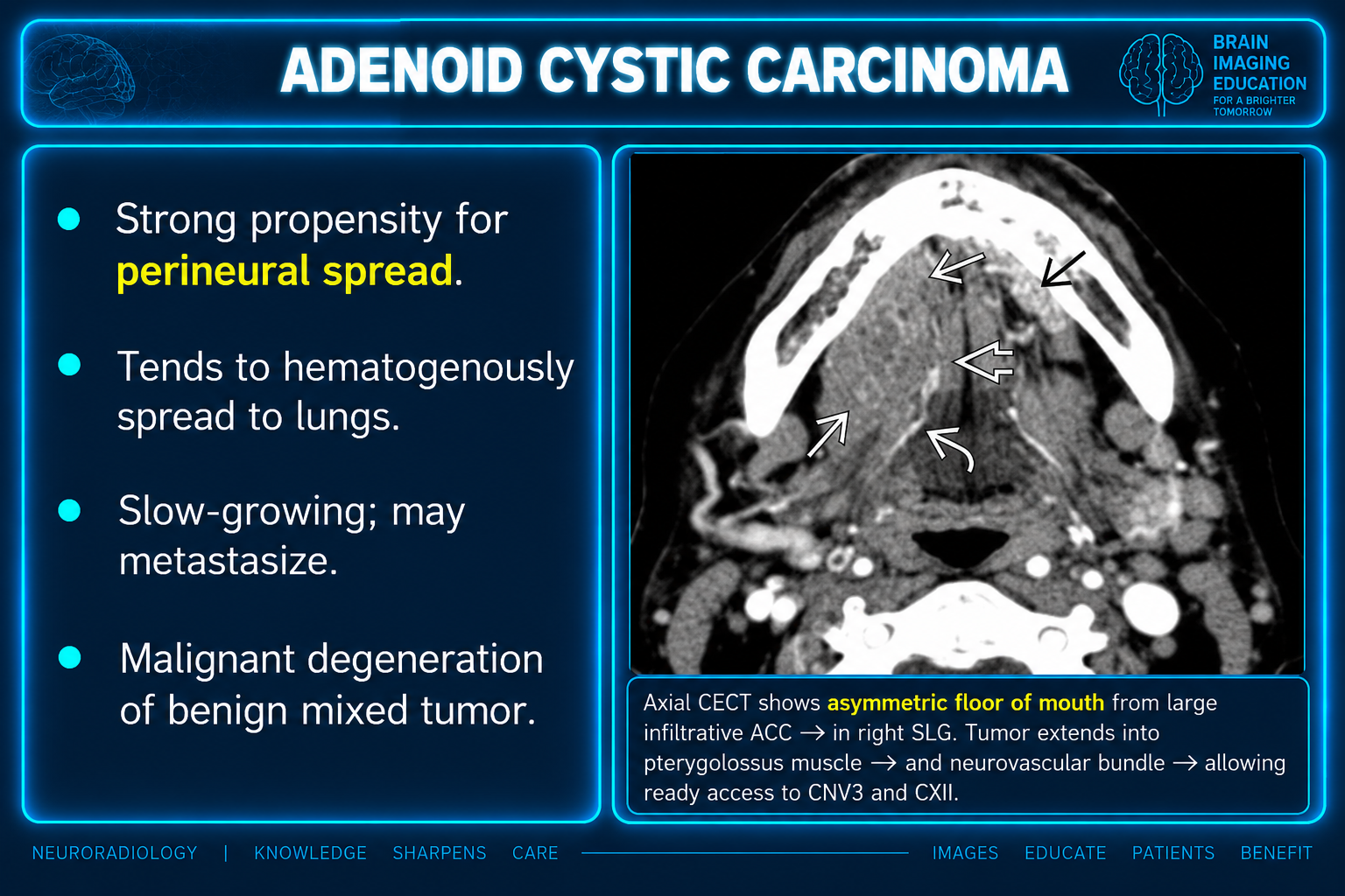
Source limitations and differing statements
The prose retains numbered and caret-prefixed reference markers, but no bibliography or reference URLs were supplied; these citations cannot be resolved from this package. The graphics are reproduced unchanged with their embedded captions. No separate caption file was supplied. The anatomy slide calls the sublingual glands “minor salivary glands”; the prose describes the sublingual gland separately from minor-salivary tumors. The adenoid cystic carcinoma slide states “malignant degeneration of benign mixed tumor,” whereas the prose presents adenoid cystic carcinoma as a primary glandular histology; the relationship is not established by the supplied material. The benign mixed tumor slide describes usually homogeneous enhancement, while the prose allows heterogeneity in larger pleomorphic adenomas. These source statements are preserved without reconciliation. Slide treatment statements and additional differential details have no accompanying references and are retained within the originals.
Executive overview
The most useful first step is compartmental localization, not histologic guessing: determine whether the lesion is centered in the sublingual gland, mucosal floor of mouth, Wharton duct, or another structure within the sublingual space; then determine its relationship to the mylohyoid muscle and adjacent submandibular space. The highest-yield diagnoses are ranula, dermoid/epidermoid cyst, odontogenic or salivary infection, Wharton-duct obstruction, vascular malformation, floor-of-mouth squamous cell carcinoma, and primary sublingual-gland carcinoma.[1][2]
Three practical rules dominate interpretation:
- A sublingual-gland mass deserves oncologic concern. Sublingual-gland tumors are rare, but approximately 70%–90% are malignant in published series; adenoid cystic carcinoma and mucoepidermoid carcinoma predominate.[3][4][^5]
- The mylohyoid is the key landmark. It separates the sublingual space above from the submandibular space below, but posterior communication and common mylohyoid defects permit disease to cross between them.[6][1]
- MRI solves most indeterminate masses; contrast CT solves most acute infections and mineralized/osseous questions. MRI is superior for tissue characterization, marrow infiltration, perineural spread, and deep local extent; CT is superior for calculi, cortical bone, gas, and urgent mapping of deep neck infection.[2][7][^8]
Applied anatomy
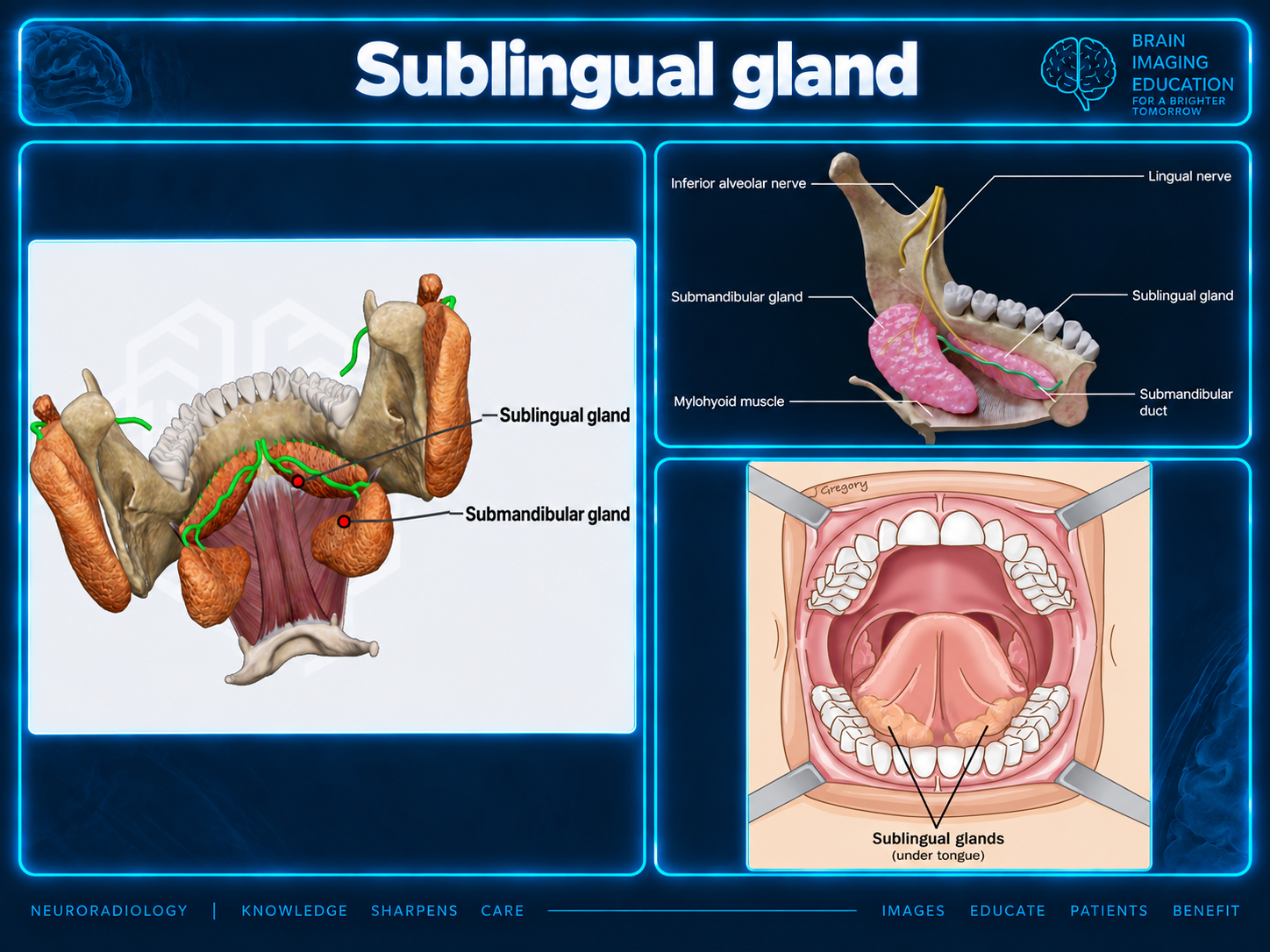
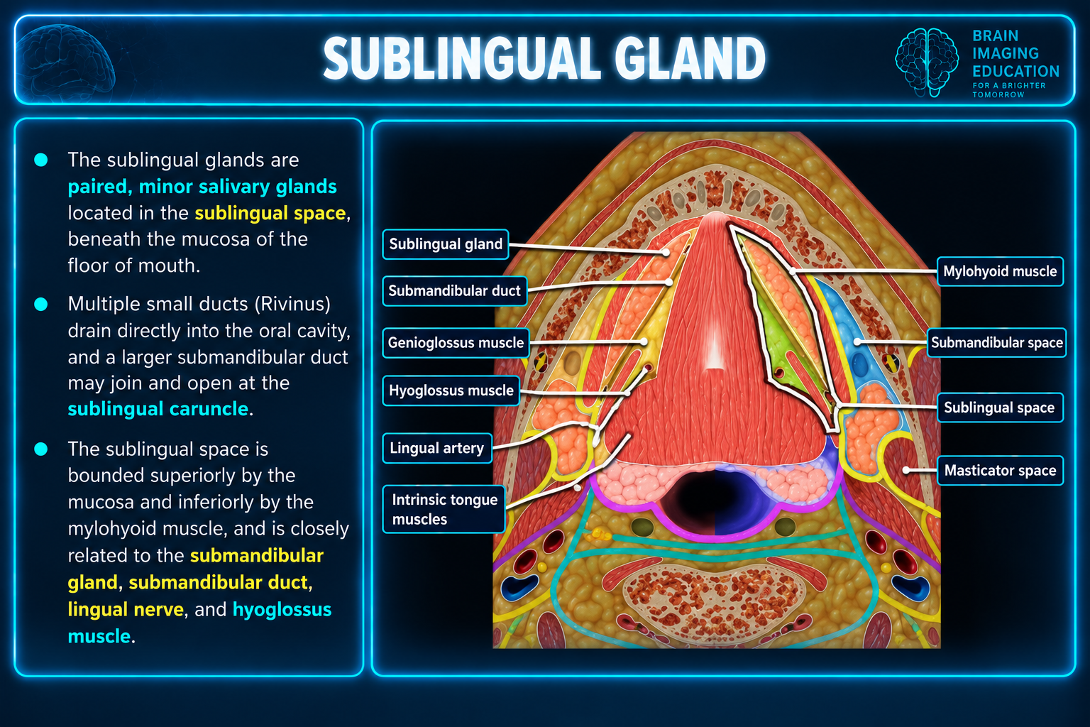
The sublingual space lies superior to the mylohyoid, lateral to the genioglossus–geniohyoid complex, medial to the mandible, and deep to the floor-of-mouth mucosa. It contains fat, the sublingual gland and ducts, Wharton duct and the deep submandibular gland component, lingual vessels, lingual nerve, portions of cranial nerves IX and XII, and part of the hyoglossus muscle.[1][9]
The right and left sublingual spaces communicate anteriorly beneath the frenulum. Each communicates with the submandibular space around the free posterior edge of the mylohyoid; disease may subsequently reach the parapharyngeal space, and an anterior mylohyoid defect provides an additional route.[2][1]
The sublingual gland is nonencapsulated and lies immediately beneath the mucosa, adjacent to the lingual nerve, Wharton duct, mandible, and floor-of-mouth muscles. This explains why even a small primary carcinoma may show extraparenchymal extension or involve neighboring structures and why localization to the gland has more clinical value than attempting definitive histology by imaging.[^3]
Mylohyoid boutonnière
Focal mylohyoid defects are common and may contain sublingual gland tissue, fat, or vessels. Continuity with the normal gland, identical attenuation/signal and enhancement, and passage through a discrete muscular defect establish a benign mylohyoid boutonnière rather than a submandibular-space mass.[10][11][^6]
A true lesion can also descend through a boutonnière. Therefore, the finding of a mylohyoid defect does not by itself make a process benign; confirm whether the tissue is normal gland or a distinct cystic, inflammatory, or solid lesion.[^6]
Modality strategy
| Clinical problem | Preferred examination | Main objective |
|---|---|---|
| Acute painful swelling, fever, airway concern | Contrast-enhanced neck CT; include mandible/dentition and extend through the superior mediastinum when deep infection is suspected | Detect cellulitis, drainable abscess, gas, odontogenic source, vascular complication, osteomyelitis, and airway compromise.[2][12] |
| Suspected calculus or distal Wharton-duct obstruction | Thin-section noncontrast CT or ultrasound; add contrast CT if infection/abscess is suspected | Identify and localize mineralized calculi, duct dilation, and secondary adenitis.[8][13] |
| Indeterminate cystic floor-of-mouth lesion | MRI with T1, fluid-sensitive fat-suppressed imaging, DWI/ADC, and postcontrast fat-suppressed T1 | Separate ranula, epidermoid, dermoid, abscess, vascular/lymphatic lesion, and cystic tumor.[1][2] |
| Solid sublingual-gland or floor-of-mouth mass | Contrast-enhanced MRI from floor of mouth through skull base; CT bone algorithm when mandibular cortex is in question | Define mucosal versus glandular origin, muscle/duct/mandibular involvement, marrow disease, perineural spread, and nodal disease.[3][14] |
| Suspected vascular malformation | Doppler ultrasound plus contrast MRI; CTA/MRA or catheter angiography for suspected high-flow lesion | Establish flow category, full trans-spatial extent, feeding arteries, nidus, and drainage.[^1] |
| Suspected ductal stenosis without visible stone | MR sialography or sialendoscopy according to local practice | Demonstrate duct stenosis and upstream ectasia without duct cannulation.[^8] |
Dental amalgam commonly degrades CT evaluation of the floor of mouth. If the critical region is obscured, consider metal-artifact reduction, gantry/plane adjustment parallel to the mandible, complementary MRI, or targeted ultrasound rather than accepting a nondiagnostic primary site.[9][2]
Localization algorithm
- Confirm epicenter. Decide whether the process originates in the sublingual gland, Wharton duct, mucosal floor of mouth, mandible/dentition, neurovascular bundle, or merely extends into the space.
- Use mylohyoid geography. Above the muscle favors a sublingual-space origin; below favors submandibular/submental origin. Trace extension around the posterior free edge or through a boutonnière.
- Classify composition. Simple fluid, diffusion-restricting keratin/pus, fat-containing, vascular, infiltrative soft tissue, or circumscribed solid mass.
- Interrogate the duct. Follow Wharton duct from the hilum to the papilla; look for a stone, abrupt cutoff, upstream dilation, and secondary submandibular adenitis.
- Map danger features. Airway narrowing, drainable collection, mandibular invasion, perineural spread, nodal disease, and trans-spatial extension should drive the impression.
Cystic lesions
| Entity | Typical location and morphology | Key CT/MRI findings | Best discriminator |
|---|---|---|---|
| Simple ranula | Usually unilateral and lateral in the sublingual space; closely related to the sublingual gland | Thin-walled unilocular fluid lesion, T2 hyperintense, usually T1 hypointense, without a solid enhancing nodule; protein or hemorrhage may increase T1 signal.[1][15] | Sublingual epicenter and no marked diffusion restriction |
| Plunging ranula | Dominant submandibular component with connection to the sublingual space | Fluid lesion extending around the posterior mylohyoid edge or through a defect; tapered sublingual component forms the tail sign.[16][2] | Definite communication with the ipsilateral sublingual space |
| Epidermoid cyst | Commonly midline or paramedian; often displaces/splays floor-of-mouth muscles | Well-circumscribed nonenhancing cyst with pronounced DWI hyperintensity and low ADC from keratin.[1][17] | Diffusion restriction in an otherwise simple cyst |
| Dermoid cyst | Usually midline floor of mouth | Variable fluid–fat lesion; coalescent intracystic fat globules produce the sack-of-marbles appearance.[1][15] | Macroscopic fat globules |
| Lymphatic malformation | Infiltrative, multiloculated, trans-spatial; often crosses expected boundaries | Very T2 hyperintense cystic spaces, septal enhancement, possible fluid-fluid levels after hemorrhage; little or no solid enhancement.[^1] | Multispatial, multiloculated morphology without a sublingual tail |
| Abscess | Centered on dental, ductal, or glandular source; surrounding inflammatory change | Thick/irregular rim enhancement, central restricted diffusion, edema, fat stranding, and sometimes gas.[18][8] | Inflammatory context plus enhancing wall and restricted pus |
| Cystic/necrotic tumor | Irregular solid-cystic mass, often mucosal or gland-centered | Nodular or irregular enhancing tissue, invasion, abnormal nodes, possible duct obstruction | Enhancing mural/solid component or invasive behavior |
Ranula pearls
A ranula is a mucus extravasation phenomenon arising from the sublingual gland/ductal system rather than a true epithelial-lined cyst. A simple ranula remains above the mylohyoid; a plunging ranula extends into the submandibular space by the posterior route or a mylohyoid defect.[2][5]
The tail sign is highly supportive but should not be treated as universally visible. Large ranulas can extend into the parapharyngeal or upper cervical spaces, but their smooth, homogeneous fluid character and persistent tapered communication with the sublingual space help distinguish them from lymphatic malformations, which tend to be lobulated, septated, heterogeneous, and trans-spatial.[^16]
When reporting a ranula, state: simple versus plunging; side; maximum dimensions; route through/around the mylohyoid; tail sign; complexity from infection or hemorrhage; mass effect on tongue/airway; and any unexpected enhancing solid component.
Dermoid versus epidermoid
The surgical approach depends partly on position relative to the mylohyoid: lesions above it are usually approached intraorally, whereas those below it may require an external approach. This makes the muscular relationship a required reporting element, not merely an anatomic description.[^1]
Restricted diffusion is a powerful discriminator for epidermoid, but an abscess can also restrict. The absence of surrounding inflammatory change and rim enhancement, together with a chronic midline nonenhancing morphology, favors epidermoid.[18][1]
Infection and inflammation
Cellulitis and abscess
Most acute sublingual-space infections are odontogenic or salivary/ductal. Contrast CT findings include fat stranding, edema and enhancement of fascial planes; progression produces a rim-enhancing fluid collection, while MRI can increase confidence in small abscesses through central diffusion restriction.[1][18][^8]
The mylohyoid attachment helps predict dental spread. Infection from mandibular roots above the attachment tends to enter the sublingual space, whereas a source below the attachment—classically posterior mandibular molars—tends to enter the submandibular space; established infection may then become trans-spatial.[18][1]
The report should identify the diseased tooth or duct when visible, distinguish phlegmon from drainable abscess, measure collections in three planes, and evaluate mandibular osteomyelitis, major-vessel complications, nodal disease, and inferior extension.
Ludwig angina
Ludwig angina is rapidly progressive cellulitis involving the bilateral sublingual, submandibular, and submental spaces, most often from a mandibular odontogenic source. Imaging is performed after immediate airway priorities and should assess tongue/floor-of-mouth elevation, airway narrowing, abscess, gas, odontogenic source, parapharyngeal/retropharyngeal spread, and mediastinal extension.[12][19]
Diffuse edema and loss of fat planes may predominate without a discrete abscess. A report should not require a collection before raising the diagnosis when the bilateral compartment pattern and clinical setting are compelling.[2][12]
Obstructive disease
Wharton-duct disease is far more common than a true sublingual-gland calculus. Noncontrast CT is most sensitive for calcified stones; ultrasound demonstrates an echogenic focus with posterior shadowing and can show duct ectasia, while MR sialography is useful for a noncalcified obstruction or stricture.[8][20]
An obstructing floor-of-mouth tumor can mimic stone-related sialadenitis by occluding Wharton duct. In an adult with unexplained unilateral duct cutoff, recurrent ipsilateral submandibular adenitis, or disproportionate soft tissue at the duct papilla, scrutinize the mucosal floor of mouth and sublingual gland and consider MRI.[2][21]
Acute sialadenitis causes gland enlargement, hyperenhancement, duct dilation, and surrounding edema; abscess appears as a rim-enhancing collection with restricted diffusion. Chronic disease may produce irregular duct ectasia, parenchymal heterogeneity, fatty replacement, calcification, and gland volume loss.[^8]
Systemic disease
Sjögren disease, IgG4-related disease, granulomatous infection, and treatment-related sialadenitis can involve major salivary glands, but isolated sublingual-gland involvement is uncommon. Their recognition usually depends on a multiglandular pattern and systemic context rather than a solitary sublingual lesion; a unilateral focal mass should not be attributed to systemic sialadenitis without appropriate evidence.[8][22][^23]
Vascular lesions
Venous malformations are compressible slow-flow lesions that may contain phleboliths and demonstrate gradual or diffuse enhancement. MRI shows lobulated T2-hyperintense channels; Doppler usually demonstrates slow monophasic flow, although very slow flow may be difficult to detect.[1][15]
Lymphatic malformations are multicystic and trans-spatial with enhancing septa and possible fluid-fluid levels. Venolymphatic lesions combine T2-bright cystic spaces with enhancement of venous components.[^1]
Arteriovenous malformations show enlarged feeding arteries, a vascular nidus, early draining veins, and serpiginous flow voids. Avoid biopsy or aspiration of an unexplained enhancing or flow-containing floor-of-mouth lesion before vascular characterization.[^1]
Benign solid lesions
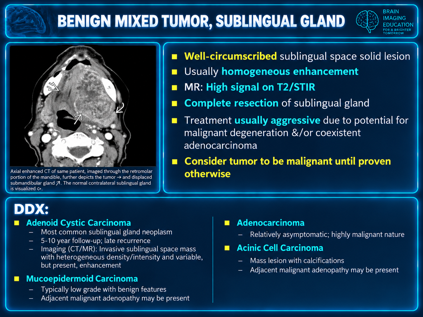
A pleomorphic adenoma is usually circumscribed, strongly T2 hyperintense, and enhancing; larger lesions may develop hemorrhagic, cystic, or necrotic heterogeneity. In the sublingual gland, however, benign imaging morphology cannot override the high site-specific probability of malignancy, so tissue diagnosis and complete oncologic mapping remain necessary.[1][5]
Lipoma follows fat on all sequences and should suppress with fat saturation; internal nonfat nodularity, thick irregular septa, or infiltrative behavior raises concern for an atypical lipomatous tumor rather than a simple lipoma.[^1]
Schwannoma or neurofibroma can arise from the lingual or hypoglossal nerve and is typically circumscribed, T1 isointense to muscle, T2 hyperintense, and enhancing. Nerve displacement, denervation change, or extension along a neural pathway can suggest the nerve of origin.[24][1]
Malignant disease
Primary sublingual-gland tumors
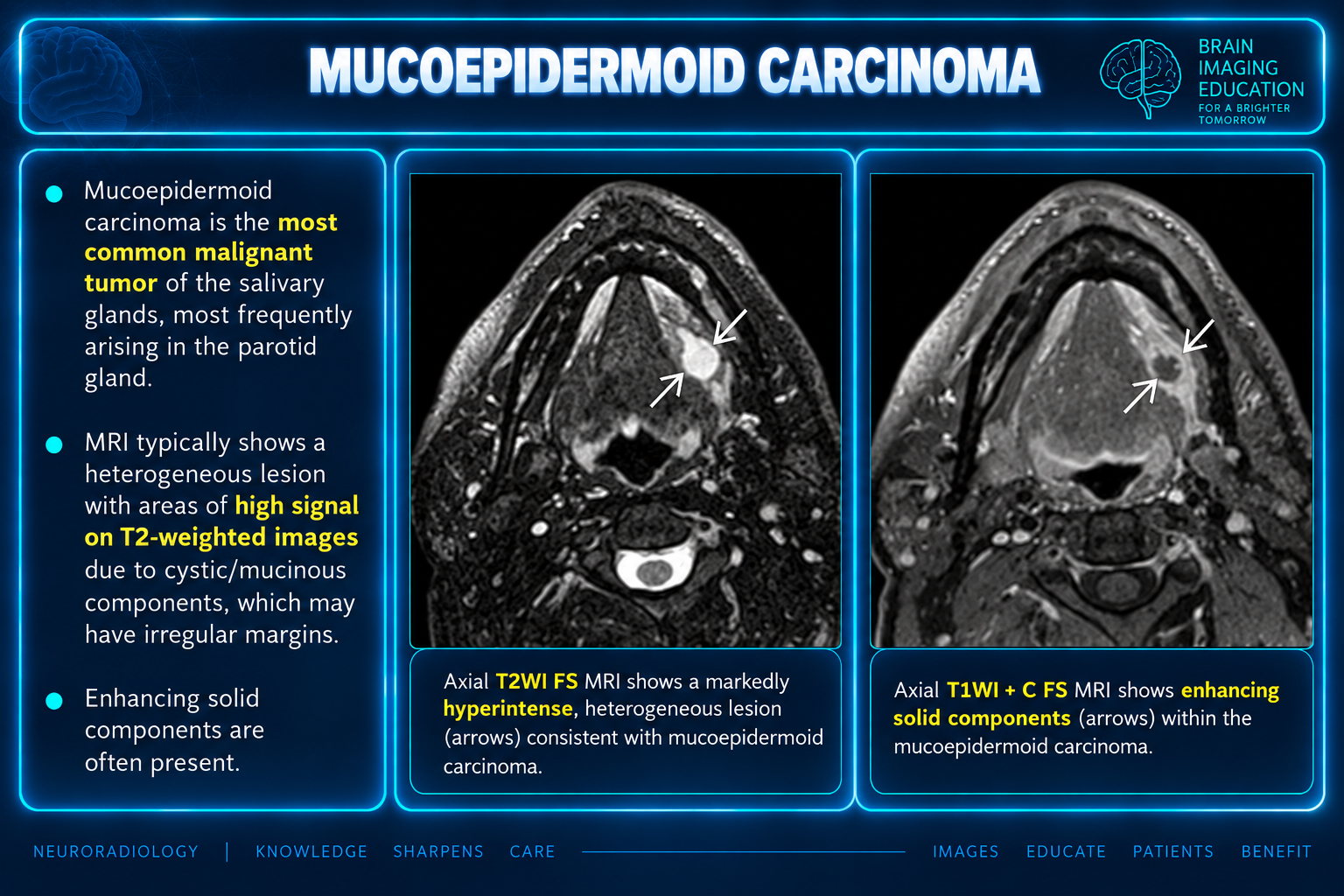
Sublingual-gland neoplasms account for about 1% of epithelial salivary tumors, but most reported sublingual tumors are malignant. Adenoid cystic carcinoma is generally the leading histology, followed by mucoepidermoid carcinoma; published proportions vary because case series are small and referral-biased.[4][5][^25]
Imaging often cannot reliably assign salivary histology. Features favoring malignancy include an ill-defined or infiltrative margin, intermediate/low rather than very high T2 signal, heterogeneous enhancement or necrosis, adjacent muscle/duct/mandibular invasion, perineural spread, and abnormal nodes; a well-defined lesion can nevertheless be malignant.[26][7]
Adenoid cystic carcinoma deserves special scrutiny for perineural spread, particularly along the lingual nerve/V3 pathway and, less commonly, the inferior alveolar nerve. MRI should cover the floor of mouth through the skull base and evaluate asymmetric nerve enhancement/enlargement, denervation change, foramen enlargement, and loss of expected fat at neural foramina.[3][14]
For any suspected sublingual-gland carcinoma, report relationship to the lingual nerve, Wharton duct and submandibular gland, mylohyoid, intrinsic/extrinsic tongue musculature, mandible, and neurovascular bundle. CT bone windows best show cortical erosion; MRI better detects marrow invasion and perineural spread.[^3]
Floor-of-mouth SCC
Floor-of-mouth squamous cell carcinoma is mucosal in origin but commonly invades the sublingual gland/space. It typically appears as an ill-defined enhancing soft-tissue mass; it may cross the anterior midline, obstruct Wharton duct, invade tongue and floor-of-mouth musculature, traverse the mylohyoid into the submandibular space, or invade the mandible.[2][21]
MRI is preferred for local soft-tissue extent and provides an imaging estimate of tumor thickness, but pathologic depth of invasion is measured from the reconstructed basement membrane rather than the visible mucosal surface. The report should avoid treating imaging thickness and pathologic depth of invasion as interchangeable.[27][28]
Required staging observations include maximum tumor dimensions; crossing of midline; tongue and floor-of-mouth muscle involvement; mylohyoid/submandibular extension; mandibular cortical and marrow invasion; perineural spread; and cervical nodal metastases. Very superficial mucosal tumors may remain occult on MRI, so a negative study does not exclude clinically evident early disease.[28][2]
Lymphoma and other malignancy
Lymphoma generally appears as a bulky relatively homogeneous mass with pronounced diffusion restriction and less necrosis than SCC, although mandibular infiltration can occur. Salivary MALT lymphoma is associated with chronic autoimmune inflammation, particularly Sjögren disease.[1][2]
Metastatic, sarcomatous, minor-salivary, and mandibular tumors may secondarily involve the sublingual space. The epicenter, pattern of bone destruction, mucosal abnormality, nodal distribution, and neural/vascular relationships are more reliable than nonspecific signal characteristics for determining origin.[^2]
Pattern-based differential
| Imaging pattern | Leading considerations | Red flags / tie-breakers |
|---|---|---|
| Lateral unilocular sublingual cyst | Simple ranula | Enhancing nodule, thick irregular wall, marked restriction, or no relationship to sublingual gland suggests another process |
| Dominant submandibular cyst with sublingual tract | Plunging ranula | Multiloculated trans-spatial morphology favors lymphatic malformation; irregular restricted collection favors abscess |
| Midline cyst | Epidermoid, dermoid, thyroglossal-duct variant | Restriction = epidermoid; fat globules = dermoid; tract toward hyoid/tongue base favors thyroglossal remnant |
| Diffuse edematous bilateral floor of mouth | Ludwig angina | Airway compromise, gas, odontogenic source, or mediastinal extension requires urgent communication |
| Focal calcification along Wharton duct | Sialolith | Confirm duct-centered position; phleboliths plus vascular channels favor venous malformation |
| Circumscribed solid sublingual-gland mass | Salivary neoplasm | High malignancy prior probability at this site; MRI and tissue diagnosis required even if margins are smooth |
| Infiltrative enhancing mass | SCC, salivary carcinoma, lymphoma, aggressive infection | Mucosal epicenter favors SCC; perineural spread favors adenoid cystic carcinoma; homogeneous marked restriction favors lymphoma; inflammatory source favors infection |
| Tissue protruding through mylohyoid defect | Herniated sublingual gland versus lesion traversing boutonnière | Identical gland signal/enhancement and direct continuity indicate pseudolesion |
Structured reporting checklist
General
- Clinical indication, symptom duration, pain/fever, dental history, and prior surgery or trauma.
- Exact epicenter and side; mucosal, glandular, ductal, mandibular, neural, or vascular origin.
- Composition: simple fluid, proteinaceous/hemorrhagic, keratinous, fatty, vascular, solid, or mixed.
- Size in three orthogonal planes and effect on tongue, oral cavity, and airway.
- Relationship to mylohyoid; posterior communication versus discrete boutonnière; simple versus trans-spatial.
- Sublingual gland, Wharton duct, deep submandibular gland, lingual neurovascular bundle, tongue muscles, and mandible.
- Diffusion restriction, enhancement pattern, calcification, gas, hemorrhage, and phleboliths.
- Cervical lymph nodes and distant/second-primary findings when staging is performed.
Infection
- Cellulitis/phlegmon versus organized abscess, including dimensions and accessibility.
- Odontogenic or salivary source; calculus and duct dilation.
- Airway narrowing and tongue elevation.
- Parapharyngeal, retropharyngeal, carotid-space, and mediastinal spread.
- Mandibular osteomyelitis, venous thrombosis, arterial complication, and gas.
Neoplasm
- Mucosal versus sublingual-gland epicenter.
- Three-dimensional size and imaging tumor thickness when appropriate.
- Midline crossing and involvement of mylohyoid, geniohyoid, genioglossus, hyoglossus, oral tongue, and submandibular space.
- Wharton-duct involvement/obstruction and secondary submandibular sialadenitis.
- Mandibular cortex and marrow.
- Perineural spread along lingual/V3, inferior alveolar, and hypoglossal pathways; include skull-base foramina.
- Cervical nodal disease, extranodal extension, and distant disease when covered.
Impression templates
Plunging ranula
Unilocular cystic lesion centered in the [right/left] submandibular space with a tapered component extending into the ipsilateral sublingual space through [the posterior mylohyoid gap/a focal mylohyoid defect], compatible with a plunging ranula. [No enhancing solid component or restricted diffusion.] Mild mass effect on the floor of mouth without airway compromise.
Sublingual-space abscess
Rim-enhancing, diffusion-restricting collection in the [right/left] sublingual space measuring ___, compatible with abscess, with surrounding cellulitis extending into ___. Probable source is [mandibular dental infection/obstructing distal Wharton-duct calculus]. [No/Present] airway narrowing, deep-space extension, gas, or mandibular osteomyelitis.
Suspicious sublingual-gland mass
Enhancing solid mass centered in the [right/left] sublingual gland measuring ___, suspicious for primary salivary malignancy given the site. Tumor [does/does not] involve Wharton duct, mylohyoid, tongue musculature, mandible, or submandibular space. [No/Present] MRI evidence of perineural spread along the lingual/inferior alveolar nerve and [no/present] cervical nodal metastasis. Tissue diagnosis is recommended.
Mylohyoid boutonnière
Focal defect in the [right/left] mylohyoid containing tissue continuous with and identical in signal/enhancement to the sublingual gland, consistent with benign sublingual-gland herniation (mylohyoid boutonnière); no discrete mass.
High-yield pitfalls
- Calling every floor-of-mouth cyst a ranula: use location, gland communication, DWI, fat, septation, and inflammatory change.
- Assuming smooth margins imply benignity in a sublingual-gland mass: this site has a high proportion of malignancy.[3][4]
- Mistaking herniated sublingual gland through a mylohyoid boutonnière for a level Ib node or neoplasm.[10][6]
- Missing an anterior floor-of-mouth tumor when the presenting finding is recurrent unilateral submandibular sialadenitis from Wharton-duct obstruction.[^21]
- Equating diffusion restriction with epidermoid: pus and highly cellular lymphoma also restrict; enhancement and surrounding findings resolve the distinction.
- Failing to extend an adenoid cystic carcinoma MRI to the skull base despite the risk of perineural spread.[^14]
- Under-calling Ludwig angina because no drainable abscess is present; it is fundamentally a rapidly progressive cellulitis.[12][19]
- Reporting “depth of invasion” as a direct imaging measurement; imaging estimates tumor thickness, while final depth of invasion is pathologic.[27][28]