Sublingual spaces

The sublingual spaces are paired suprahyoid deep spaces of the head and neck located below the tongue. They form part of the floor of the mouth.

Reference Images

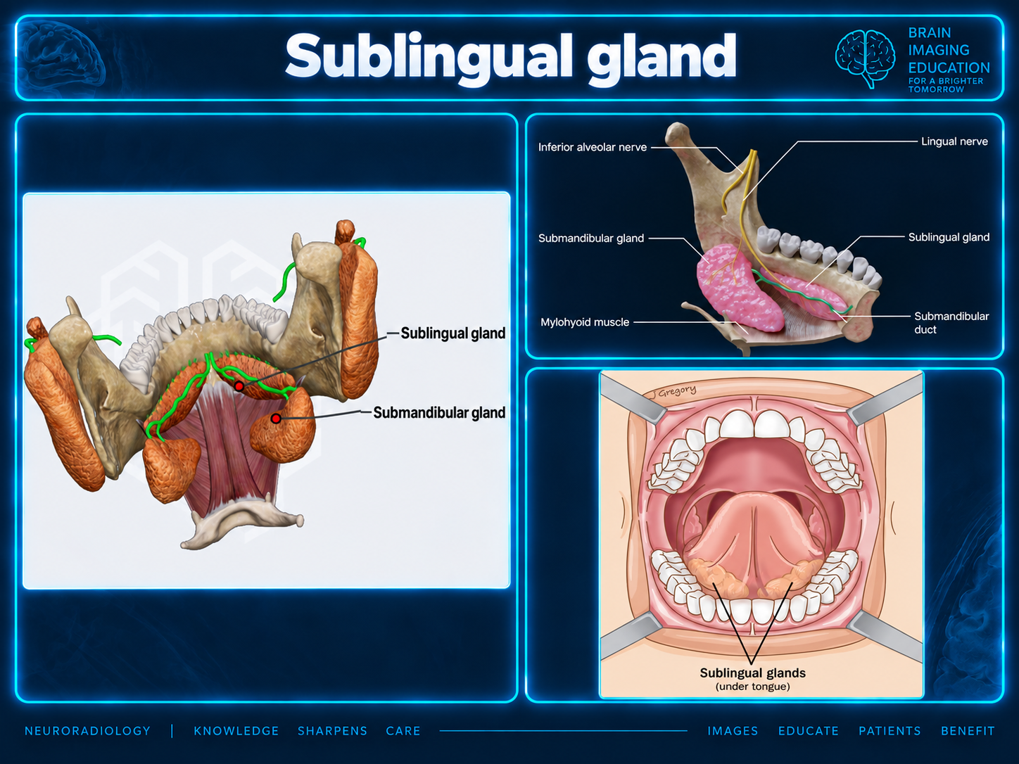
Teaching illustration showing sublingual and submandibular glands, their ducts, the lingual nerve, and the floor of mouth.
Sublingual gland: anatomic relationshipsSelect to enlarge · supplied teaching graphic
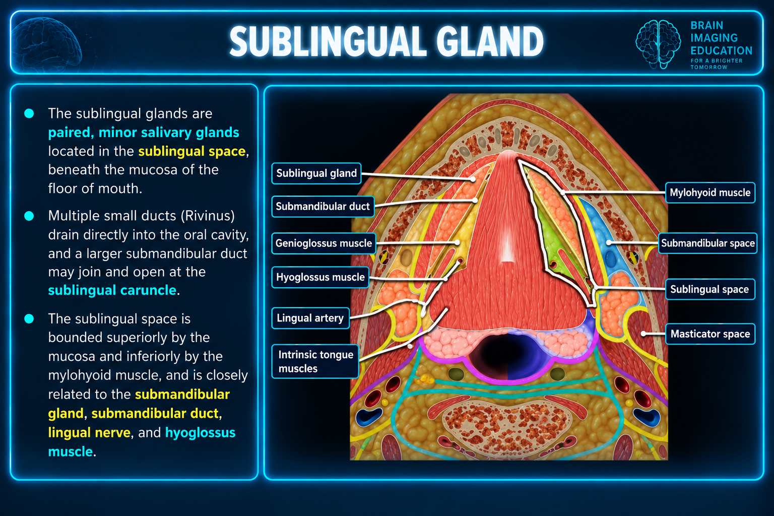
Axial teaching diagram labeling the sublingual gland and space, submandibular duct and space, mylohyoid, genioglossus, hyoglossus, lingual artery, and intrinsic tongue muscles.
Sublingual space: axial anatomic referenceSelect to enlarge · supplied teaching graphic

Source notes and limitations

The second graphic calls the sublingual glands “minor salivary glands”; the supplied prose describes them as the smallest of the three paired major salivary glands. Its superior boundary is described as mucosa, whereas the prose lists intrinsic tongue muscles. Its duct wording also differs: the prose describes an occasional major sublingual duct (of Bartholin) opening together with the submandibular duct, while the graphic describes a larger submandibular duct that may join. Both graphics are preserved unchanged; the text below follows the supplied prose without reconciling these differences.

Numbered citations (1–5) are retained from the supplied prose, but no bibliography was supplied. The contents list includes glossopharyngeal (CN IX) and hypoglossal (CN XII) nerves as provided; the source gives no further explanation of their course. The material supplies anatomy and a pathology list, without diagnostic imaging criteria or management guidance.

Space and communications

The sublingual space is part of the floor of mouth 1.

It is not bounded by fascia posteriorly. Some authors therefore consider it a component of the submandibular space 2; more commonly, the two spaces are discussed separately 3–5.

Each space communicates with the contralateral sublingual space through a small isthmus just below the frenulum.

Contents

  • Sublingual gland and duct
  • Lingual nerve
  • Lingual artery and vein
  • Glossopharyngeal (CN IX) and hypoglossal (CN XII) nerves
  • Deep portion of the submandibular gland and duct

Related pathology

  • Ranula
  • Sialadenitis
  • Ludwig angina
  • Salivary gland tumors
  • Hemangioma
  • Schwannoma

Boundaries

The space is shaped like an inverted V, with its apex pointing anteriorly.

Boundaries as described in the supplied prose
DirectionBoundary or communication
SuperiorIntrinsic muscles of the tongue
InferolateralMylohyoid muscle
PosteriorFreely communicates along the posterior border of the mylohyoid muscle with the submandibular and parapharyngeal spaces
AnteriorMandible
MedialGeniohyoid/genioglossus complex

Sublingual glands

Gross anatomy

The sublingual glands are the smallest of the three paired major salivary glands. They lie in the sublingual space on the floor of the mouth, anterior to the submandibular glands.

These small, almond-shaped glands lie against the medial surface of the mandible at the sublingual fossa, immediately lateral to the submandibular duct and lingual nerve.

The superior surface indents the floor-of-mouth mucosa, creating the sublingual fold. This extends to the sublingual papilla at the base of the tongue frenulum.

The sublingual gland is the only major salivary gland that lacks a capsule 5. The parotid and submandibular glands have a fibrous capsule derived from the superficial (investing) layer of the deep cervical fascia 4.

Secretion and ducts

The glands secrete predominantly mucous saliva, which drains through numerous minor sublingual ducts (of Rivinus).

Occasionally, the anteriormost duct is enlarged, forming the major sublingual duct (of Bartholin). It opens together with the submandibular duct at the sublingual papilla.

Innervation

Autonomic innervation is from the lingual nerve via the submandibular ganglion. Parasympathetic fibers arise from the chorda tympani; sympathetic fibers arise from the superior cervical ganglion.
Arterial supply

  • Sublingual artery: branch of the lingual artery
  • Submental artery: branch of the facial artery

Venous drainage

  • Sublingual vein: tributary of the lingual vein
  • Submental vein: tributary of the facial vein

Secondary Links