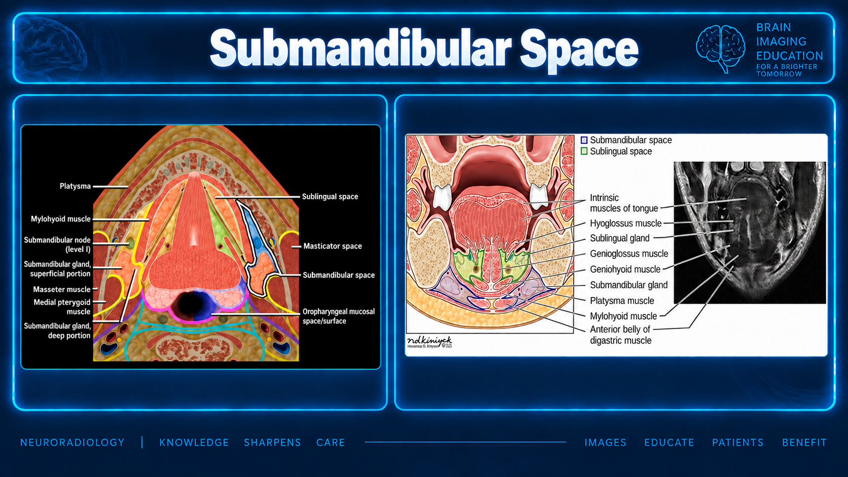
Submandibular Space
Normal anatomy. This reference uses the narrower definition of the submandibular space: the submylohyoid (submaxillary) space, with the sublingual and submental spaces treated as separate compartments.
Definition
Authors use the term in two ways. The narrower definition, used by Radiopaedia, treats the submental and sublingual spaces as separate compartments. Older surgical writing sometimes uses the term more broadly to cover these spaces as well.
This page uses the narrower definition. [radiopaedia]
Fascial enclosure
The superficial layer of the deep cervical fascia surrounds the space and forms its floor between the mandible and the hyoid.
The only opening in this wrapping is at the posterior free edge of the mylohyoid. [pmc.ncbi.nlm.nih]
Gland and duct
The main structure is the superficial (main) part of the submandibular gland. The gland wraps around the posterior free edge of the mylohyoid, joining its deep part in the sublingual space.
Wharton duct comes off the deep part and runs forward to the sublingual caruncle.
Boundaries
Boundaries described in the supplied anatomy summary| Aspect | Structure |
|---|
| Anterolateral | Mandible: front and outer wall |
|---|
| Medial | Anterior belly of the digastric muscle; separates the submandibular and submental spaces |
|---|
| Roof | Mylohyoid muscle |
|---|
| Floor | Hyoid bone |
|---|
| Posterior relationship | Tongue muscles lie behind the space |
|---|
| Outer surface | Platysma overlies the space |
|---|
Source markers: PMC review; Radiopaedia. See source note.
Contents and surgical relationships
Additional contents
- Submandibular lymph nodes (level IB)
- Facial artery and vein
- Lower loop of the hypoglossal nerve
- Fat surrounding these structures
[radiopaedia]
Vessels and facial nerve
Classically, the facial artery runs deep and grooves the back and top of the gland before curving over the lower edge of the mandible. The facial vein runs more superficially, along the outer side of the gland.
The marginal mandibular branch of the facial nerve runs just beneath the platysma, over the outer surface of the space. This relationship matters in surgery around the gland.
Communications
The space opens into the sublingual space behind the posterior edge of the mylohyoid. It is related to the submental space on its inner side and can communicate posteriorly with the parapharyngeal space through a gap created by the styloglossus muscle.
The two submandibular spaces connect across the midline through the submental space.
Mylohyoid gaps (boutonnières) are a normal variant that provides another direct path to the sublingual space. They sometimes contain fat, vessels, or herniated salivary tissue. [pmc.ncbi.nlm.nih] [radiopaedia]
Normal imaging appearance
CT and MRI
On CT, the normal gland is usually slightly denser than muscle and enhances evenly. It tends to become fattier and lower in density with age.
On MRI, the gland is intermediate to slightly bright on T2 and enhances evenly. The two sides should look similar.
Nodes and mylohyoid sling
Normal level IB nodes are small and oval, have a fatty hilum, and lie next to the gland and facial vessels.
On coronal images, the mylohyoid appears as a sling running from the inner surface of the mandible to the hyoid. The edge of this sling marks the boundary between the submandibular and sublingual spaces.
Source note
The supplied text cites Radiopaedia and a PMC review using the markers [radiopaedia] and [pmc.ncbi.nlm.nih], but provides no article titles, full citations, or URLs. These attributions are retained; the exact references cannot be identified from the supplied material.
The prose identifies submandibular nodes as level IB; the supplied slide labels them more broadly as level I. Both are preserved as supplied.