Thyroid nodules
Reference Images
Look for aggressive features
Extrathyroidal invasion or suspicious cervical lymphadenopathy overrides the usual size and age thresholds.
Apply age and size thresholds
With normal life expectancy and no high-risk features: ultrasound at ≥1.0 cm if younger than 35 years; ≥1.5 cm if 35 years or older. Use maximum axial dimension.
Limit non-actionable workup
Consider life expectancy and comorbidities. The supplied ACR guidance advises omitting non-actionable nodules from the Impression.
CT/MRI triage at a glance
| Clinical / imaging context | Threshold | Supplied recommendation |
|---|---|---|
| Invasion or suspicious lymphadenopathy | Any nodule size | Dedicated evaluation; aggressive features override standard cutoffs |
| Normal life expectancy; age <35 years; no high-risk features | Maximum axial dimension ≥1.0 cm | Dedicated thyroid ultrasound |
| Normal life expectancy; age ≥35 years; no high-risk features | Maximum axial dimension ≥1.5 cm | Dedicated thyroid ultrasound |
| Limited life expectancy or severe comorbidity | Consider competing clinical risks | No further evaluation unless specifically requested by managing clinician or patient, per supplied prose |
| Multinodular gland without aggressive features | Largest dominant nodule | Apply the same age and size criteria; see detailed qualifications below |
Clinical Context and Scope
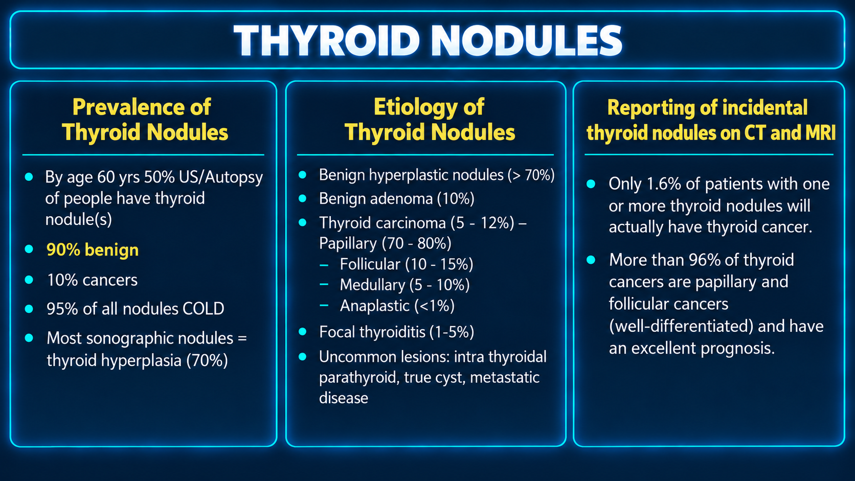
Incidental thyroid nodules are among the most frequent incidental findings encountered on cross-sectional imaging, identified on up to 25% of contrast-enhanced chest CT examinations and 16% to 18% of neck CT and MRI examinations according to the American College of Radiology Incidental Thyroid Findings Committee White Paper. While autopsy studies demonstrate occult thyroid carcinoma in more than one-third of the general population, the vast majority of incidentally discovered thyroid carcinomas are subclinical, indolent papillary microcarcinomas that carry a 10-year survival rate exceeding 99% (Hoang et al., JACR; AJNR practice review).[geiselmed.dartmouth][ajnr]
Unselective recommendation of diagnostic workup for incidental nodules on CT and MRI leads to substantial downstream harms, including defensive ordering, diagnostic cascades, patient anxiety, indeterminate fine-needle aspiration cytology, and diagnostic hemithyroidectomies for ultimately benign lesions. Standard cross-sectional guidance exists to distinguish truly suspicious lesions from benign background disease without triggering unnecessary downstream evaluation. These guidelines apply specifically to asymptomatic, low-risk adult patients and exclude pediatric populations, patients with palpable thyroid masses, or individuals with known high-risk syndromes such as multiple endocrine neoplasia type 2 or prior therapeutic childhood head and neck radiation (Hoang et al., JACR; CMS Quality Payment Program).[qpp.cms][geiselmed.dartmouth]
Imaging Strengths and Physical Limits
Normal thyroid tissue possesses an intrinsically high iodine concentration, yielding high unenhanced CT attenuation ranging from 70 to 120 Hounsfield units, followed by avid, uniform contrast enhancement on contrast-enhanced scans. Consequently, most thyroid nodules present on CT as focal hypoattenuating areas against dense, enhancing parenchyma. On MRI, the normal thyroid exhibits intermediate T1 signal and intermediate to slightly hyperintense T2 signal with brisk gadolinium enhancement.[appliedradiology][radlines]
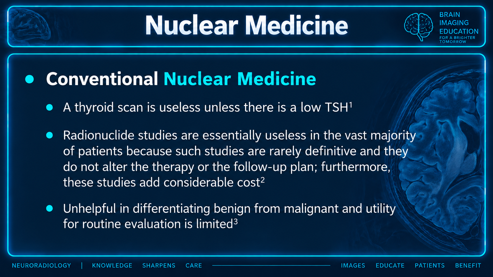
Cross-sectional imaging has fundamental physical limitations that preclude definitive benign versus malignant differentiation for the internal architecture of a nodule. Neither CT nor MRI can resolve submillimeter psammomatous microcalcifications, assess fine margin microlobulation, or reliably differentiate a benign colloid cyst from a solid, non-calcified papillary carcinoma based solely on attenuation. On MRI, both benign hyperplastic nodules and malignant neoplasms often demonstrate T2 hyperintensity and variable enhancement. While high intrinsic T1 signal on noncontrast MRI typically reflects concentrated colloid or subacute proteinaceous/hemorrhagic degradation products in a benign cyst or resolving adenoma, it remains non-specific. Furthermore, cross-sectional examinations covering the thoracic inlet frequently suffer from beam-hardening and streak artifacts caused by dense intravenous contrast within the brachiocephalic and subclavian veins, as well as photon starvation between the shoulders, degrading nodule visualization at the inferior thyroid poles.[pmc.ncbi.nlm.nih][geiselmed.dartmouth]
High-Risk Imaging Features
The initial step when evaluating any incidental thyroid nodule on CT or MRI is searching for secondary imaging signs of invasive malignancy or metastatic disease. When present, these findings override standard size and age cutoffs and mandate dedicated evaluation regardless of how small the primary thyroid lesion appears:[geiselmed.dartmouth]
Extrathyroidal extension and local invasion manifest as gross disruption of the thyroid capsule with loss of surrounding perilobular fat planes. Frank invasion of the strap muscles, esophagus, or trachea indicates aggressive pathology. Tracheal invasion is identified on CT or MRI when a nodule forms an obtuse angle with the outer tracheal margin, deforms the cartilaginous rings, or projects as endotracheal soft tissue. Extension into the tracheoesophageal groove warrants scrutiny for involvement of the recurrent laryngeal nerve, which may be signaled clinically by hoarseness or radiographically by medialization and atrophy of the ipsilateral vocal cord.[jksronline][ncbi.nlm.nih]
Suspicious regional lymphadenopathy strongly predicts metastatic thyroid carcinoma. Papillary thyroid carcinoma metastases frequently demonstrate internal cystic change, punctate psammomatous calcifications, or hypervascular enhancement matching the thyroid gland itself. Cervical nodes in levels IV and VI represent primary drainage stations for thyroid malignancies and are particularly concerning when altered. While size alone is less specific than internal architecture, isolated lymph node enlargement warrants suspicion when the short-axis diameter exceeds 1.5 cm for level IIA jugulodigastric nodes or 1.0 cm for other cervical stations.[pmc.ncbi.nlm.nih][geiselmed.dartmouth]
ACR Triage Framework
When a nodule lacks invasive features or suspicious lymphadenopathy, further workup is dictated by patient life expectancy, patient age, and axial nodule size, as outlined by the ACR Incidental Thyroid Findings Committee White Paper:[geiselmed.dartmouth]
Patients with severe medical comorbidities, active advanced non-thyroid malignancies, severe organ failure, or limited life expectancy should not undergo further evaluation. Because incidentally detected small thyroid cancers rarely progress within a shortened lifespan, diagnostic and surgical interventions pose greater morbidity and mortality risks than the thyroid disease itself. Further imaging should only be pursued in this cohort if specifically requested by the managing clinician or patient.
In patients with normal life expectancy, nodule size should be measured in the maximum dimension on axial images. The management recommendations fall into three categories:
For patients younger than 35 years of age, dedicated thyroid ultrasound is recommended for nodules measuring 1.0 cm or greater in the axial plane. Benign hyperplastic nodules are less prevalent in younger adults, conferring a higher relative malignancy ratio, and young patients face a longer cumulative lifetime risk for potential disease progression (Hoang et al., JACR; PMC Incidental Thyroid Review).[pmc.ncbi.nlm.nih][geiselmed.dartmouth]
For patients 35 years of age and older, the threshold to recommend dedicated thyroid ultrasound is raised to 1.5 cm or greater. Below 1.5 cm, incidental nodules in older adults carry a negligible risk of clinically meaningful adverse outcomes.
For multinodular thyroid glands, the same size and age thresholds are applied strictly to the single largest dominant nodule, provided no individual nodule exhibits aggressive features. If an enlarged, heterogeneously multinodular gland is encountered without a distinct dominant nodule, dedicated ultrasound is recommended only if the patient has normal life expectancy and no prior characterization.[geiselmed.dartmouth]
Thoracic Inlet and Mass Effect
Incidental thyroid lesions frequently produce mechanical complications that warrant surgical or clinical attention independent of oncologic risk. When evaluating thyroid enlargement on neck or chest CT and MRI, the report must systematically assess mediastinal extension and aerodigestive compromise:[ncbi.nlm.nih]
Retrosternal and substernal extension should be defined by the relationship of the inferior thyroid border to the thoracic inlet. Lesions extending below the level of the clavicles and first ribs into the anterior or posterior mediastinum should be quantified by craniocaudal descent and spatial relationship to the aortic arch and great vessels.
Tracheal compression and displacement require quantitative description. The report should document the narrowest transverse and anteroposterior internal luminal diameters of the trachea, the percentage of luminal stenosis, and whether airway narrowing is accompanied by dynamic tracheomalacia or cartilaginous ring distortion.
Mass effect on the esophagus and major cervical vasculature, such as displacement or compression of the internal jugular veins or common carotid arteries, should be documented to assist surgical planning and clinical airway management.
Quality Standards and MIPS 406
Adherence to evidence-based cross-sectional thyroid recommendations directly impacts Medicare reimbursement and clinical quality benchmarks through MIPS Quality Measure #406: Appropriate Follow-up Imaging for Incidental Thyroid Nodules.[mdinteractive]
Measure #406 evaluates diagnostic CT, CTA, MRI, and MRA reports of the chest, neck, and cervical spine in adults aged 18 years and older with no prior diagnosis of thyroid disease in whom an incidental thyroid nodule under 1.0 cm is documented. Because this is an inverse measure, recommending follow-up imaging for a subcentimeter nodule constitutes a performance failure unless a documented medical exception applies. Valid clinical exceptions include documented cervical lymphadenopathy, personal or family history of thyroid carcinoma, or known multiple endocrine neoplasia. Radiologists who routinely suggest ultrasound for subcentimeter incidental nodules on CT or MRI directly degrade their practice quality scores and expose patients to low-value care.[qpp.cms]
Practical Reporting and Structuring
The ACR White Paper provides explicit instructions regarding report structure to minimize non-actionable consultations:[geiselmed.dartmouth]
For nodules that do not meet criteria for dedicated workup (nodules under 1.0 cm in patients younger than 35, nodules under 1.5 cm in patients 35 and older, or nodules in patients with limited life expectancy), the ACR recommends omitting the finding from the Impression or Conclusion section entirely. Subcentimeter or small non-actionable nodules may be noted as an incidental finding within the body Findings section or omitted altogether if trivial. Including an incidental subcentimeter nodule in the final impression frequently triggers defensive ordering of follow-up ultrasound by primary care clinicians, even when the radiologist notes that the finding is likely benign.
When a nodule does not meet workup criteria but is noted in the Findings, phrasing should emphasize lack of clinical significance: "Subcentimeter hypodense thyroid nodule measuring 7 mm, non-actionable by ACR Incidental Findings Committee criteria."
When a nodule meets size and age thresholds without high-risk features, the Impression should recommend non-emergent sonographic correlation: "Dominant 1.8 cm hypoattenuating nodule in the left thyroid lobe. In a patient 35 years or older with normal life expectancy, dedicated thyroid ultrasound is recommended for further characterization per ACR Incidental Findings Committee guidelines."
When high-risk imaging features are identified, the Impression should highlight the aggressive characteristics immediately: "Heterogeneous 2.2 cm right thyroid mass demonstrating invasion of the adjacent right strap muscles and tracheoesophageal groove, accompanied by an ipsilateral level IV lymph node with internal cystic changes and punctate calcification. Dedicated thyroid ultrasound and surgical evaluation are recommended."