Describe the depth
Distinguish inner thyroid-cartilage involvement from outer cortical breakthrough and extralaryngeal extension. State the cartilage, side, and confidence.
Distinguish inner thyroid-cartilage involvement from outer cortical breakthrough and extralaryngeal extension. State the cartilage, side, and confidence.
CT is usually first-line for staging. MRI is useful when subtle, nonossified, or equivocal cartilage involvement will change management.
Sclerosis is nonspecific. Structural destruction, tumor on both sides, and tumor-matched marrow replacement provide stronger evidence.
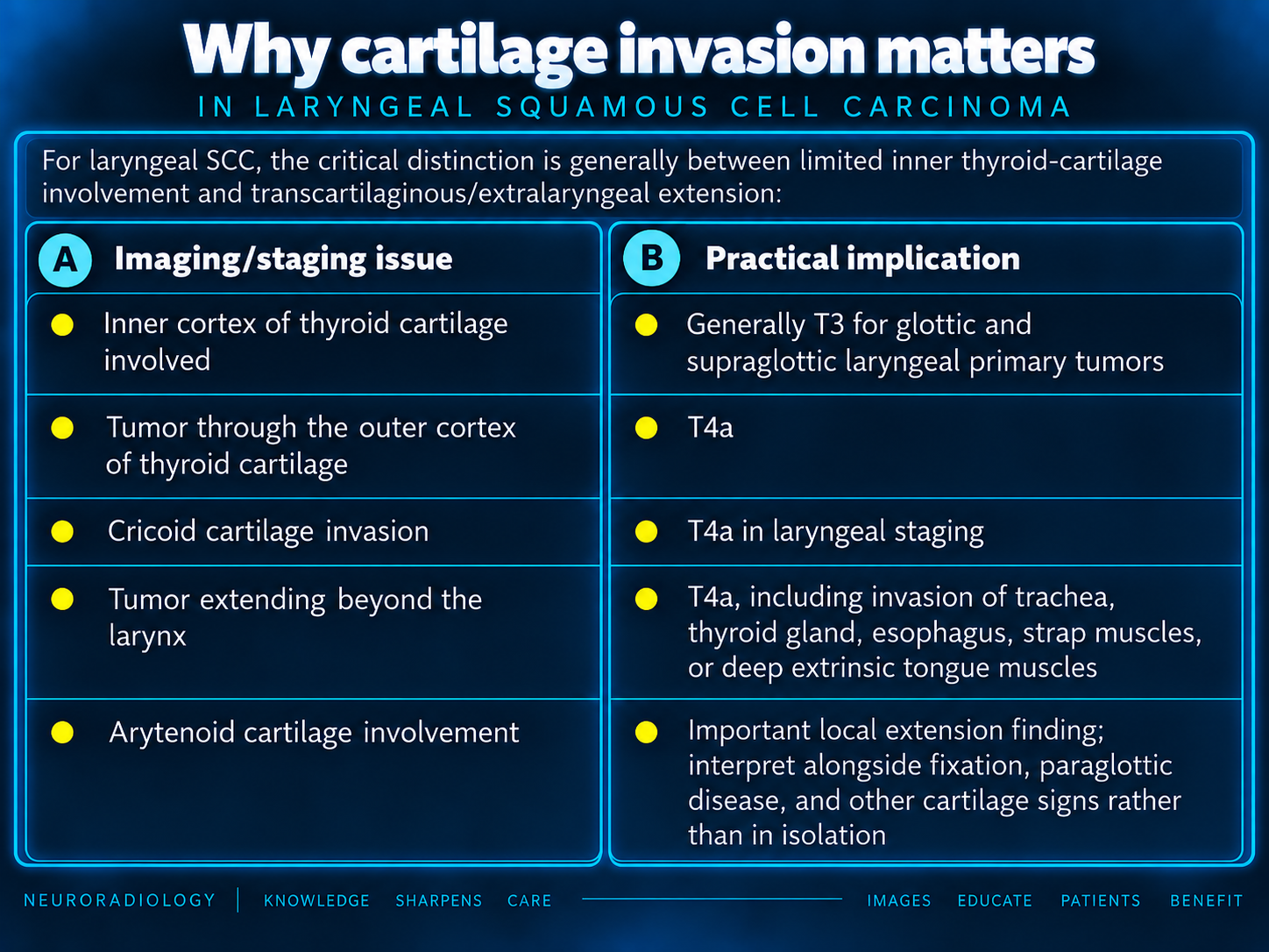
For laryngeal SCC, the critical distinction is generally between limited inner thyroid-cartilage involvement and transcartilaginous/extralaryngeal extension:
| Imaging/staging issue | Practical implication |
|---|---|
| Inner cortex of thyroid cartilage involved | Generally T3 for glottic and supraglottic laryngeal primary tumors |
| Tumor through the outer cortex of thyroid cartilage | T4a |
| Cricoid cartilage invasion | T4a in laryngeal staging |
| Tumor extending beyond the larynx | T4a, including invasion of trachea, thyroid gland, esophagus, strap muscles, or deep extrinsic tongue muscles |
| Arytenoid cartilage involvement | Important local extension finding; interpret alongside fixation, paraglottic disease, and other cartilage signs rather than in isolation |
The NCI summary describes T3 disease as tumor limited to the larynx with vocal-cord fixation, paraglottic-space invasion, and/or inner thyroid-cartilage cortex involvement; extension through the outer thyroid-cartilage cortex or into extralaryngeal tissues is T4a. For radiology reporting, it is therefore much more useful to state the depth and side of cartilage involvement than simply writing “cartilage invasion present.”[cancer]
The thyroid cartilage is the key cartilage for imaging staging, especially for glottic/anterior commissure and supraglottic cancers.
Important routes include:
Be cautious with the common shorthand that “anterior commissure involvement equals cartilage invasion.” It increases risk, but invasion requires direct imaging evidence of abnormal cartilage signal/density, cortical breakthrough, or tumor on both sides of the cartilage.
Cricoid involvement is particularly relevant in subglottic and postcricoid/hypopharyngeal tumors. It may be subtle because of variable mineralization and because tumor can track in the subglottic paraglottic region. In laryngeal staging, cricoid-cartilage invasion is T4a.[radiopaedia]
Arytenoid sclerosis can be reactive or related to altered stress/loading, particularly in the setting of vocal-fold paralysis. A combination of findings is more convincing than sclerosis alone: tumor contact/encasement, focal erosion or lysis, abnormal marrow replacement, loss of contour, and additional abnormal cartilage findings. An AJNR study found that impaired arytenoid mobility and more than one CT cartilage abnormality were relatively reliable indicators of neoplastic cartilage invasion.[ajnr]
The suprahyoid epiglottis is fibroelastic rather than hyaline cartilage. For supraglottic tumors, report the involvement of:
“Epiglottic cartilage invasion” should not be treated as equivalent to thyroid or cricoid cartilage invasion for T staging.
For primary laryngeal staging, use thin-section contrast-enhanced multidetector CT with reconstructions in axial, coronal, and sagittal planes. Images should be obtained during quiet respiration rather than phonation or swallowing, with the patient avoiding motion and swallowing during acquisition.
A practical approach:
CT is fast, accessible, less affected by swallowing artifacts than MRI, and excellent for cortical erosion, gross lysis, mineralized cartilage, extralaryngeal extension, nodal disease, and chest staging.[thieme-connect]
Use a hierarchy of confidence rather than treating every sclerosis as invasion.
Historically, sclerosis has been sensitive but nonspecific—particularly in the thyroid cartilage, where mineralization is highly variable. Erosion, lysis, and especially extralaryngeal tumor are more specific but generally later findings.[academic.oup][pmc.ncbi.nlm.nih]
MRI is particularly valuable for:
A practical laryngeal MRI protocol includes:
The classic concept is to compare cartilage signal/enhancement directly with the adjacent tumor:
A commonly used MRI pattern is: tumor abuts cartilage with reduced intrachondral T1 signal, increased T2 signal matching tumor, and postcontrast enhancement matching tumor.[epos.myesr]
MRI is more sensitive for cartilage invasion, particularly early intrachondral tumor spread and nonossified cartilage. A systematic review/meta-analysis of 14 studies and 776 patients found pooled sensitivity of 88% for MRI versus 66% for CT; pooled specificity was 81% for MRI versus 90% for CT, without a statistically significant specificity difference in that analysis.[pubmed.ncbi.nlm.nih]
However, MRI can overcall invasion because edema, inflammation, perichondritis, reactive hyperemia, biopsy-related change, and treatment effects may produce T2 hyperintensity and enhancement that resemble tumor. The key MRI false-positive pattern is diffuse or ill-defined cartilage edema/enhancement adjacent to bulky mucosal disease without a discrete tumor-matched marrow-replacement pattern or cortical/transcartilaginous extension.
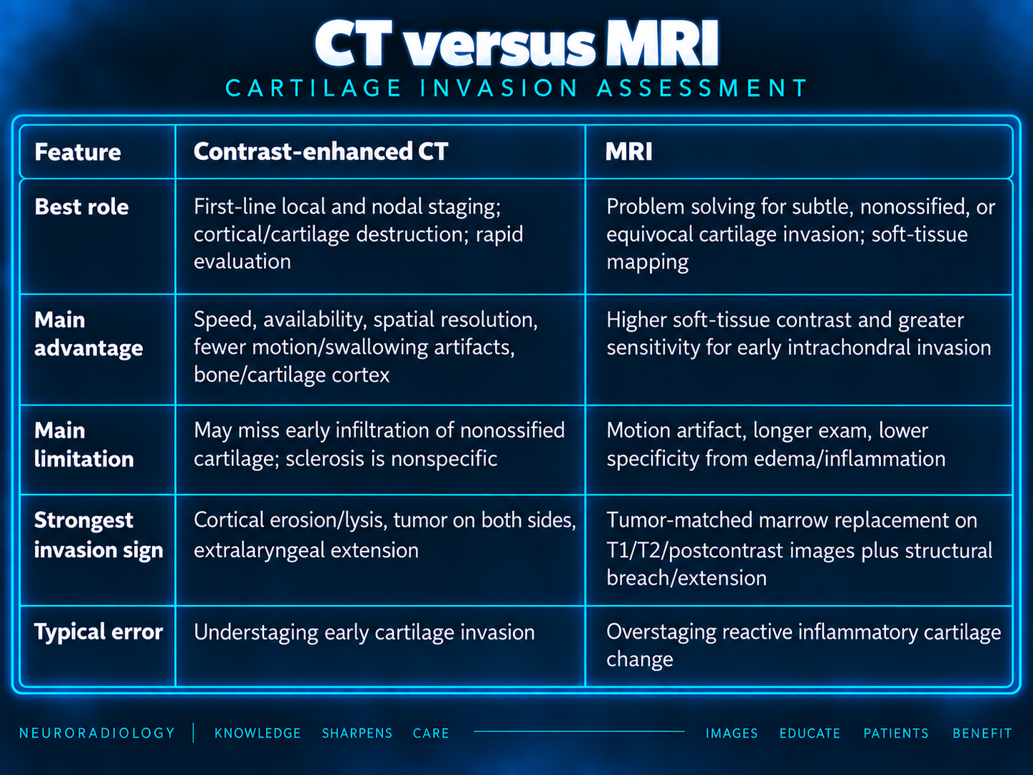
| Feature | Contrast-enhanced CT | MRI |
|---|---|---|
| Best role | First-line local and nodal staging; cortical/cartilage destruction; rapid evaluation | Problem solving for subtle, nonossified, or equivocal cartilage invasion; soft-tissue mapping |
| Main advantage | Speed, availability, spatial resolution, fewer motion/swallowing artifacts, bone/cartilage cortex | Higher soft-tissue contrast and greater sensitivity for early intrachondral invasion |
| Main limitation | May miss early infiltration of nonossified cartilage; sclerosis is nonspecific | Motion artifact, longer exam, lower specificity from edema/inflammation |
| Strongest invasion sign | Cortical erosion/lysis, tumor on both sides, extralaryngeal extension | Tumor-matched marrow replacement on T1/T2/postcontrast images plus structural breach/extension |
| Typical error | Understaging early cartilage invasion | Overstaging reactive inflammatory cartilage change |
The evidence supports a complementary strategy: CT for most patients and MRI when cartilage involvement is uncertain or would materially alter organ-preservation versus surgical decision-making.[thieme-connect][pubmed.ncbi.nlm.nih][academic.oup]
For a laryngeal or hypopharyngeal SCC, avoid a binary “cartilage invasion: yes/no” statement unless unmistakable. Instead, report the specific cartilage, imaging confidence, and whether there is outer cortical/transcartilaginous extension.
Reporting checklist limitation: The supplied checklist ends after paraglottic-space involvement. No additional checklist items were supplied.
Bracketed citation labels are reproduced from the supplied text. Full bibliographic details and article URLs were not supplied, so the cited study and staging claims require source verification before clinical use. The staging table describes laryngeal primary tumors; a separate hypopharyngeal staging table was not supplied.