Fovea ethmoidalis
The fovea ethmoidalis is the frontal-bone component of the ethmoid roof. Its radiological importance is defining the superior limit of endoscopic ethmoid surgery and identifying anatomy that increases the risk of skull-base, cerebrospinal fluid (CSF) leak, or anterior ethmoidal artery injury.
Reference Images
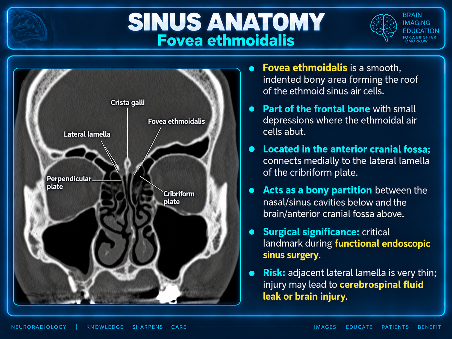
Anatomy
The supplied figure describes a smooth, indented bony area of the frontal bone forming the roof of the ethmoid sinus air cells, with small depressions where the cells abut.
It lies in the anterior cranial fossa and connects medially to the lateral lamella of the cribriform plate.
Skull-base boundary
The fovea acts as a bony partition between the nasal and sinus cavities below and the brain/anterior cranial fossa above.
The reference image labels the neighboring lateral lamella, cribriform plate, crista galli and perpendicular plate.
Surgical significance
A critical landmark during functional endoscopic sinus surgery (FESS).
The adjacent lateral lamella is very thin; injury may lead to CSF leak or brain injury, as emphasized in the supplied figure.
Pre-FESS reporting priorities
Keros type alone is insufficient. The supplied summary recommends also identifying:
- The lower ethmoid-roof side and roof height/contour asymmetry.
- Lateral-lamella slope.
- Focal dehiscence.
- Supraorbital pneumatization.
- Whether the anterior ethmoidal artery is suspended below the skull base.
These are reporting considerations from the supplied summary; no patient-specific findings or measurements are provided.
Scope of the supplied report summary
The source identifies the following topics for radiologist-level assessment:
- Fovea–lateral lamella–cribriform plate anatomy.
- Thin-section CT evaluation.
- Keros and Gera classifications.
- Ethmoid-roof height and contour asymmetry.
- Anterior ethmoidal artery risk anatomy.
- Pre-FESS reporting recommendations.
- Trauma, CSF leak, encephalocele and tumor involvement.
- Reporting language and interpretive pitfalls.
Source limitations
The supplied prose states that a detailed report was created, but only its summary was supplied. Classification definitions and thresholds, CT acquisition details, copy-ready reporting language, interpretive pitfalls and detailed disease-specific discussion are absent. This page preserves the available summary and teaching figure; it does not reconstruct the missing report.
Reference
Source-linked article on PubMed Central — retained from the supplied content.