Drainage corridors
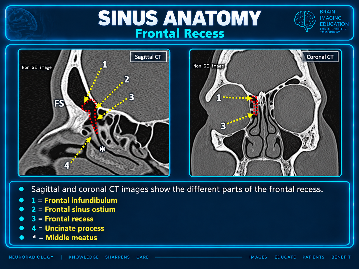
Frontal, maxillary and anterior ethmoid drainage converge in the middle meatus. Follow the frontal recess and ethmoid infundibulum in three dimensions.
Radiology-oriented reference for CT/MR interpretation and functional endoscopic sinus surgery (FESS) planning. The basal lamella of the middle turbinate divides the anterior and posterior sinonasal compartments.
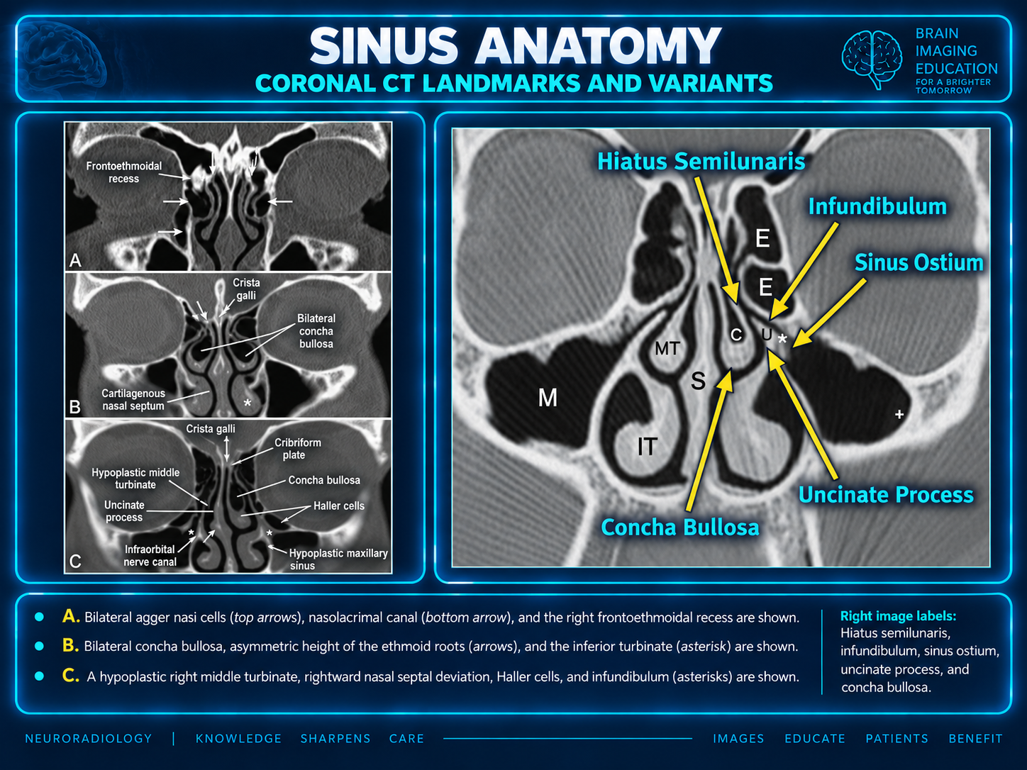
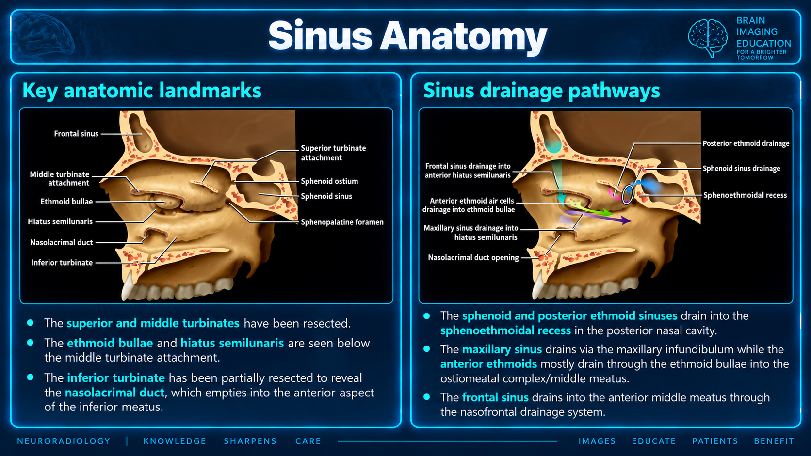
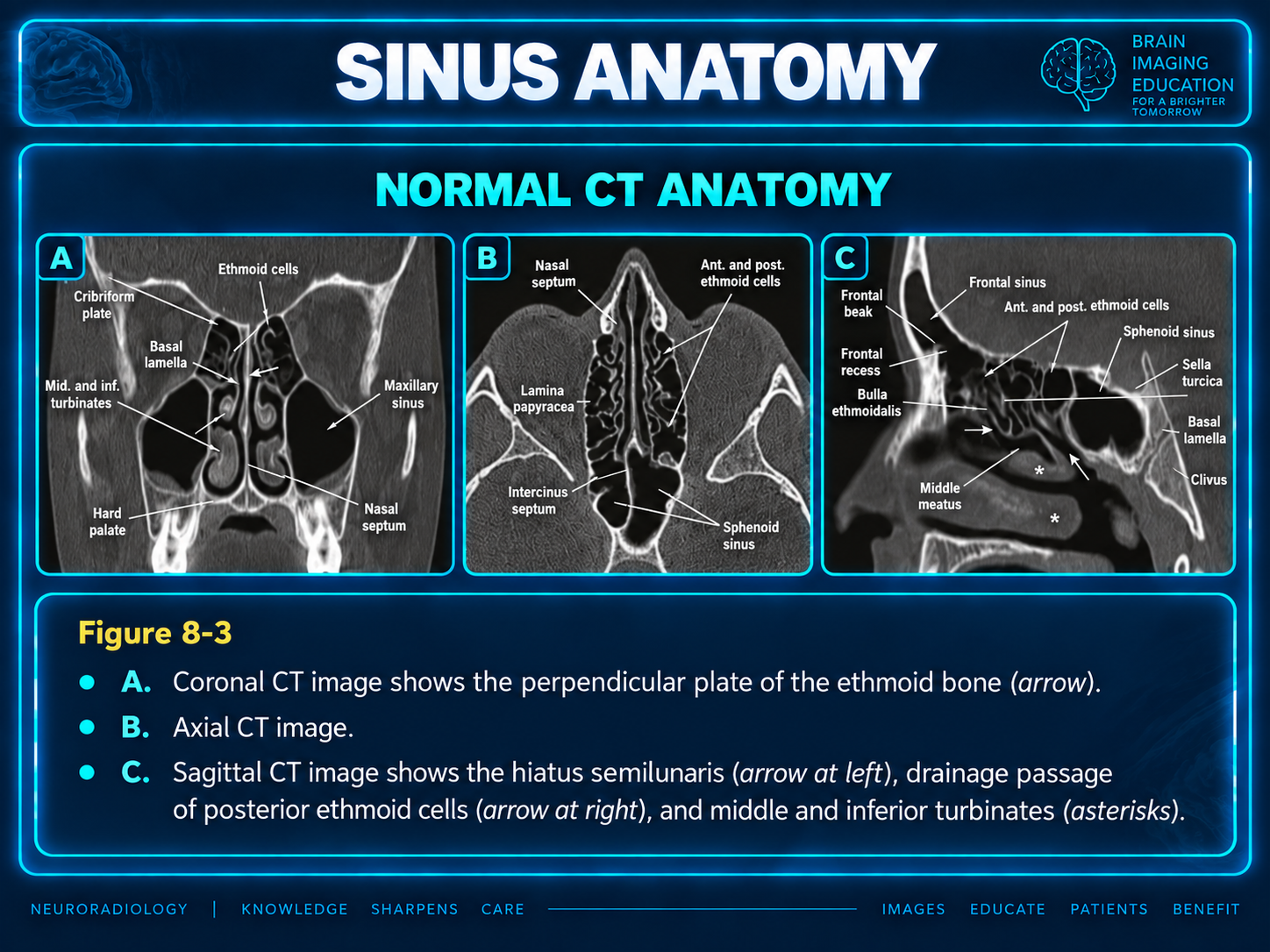
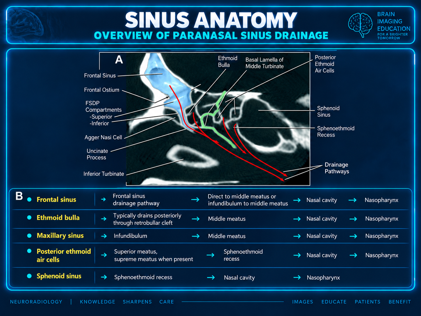
Frontal, maxillary and anterior ethmoid drainage converge in the middle meatus. Follow the frontal recess and ethmoid infundibulum in three dimensions.
Assess the ethmoid roof, lateral lamella and lamina papyracea; map sphenoid relationships to the optic canals and carotid arteries.
Define disease distribution, impaired drainage, access-limiting variants and vulnerable adjacent structures. Use the supplied CLOSE checklist below.
The supplied prose places posterior ethmoid drainage in the superior meatus; Figures 2 and 4 also associate it with the sphenoethmoidal recess. These source descriptions are retained without reconciling them.
The uncinate-attachment statements below are reproduced from the supplied report. The figures do not establish the attachment-dependent direction of frontal drainage; this relationship requires source verification before clinical use.
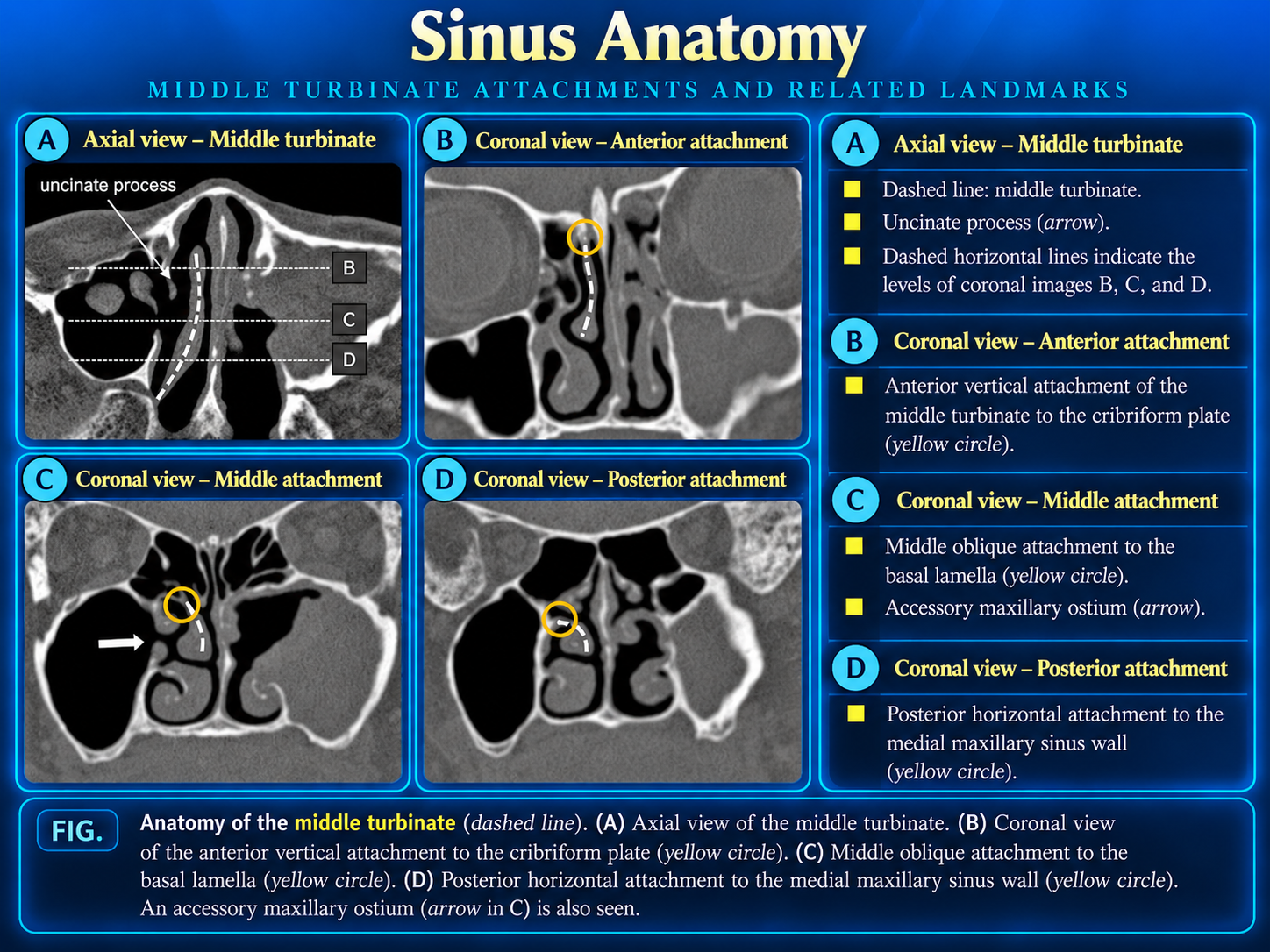
Figure 9 describes the posterior middle-turbinate attachment at the medial maxillary wall, while the prose describes its role in the roof of the posterior middle meatus. Both descriptions are preserved. The supplied complication-rate range has no accompanying citation or study context. No bibliographic references were supplied.
The paranasal sinuses are a variable system of drainage corridors bounded by the orbit, anterior skull base, optic apparatus, carotid arteries, and pterygopalatine-region neurovascular structures. For CT/MR interpretation and endoscopic-surgical planning, the most useful framework is the anterior versus posterior sinonasal compartment, divided by the basal lamella of the middle turbinate.
| Region | Principal anatomy | Drainage endpoint | Key imaging/surgical relevance |
|---|---|---|---|
| Frontal sinus | Paired frontal-bone cavities; often asymmetric, variably septated, and sometimes hypoplastic or aplastic. | Frontal ostium → frontal recess/outflow tract → middle meatus or infundibulum, depending largely on uncinate attachment. | The frontal recess/outflow tract is highly variable; agger nasi, frontal cells, bulla, and uncinate configuration may obstruct drainage. |
| Maxillary sinus | Largest sinus; pyramidal cavity in the maxilla. | Natural ostium in the superior medial wall → infundibulum/hiatus semilunaris → middle meatus. | Natural ostium is nondependent. Evaluate dental roots, orbital floor, infraorbital canal, and accessory ostia. |
| Ethmoid labyrinth | Multicellular air-cell system between nasal cavity and orbit. | Anterior/middle cells → middle meatus; posterior cells → superior meatus. | Ethmoid roof/lateral lamella and lamina papyracea are major skull-base and orbital danger zones. |
| Sphenoid sinus | Central posterior skull-base sinus with variable septa and lateral recesses. | Anterior-wall ostium → sphenoethmoidal recess. | Assess optic nerves, cavernous ICA, sellar floor, vidian canals, V2, clivus, and pituitary relationships. |
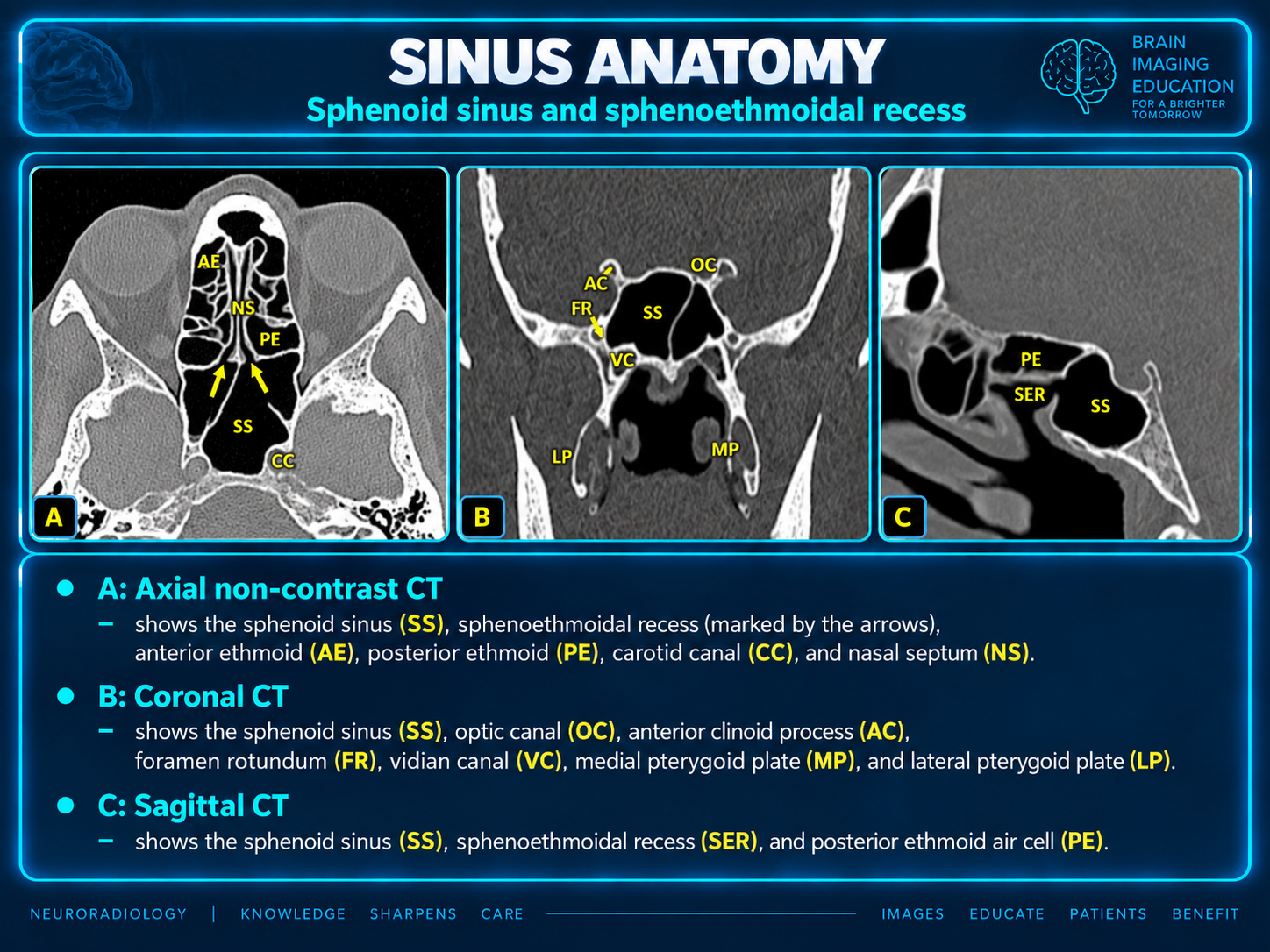
The nasal cavity extends from vestibule to choanae and is divided by the septum. Superior, middle, and inferior turbinates create the corresponding meatuses. The middle meatus receives the anterior sinonasal compartment; the superior meatus receives posterior ethmoid drainage; and the sphenoid sinus opens into the sphenoethmoidal recess above and posterior to the superior turbinate.
Preoperative CT is central to FESS planning. A useful report defines disease distribution and impaired drainage, identifies variants that narrow the frontal recess, ostiomeatal complex, or ethmoid infundibulum, and calls attention to vulnerable adjacent structures. Major FESS complications have been reported in the 0–2.25% range and include CSF leak, hemorrhage, orbital injury, optic-nerve injury, meningitis, and lacrimal-duct injury.
Asymmetric turbinate, septal, or ethmoid-region mucosal thickening may be physiologic rather than inflammatory. This periodic nasal-cycle change typically spares the frontal, maxillary, and sphenoid sinuses and should not be mistaken for sinonasal inflammatory disease on CT.
The ostiomeatal complex (OMC/OMU) is a functional bottleneck rather than a single discrete anatomic structure. It is the common drainage region for the maxillary sinus, frontal sinus, and anterior ethmoid cells. Focal mucosal disease here can create the recognizable ipsilateral anterior OMC pattern of maxillary, anterior ethmoid, and frontal sinus disease.
This relationship explains why maxillary and frontal disease may coexist or be dissociated despite adjacent anatomy.
The natural ostium lies high on the medial wall, limiting gravity-dependent clearance. An accessory ostium may occur in the posterior fontanelle. When natural and accessory ostia are both patent, mucus recirculation along the medial maxillary wall is a useful CT clue in chronic disease. Potential recesses include alveolar, zygomatic, infraorbital, and palatine recesses.
The ethmoid complex is a variable honeycomb of air cells. The third basal lamella (the basal lamella of the middle turbinate) separates anterior from posterior ethmoid cells: anterior cells drain to the middle meatus and participate in OMC disease, whereas posterior cells drain to the superior meatus. “Middle ethmoid” terminology varies but generally behaves with the anterior compartment in drainage and surgical practice.
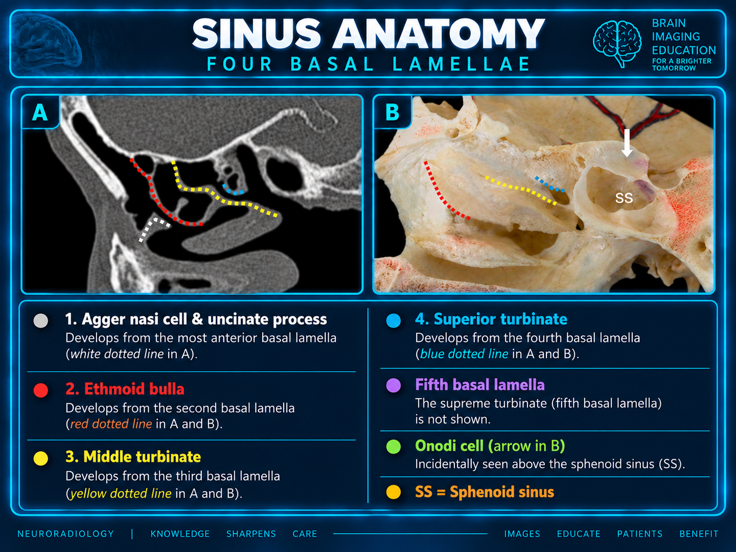
The ethmoid bone includes the perpendicular plate, cribriform plate, and paired ethmoidal labyrinths. In the Killian basal-lamella framework, the uncinate, ethmoid bulla, middle turbinate, superior turbinate, and sometimes supreme turbinate form sequential partitions. Frontal and maxillary drainage lies between the first and second lamellae; the ethmoid bulla drains between the second and third; the third separates anterior from posterior ethmoids; and the fourth separates posterior and supreme meatuses.
The frontal sinus varies widely in size, shape, septation, and laterality. The interfrontal septum is often eccentric and should not be assumed to mark the true midline. The posterior table separates the sinus from the anterior cranial fossa/dura; the floor has medial and lateral orbital components.
The frontal beak is a bony ridge formed by superior extension of the frontal process of the maxilla. The frontal ostium lies at its level; the beak separates the frontal sinus above from its drainage pathway below.
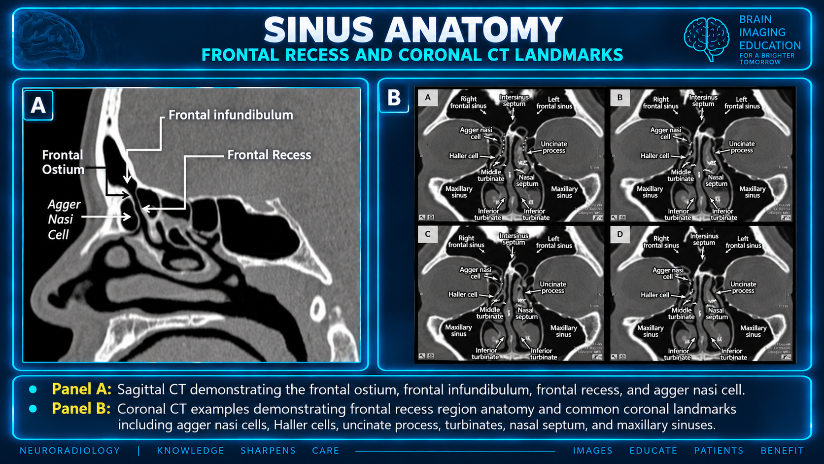
The frontal drainage pathway is bounded laterally by lamina papyracea, medially by lateral lamella of the olfactory fossa, anteriorly by frontal beak and agger nasi cell, and posteriorly by bulla lamella. It can be conceptualized as a superior frontal recess and an inferior frontal sinus outflow tract. The latter may be formed by the ethmoid infundibulum or by the middle meatus, based on uncinate attachment and adjacent anterior ethmoid cells.
Assess it as a three-dimensional channel. Enlarged agger nasi cells, frontal cells, a prominent bulla, unusual uncinate attachment, and other adjacent cells can narrow or redirect it. More than one frontal-sinus opening may be present. Lateral frontal sinus disease can be challenging to address endoscopically.
The sphenoid sinus should be assessed as a map of adjacent critical structures, not simply as an air cavity. The anterior-wall ostium is usually near the midline and opens to the sphenoethmoidal recess.
| Type | Description |
|---|---|
| Conchal | Minimal or no pneumatization beneath the sella. |
| Presellar | Pneumatization extends posteriorly but not beneath the sella. |
| Sellar | Pneumatization extends beneath and/or posterior to the sella. |
Greater pneumatization can expose critical structures through thin bone or apparent dehiscence. Sphenoid septa are frequently eccentric and may insert on an ICA or optic-canal prominence; do not assume that a septum is a safe midline landmark.
The middle turbinate and basal lamella are parts of the same embryologic structure—the third basal lamella. Its sequential skull attachments are anterior vertical (to cribriform plate, sagittal), middle oblique (to basal lamella or lamina papyracea, coronal), and posterior horizontal (forming the roof of posterior middle meatus, axial). This configuration stabilizes the turbinate; excessive posterior resection can result in anterior instability.
The cribriform plate forms the medial anterior skull base. The lateral lamella connects it to the fovea ethmoidalis and is the most vulnerable portion of the anterior skull base during ethmoid surgery. It can be extremely thin.
| Keros type | Olfactory fossa depth | Interpretation |
|---|---|---|
| I | 1–3 mm | Shallow. |
| II | 4–7 mm | Most common. |
| III | 8–16 mm | Deep; longer, more vulnerable lateral lamella. |
Side-to-side asymmetry may be more important than the absolute Keros category because it can create a unilateral low point or oblique ethmoid roof. Assess both sides independently.
The anterior ethmoidal artery arises from the ophthalmic artery and traverses the anterior ethmoid near the roof. On coronal CT, determine whether it is closely applied to the skull base in a canal, partially suspended below it, or freely coursing in a mesentery across an ethmoid cell. A low-lying or dehiscent-appearing artery is vulnerable during ethmoid dissection and may retract into the orbit if injured.
The reporting threshold should be lower for preoperative studies, disease in the corresponding drainage route, or a variant that changes risk to orbit, skull base, optic nerve, or ICA.
When sinus opacification is present, consider whether the process is centered outside the sinus with secondary invasion, such as from the nasal cavity, orbit, anterior skull base, pterygopalatine fossa, maxillary alveolus, or sinonasal neurovascular corridors.
A radiology-oriented adaptation of the CLOSE mnemonic:
Also assess frontal sinus aeration and frontal recess narrowing; agger nasi and supraorbital ethmoid cells; uncinate attachment and infundibular patency; Haller cells and maxillary ostial obstruction; concha bullosa/paradoxical middle turbinate; access-limiting septal deviation/spur; odontogenic maxillary source; and prior surgical changes such as antrostomy, ethmoidectomy, skull-base reconstruction, or retained partitions.
Illustrative report language: Adapt the findings and bracketed fields to the examination. The preoperative paragraph is a supplied example.
Sinonasal drainage pathways: Bilateral maxillary ostia/infundibula are patent. Frontal recesses are patent. No obstructing Haller cell, enlarged agger nasi cell, or substantial concha bullosa.
Ethmoid/skull base: Mild asymmetric depth of the olfactory fossae, deeper on the [side]; no focal skull-base osseous defect. Laminae papyraceae are intact.
Sphenoid region: [Sellar/presellar/conchal] sphenoid pneumatization. [No/left/right] Onodi cell. The optic canals and cavernous ICAs [do/do not] protrude into the sphenoid sinus; sphenoid septum [does/does not] attach near the [right/left] carotid canal.
Other: [Septal deviation/spur]. [Accessory maxillary ostium/Haller cells/prior surgery/odontogenic disease].
Preoperative anatomic considerations: Deep left olfactory fossa with asymmetric low left ethmoid roof; left AEA suspended below the skull base. Left Onodi cell abuts the optic canal. Sellar sphenoid pneumatization with eccentric septum inserting on the right carotid canal. Small right lamina papyracea dehiscence with orbital-fat prolapse.
The core practical goal is to answer: Where is the obstructed drainage pathway? What is the pattern of inflammatory spread? Where are the vulnerable orbit, skull-base, optic-nerve, carotid, and lacrimal landmarks?