Localize first
Identify the epicenter relative to the buccinator, masseter, Stensen duct, and accessory parotid tissue.
The major differential for a buccal space lesion includes salivary neoplasms, vascular malformations, odontogenic infection, developmental cysts, nerve sheath tumors, lymphoma, and secondary invasion from adjacent malignancy. The most useful imaging discriminators are the lesion’s epicenter, fat versus fluid versus solid composition, vascular architecture, relationship to Stensen duct, and invasive features—although a well-circumscribed mass does not exclude salivary malignancy. pmc.ncbi.nlm.nih
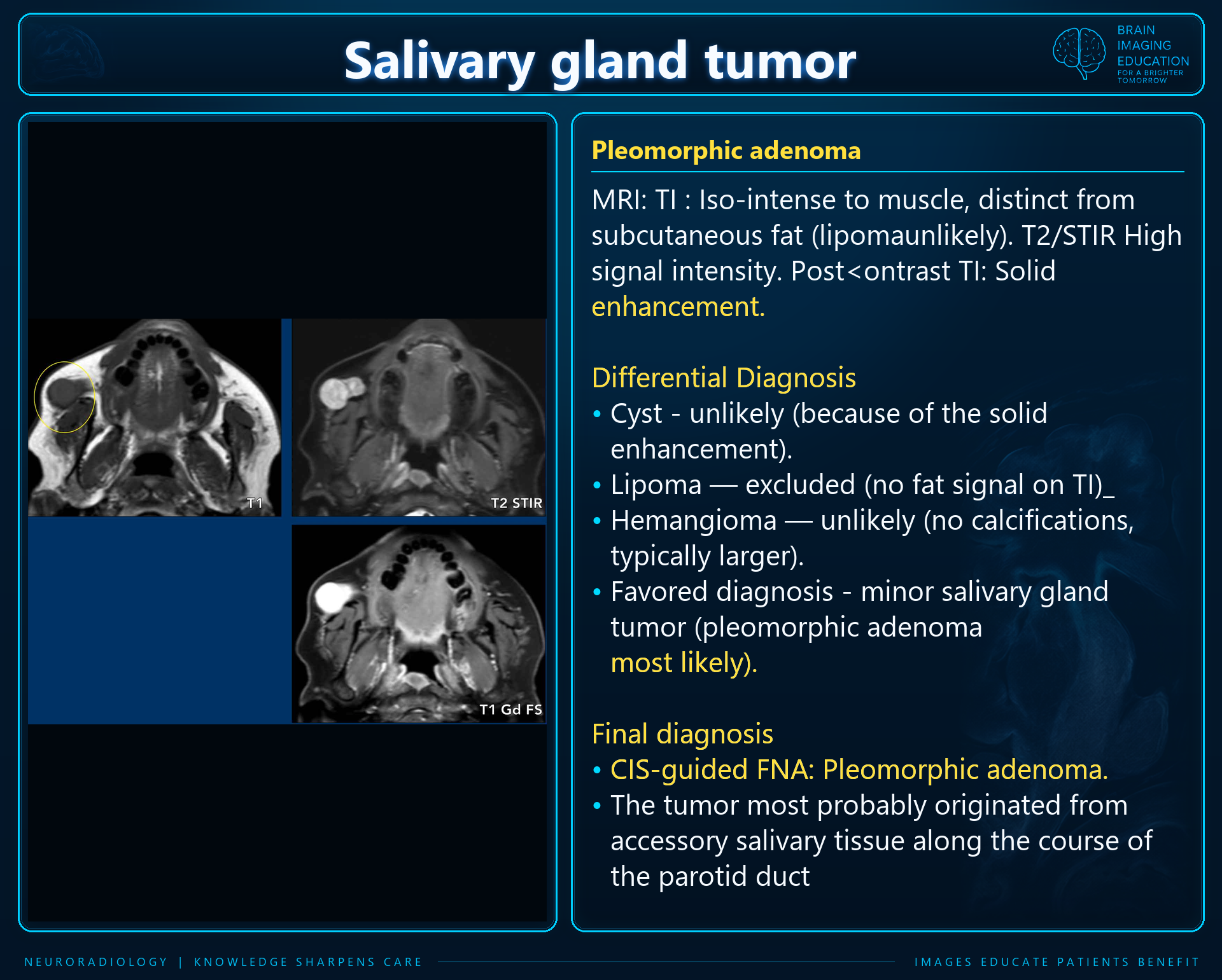
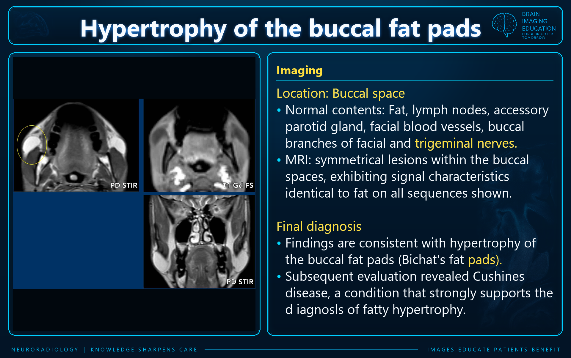
Identify the epicenter relative to the buccinator, masseter, Stensen duct, and accessory parotid tissue.
Assess fat, fluid, solid tissue, vascular architecture, enhancement, and invasive features.
A well-circumscribed buccal mass does not exclude salivary malignancy. Imaging appearances overlap.
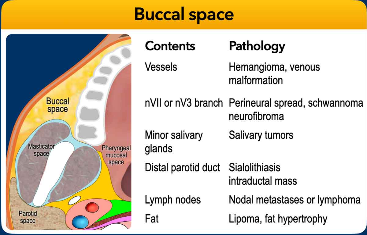
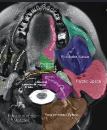
The buccal space lies lateral to the buccinator and anterior to the masseter/masticator space. It contains buccal fat, Stensen duct, minor salivary glands, accessory parotid tissue, facial vessels, lymphatics, and nerve branches. Its incomplete boundaries allow infection, vascular malformations, and tumors to extend between adjacent spaces. pmc.ncbi.nlm.nih
Practical localization points:
Salivary tumors are among the most commonly reported primary buccal space neoplasms, arising from minor salivary glands or accessory parotid tissue. Their imaging appearances overlap substantially. theajo
| Diagnosis | CT/MRI findings | Most useful discriminator |
|---|---|---|
| Pleomorphic adenoma | Rounded or lobulated, well-circumscribed enhancing mass; low T1 and usually high T2 signal, sometimes heterogeneous. | A slow-growing, circumscribed, T2-bright solid mass favors this diagnosis, but is not specific. pmc.ncbi.nlm.nih |
| Adenoid cystic carcinoma | Enhancing solid mass; T2 signal varies with cellularity. May be infiltrative, but small lesions can remain sharply marginated. | Assess carefully for invasive behavior and neural involvement; relatively low/intermediate T2 signal increases concern but is not diagnostic. pmc.ncbi.nlm.nih |
| Mucoepidermoid carcinoma | Variable solid-cystic mass; heterogeneous enhancement and central low attenuation. Low-grade tumors may be predominantly cystic. | A cystic appearance or smooth margin does not establish benignity. theajo |
| Acinic cell carcinoma | Often a circumscribed solid or solid-cystic salivary mass; nonspecific imaging appearance. | Can closely mimic a benign salivary neoplasm. pmc.ncbi.nlm.nih |
| Carcinoma ex pleomorphic adenoma | Heterogeneous enhancing mass; may contain necrotic or infiltrative components, although some remain well defined. | Clinical acceleration of a longstanding mass and invasive imaging features raise concern. theajo |
| Neurofibroma | T2-hyperintense enhancing lesion; possible target sign with central T2 hypointensity. Plexiform lesions can be ill-defined and trans-spatial. | Target sign, plexiform morphology, and neurofibromatosis context. pmc.ncbi.nlm.nih |
| Schwannoma | An additional nerve sheath differential for a circumscribed solid buccal mass; imaging is often insufficient for histologic distinction. | Consider alongside salivary tumors when a neural origin is plausible. theajo |
| Lymphoma | Often homogeneous, well-demarcated soft tissue with molding/compression rather than destructive invasion; some subtypes are infiltrative. | Homogeneity, molding, other nodal disease, or known lymphoma. pmc.ncbi.nlm.nih |
| Rhabdomyosarcoma/other sarcoma | Muscle-density mass on CT; T2 signal higher than muscle, variable heterogeneous enhancement, and possible invasion of adjacent structures. | An aggressive soft-tissue mass, particularly in a child, warrants consideration of rhabdomyosarcoma. pmc.ncbi.nlm.nih |
| Rare mesenchymal tumors | Nodular fasciitis, solitary fibrous tumor, glomus tumor, and angiomyoma can occur; enhancement and T2 appearances overlap with salivary tumors. | Usually a secondary differential when common diagnoses do not fit; tissue diagnosis is generally necessary. theajo |
| Diagnosis | CT/MRI findings | Most useful discriminator |
|---|---|---|
| Venous malformation | Lobulated or serpiginous T2-bright vascular spaces; enhancing venous lakes; may be trans-spatial. Phleboliths can be visible on CT. | Phleboliths plus venous-channel morphology strongly favor a venous malformation. pmc.ncbi.nlm.nih |
| Arteriovenous malformation | Serpiginous vascular channels with prominent MRI flow voids. | High-flow vascular architecture rather than a conventional solid mass. pmc.ncbi.nlm.nih |
| Lymphatic malformation | Multiloculated, septated cystic lesion; fluid-fluid levels may occur. Can extend across spaces. | Multicystic trans-spatial morphology, especially with hemorrhagic fluid-fluid levels. pmc.ncbi.nlm.nih |
| Hemangioma | T2-hyperintense enhancing vascular tumor; flow voids may be present. | Age and growth history matter: infantile hemangioma and vascular malformation are distinct entities. Older literature sometimes uses “hemangioma” imprecisely. pmc.ncbi.nlm.nih |
| Dermoid cyst | Well-circumscribed low-attenuation cyst; fat globules or a fat-fluid level may occur. Lipid-rich components can be T1 bright. | Intralesional fat within a cystic lesion. Infection can make it resemble an abscess. pmc.ncbi.nlm.nih |
| Epidermoid cyst | Well-defined cystic lesion, sometimes adjacent to the skin and separate from the buccinator. | Include in the differential of a superficial, circumscribed cyst; CT morphology alone is nonspecific. theajo |
| Accessory parotid tissue | Tissue over the anterior masseter with attenuation and appearance similar to the main parotid gland; unilateral or bilateral. | Matching normal parotid tissue favors an anatomic variant rather than a discrete tumor. pmc.ncbi.nlm.nih |
| Lipoma | Fat attenuation on CT; follows subcutaneous fat on MRI sequences. | A discrete mass composed of macroscopic fat. theajo |
| Diagnosis | CT/MRI findings | Most useful discriminator |
|---|---|---|
| Odontogenic abscess | Single or multiloculated low-attenuation collection with peripheral enhancement; surrounding fat stranding, skin thickening, and adjacent muscle edema. | Dental source plus rim-enhancing collection and inflammatory change. pmc.ncbi.nlm.nih |
| Cellulitis/phlegmon | Infiltrative edema and fat stranding without a definite low-attenuation collection. | No organized drainable collection identified. pmc.ncbi.nlm.nih |
| Parotid duct obstruction/inflammation | Buccal inflammatory change may arise from duct stenosis or calculi; duct dilatation can also present as a cheek mass. | Relationship to Stensen duct and an obstructing calculus or upstream duct abnormality. pubs.rsna |
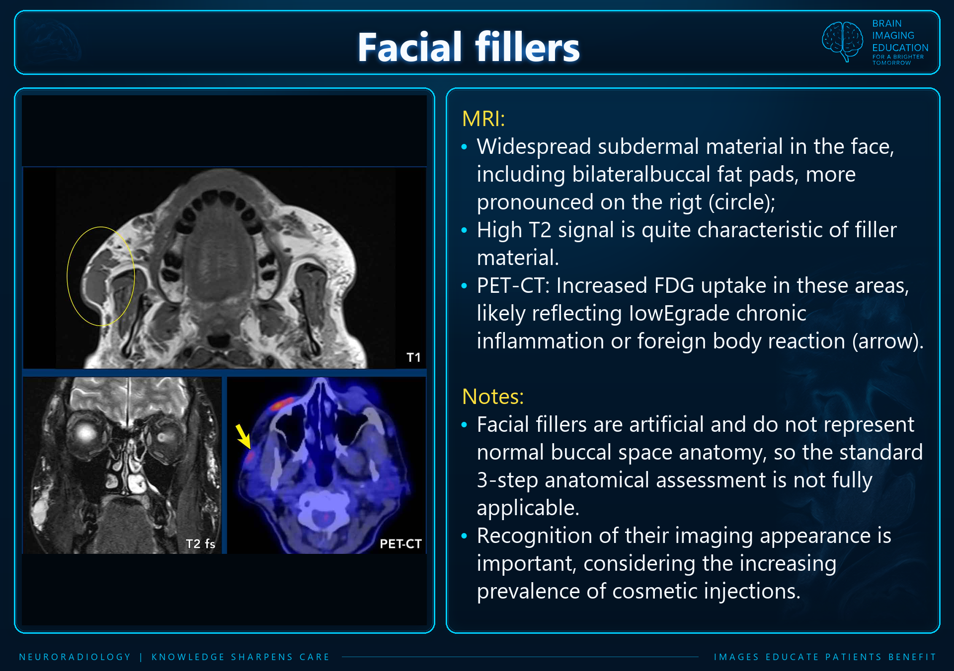
| Foreign-body/filler granuloma | Ill-defined buccal and subcutaneous infiltration, nodules, and sometimes punctate calcifications; may be bilateral. | Cosmetic injection history and bilateral distribution. Findings depend on the injected material. pmc.ncbi.nlm.nih |
| Kimura disease | Enhancing subcutaneous/buccal soft tissue with variable T2 signal; associated regional lymphadenopathy. | Demographic and clinical context, particularly an asymptomatic mass with parotid/submandibular-region nodes. pmc.ncbi.nlm.nih |
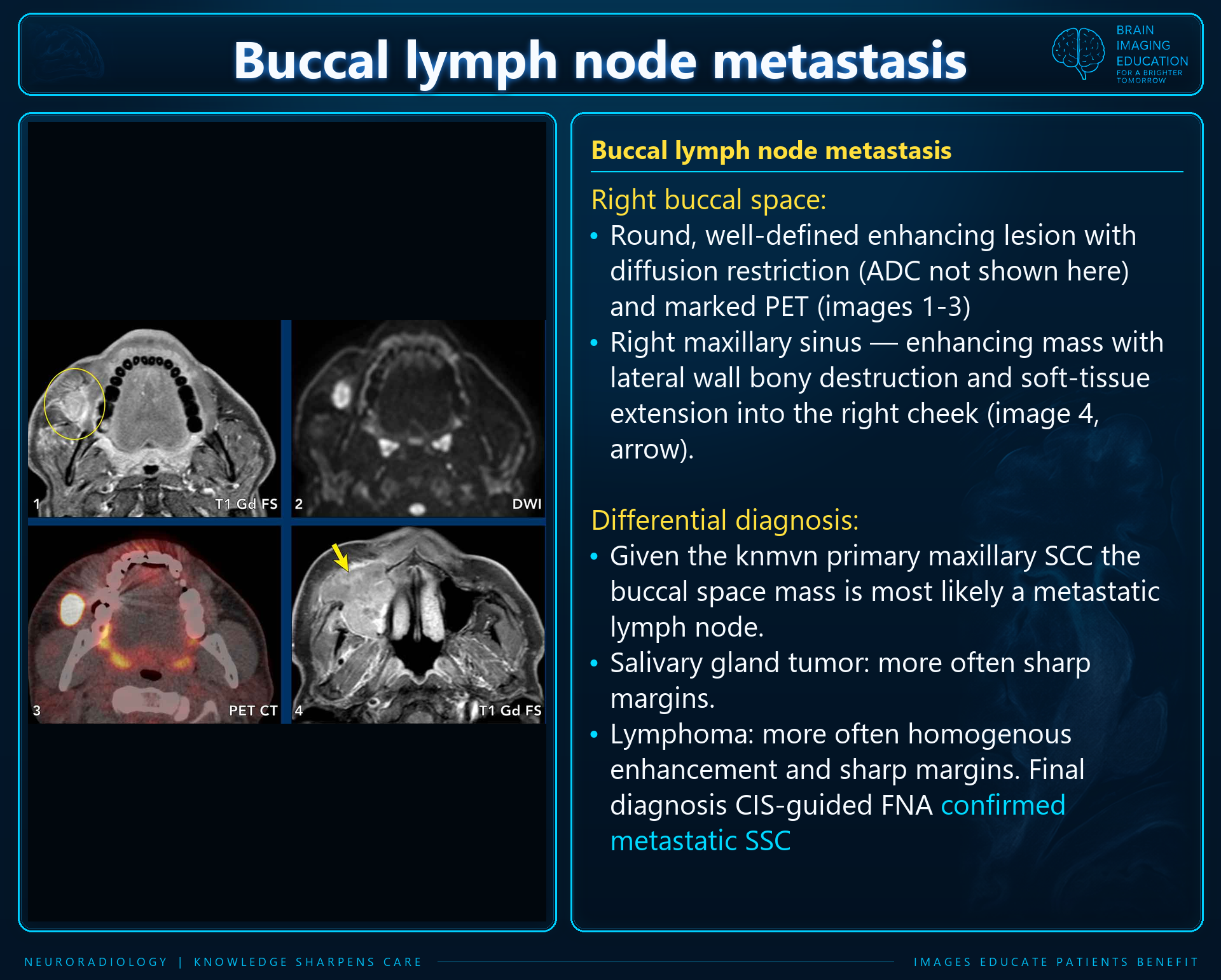
| Metastatic buccal node | Discrete node or mass with central low attenuation and rim enhancement when necrotic. | Search for a facial skin or adjacent mucosal primary; metastatic buccal nodes are described with facial SCC. pmc.ncbi.nlm.nih |
| Direct extension of SCC | A mucosal or cutaneous primary extending into buccal fat; possible fascial-plane breach, bone destruction, and associated adenopathy. | Establish the primary epicenter instead of labeling it a primary buccal space lesion. theajo |
These are useful reading-room differentials rather than reliable histologic diagnoses:
| Dominant pattern | Prioritized differential |
|---|---|
| Circumscribed enhancing solid mass | Pleomorphic adenoma; other benign or malignant salivary tumor; nerve sheath tumor; lymphoma. Smooth margins do not exclude malignancy. pmc.ncbi.nlm.nih |
| Very T2-bright lesion with phleboliths | Venous malformation. pmc.ncbi.nlm.nih |
| Serpiginous flow voids | Arteriovenous malformation/high-flow vascular lesion. pmc.ncbi.nlm.nih |
| Multiloculated cystic trans-spatial lesion | Lymphatic malformation. pmc.ncbi.nlm.nih |
| Rim-enhancing collection with marked stranding | Abscess, especially odontogenic; infected developmental cyst is an alternative. pmc.ncbi.nlm.nih |
| Cyst containing fat globules | Dermoid cyst. pmc.ncbi.nlm.nih |
| Homogeneous solid tissue with molding | Lymphoma, with salivary neoplasm still possible. pmc.ncbi.nlm.nih |
| Bilateral infiltrative nodules with calcifications | Foreign-body granulomas after cosmetic injections. pmc.ncbi.nlm.nih |
| Infiltrative mass with bone destruction or adenopathy | Malignant salivary tumor, secondary SCC, or sarcoma; inflammation can also be infiltrative. pmc.ncbi.nlm.nih |
Contrast-enhanced MRI is generally preferred for an indeterminate solid mass because it better defines soft-tissue extent. Contrast-enhanced CT is particularly useful when infection, a dental source, calcification, or bone involvement is suspected. Ultrasound with Doppler can characterize accessible superficial lesions and vascularity, but should not be the sole study when malignancy is suspected. theajo
A useful report should address:
The key pitfall is equating “well circumscribed” with “benign.” Buccal space salivary malignancies may look innocuous, and imaging is often more reliable for localization and extent than for histologic classification; an indeterminate solid lesion generally requires clinical evaluation and tissue diagnosis. pmc.ncbi.nlm.nih
The uploaded slides and their embedded captions are reproduced unchanged. The fat-pad hypertrophy slide states that the lesions follow fat on all sequences, but labels the displayed images “PD STIR” and “T1 Gd FS.” This apparent sequence/description inconsistency cannot be resolved from the supplied composite alone; confirm the original sequences before using this example as evidence of fat signal.
The pleomorphic adenoma slide uses absence of calcifications and lesion size to argue against “hemangioma.” These slide-specific statements should not be treated as exclusion criteria; the supplied reference text distinguishes vascular tumors from malformations and emphasizes vascular architecture and clinical context. “CIS-guided FNA” is retained in the source slides; the abbreviation is not defined in the supplied material. Diffusion restriction in the metastatic-node slide is stated by the source, but the ADC image is not provided.