Submandibular Space Pathology
A reference to submandibular space anatomy, congenital lesions, infection, neoplasms and pseudolesions. Reading approach: gland or node, cystic or solid, then chase the source.
Anatomy in brief
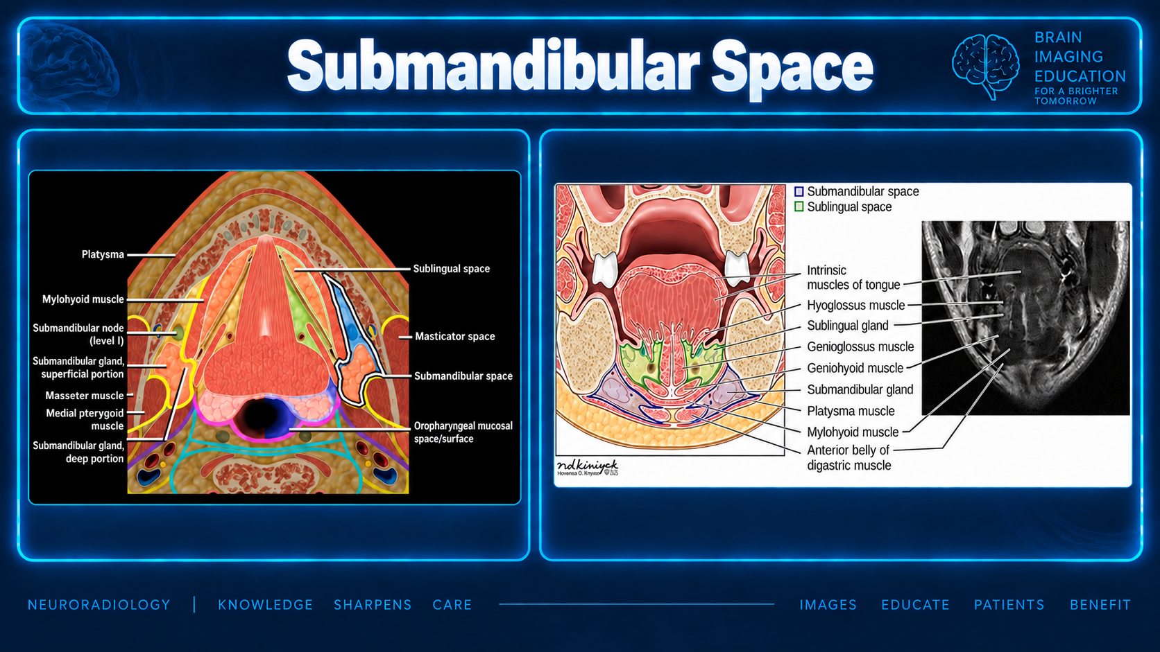
The submandibular space (SMS) lies inferolateral to the mylohyoid and above the hyoid, enclosed by the superficial layer of deep cervical fascia. It contains the superficial submandibular gland (SMG), level IA/IB nodes, the facial artery and vein, the anterior digastric bellies, and fat. It has no fascial barrier to the posterior sublingual space (SLS) or the inferior parapharyngeal space, which is how disease crosses between them.
Congenital / developmental
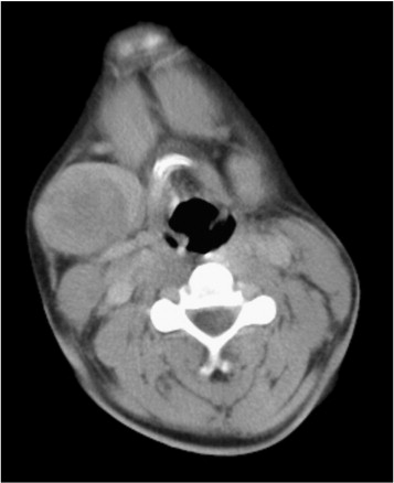
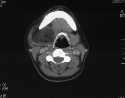
- Second branchial cleft cyst: at the mandibular angle, posterolateral to the SMG, lateral to the carotid space, anteromedial to the SCM.
- Lymphatic malformation: trans-spatial, multilocular, fluid-fluid levels.
- Venous malformation: phleboliths, slow progressive enhancement.
- Dermoid/epidermoid: more often SLS; fat globules ("sack of marbles") in dermoid, restricted diffusion in epidermoid.
- Suprahyoid thyroglossal duct cyst: midline, submental.
Inflammatory / infectious
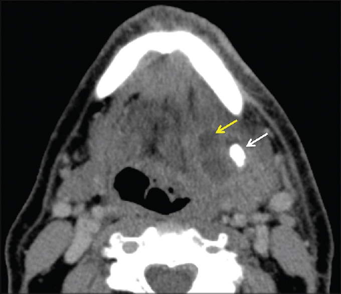
- Sialolithiasis and sialadenitis: the SMG accounts for 80–90% of salivary stones. Look for a Wharton duct calculus, ductal dilatation, and an enlarged, hyperenhancing gland.
- Chronic sclerosing sialadenitis (Küttner tumor): usually IgG4-related; mimics neoplasm.
- Diving ranula: sublingual gland extravasation pseudocyst extending into the SMS, with a "tail" in the SLS.
- Odontogenic abscess: second and third mandibular molar roots sit below the mylohyoid line, so infection drains to the SMS.
- Ludwig angina: bilateral SMS, SLS, and submental cellulitis with airway risk.
- Lymphadenitis: reactive, suppurative, nontuberculous mycobacterial (children), TB, cat-scratch.
Benign neoplasm
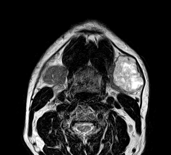
- Pleomorphic adenoma: the most common SMG tumor; well-defined, lobulated, markedly T2 bright, delayed enhancement.
- Lipoma and nerve sheath tumor: uncommon.
Malignant
- Nodal metastasis: far more common than primary gland malignancy. SCC from the lip, anterior floor of mouth, and tongue tip goes to IA; the rest of the oral cavity, anterior nasal cavity, and mid-face skin go to IB.
- SMG carcinoma: roughly 40–50% of SMG tumors are malignant, versus about 20–25% in the parotid. Adenoid cystic (perineural spread along the lingual nerve/V3 and CN XII) and mucoepidermoid lead, then carcinoma ex pleomorphic adenoma.
- Lymphoma: nodal NHL, or primary glandular MALT lymphoma associated with Sjögren.
- Direct invasion: floor-of-mouth or oral tongue SCC, or mandibular lesions.
Pseudolesions
- Motor denervation: V3 (mylohyoid nerve) injury causes atrophy of the anterior digastric and mylohyoid, making the normal side look like a mass.
- Mylohyoid boutonnière: herniated sublingual gland or fat through a mylohyoid defect.
- Gland asymmetry: compensatory SMG hypertrophy after contralateral resection, or surgical SMG transfer to the submental space before radiation.
Reading approach
Gland or node? A glandular mass displaces the facial vein laterally. A fat plane or the vein interposed between the mass and the gland favors a node.
Cystic or solid? The cystic differential is diving ranula, branchial cleft cyst, lymphatic malformation, dermoid/epidermoid, abscess, and necrotic node.
Chase the source.
Necrotic node: look for an oral cavity primary.
Abscess: check the molars on bone windows.
Adenoid cystic: trace perineural spread to the skull base.
Source and case limitations
Medical text and case captions are supplied educational material. No clinical histories, complete imaging examinations, pathology confirmation or bibliographic references were supplied. Case labels are retained as provided.