TI-RADS
Reference Images
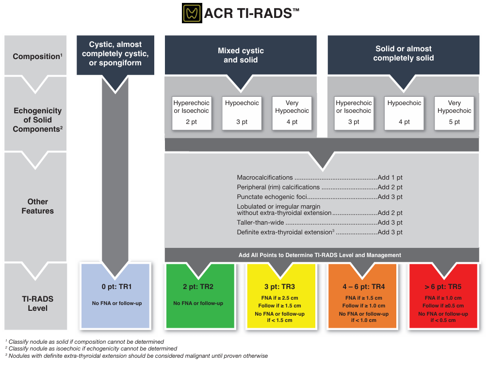
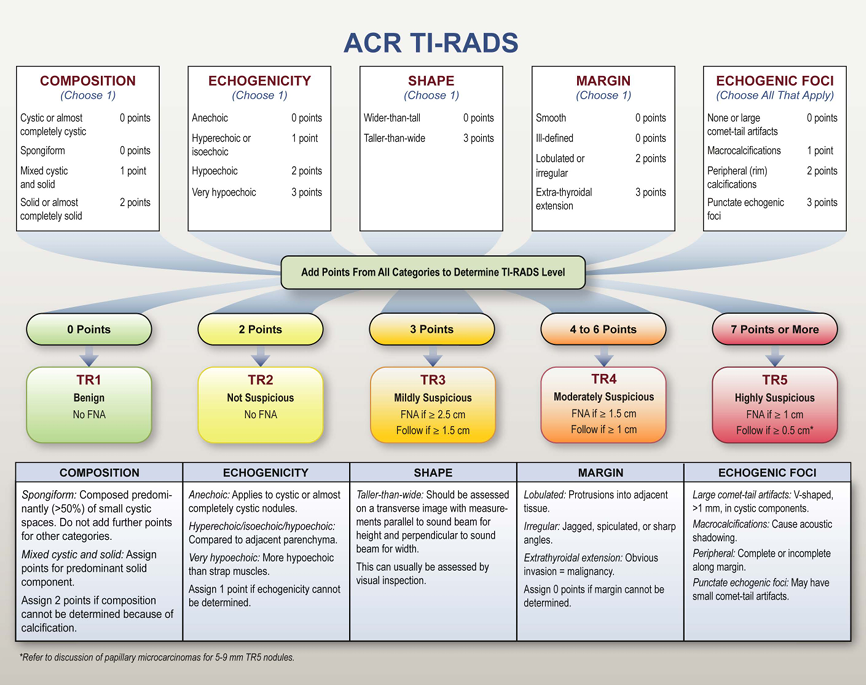
Five categories
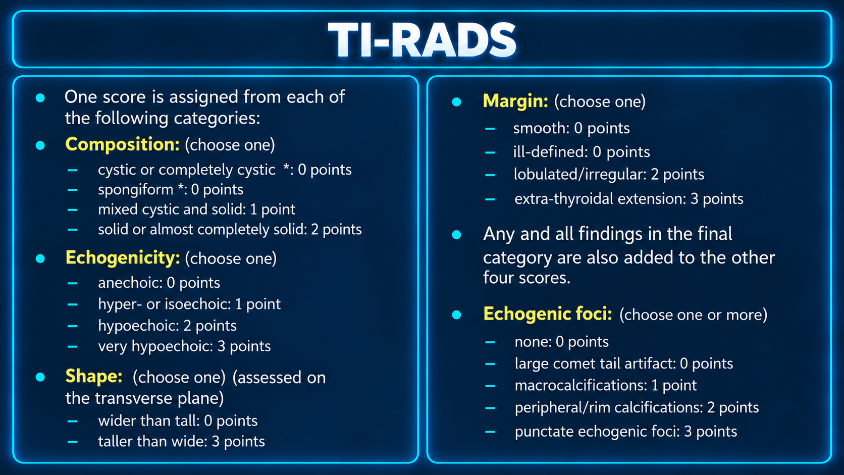
Choose one score each for composition, echogenicity, shape and margin. Add all applicable echogenic-foci findings.
Multiple nodules
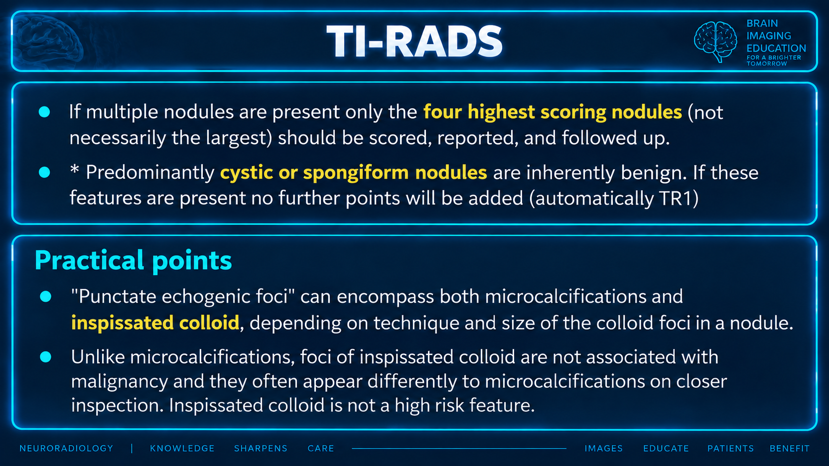
Score, report and follow the four highest-scoring nodules, which are not necessarily the largest.
FNA selection
Apply the TR3–TR5 size criteria. Sample the two highest-scoring nodules; use largest size to break ties within the same classification.
Radiographic features: ultrasound
The higher the cumulative score, the higher the TR level and likelihood of malignancy. Add all findings from the final category to the other four scores.
| Category | Finding | Points |
|---|---|---|
| Composition — choose one | Cystic or completely cystic* | 0 |
| Composition — choose one | Spongiform* | 0 |
| Composition — choose one | Mixed cystic and solid | 1 |
| Composition — choose one | Solid or almost completely solid | 2 |
| Echogenicity — choose one | Anechoic | 0 |
| Echogenicity — choose one | Hyper- or isoechoic | 1 |
| Echogenicity — choose one | Hypoechoic | 2 |
| Echogenicity — choose one | Very hypoechoic | 3 |
| Shape — choose one; transverse plane | Wider than tall | 0 |
| Shape — choose one; transverse plane | Taller than wide | 3 |
| Margin — choose one | Smooth | 0 |
| Margin — choose one | Ill-defined | 0 |
| Margin — choose one | Lobulated/irregular | 2 |
| Margin — choose one | Extrathyroidal extension | 3 |
| Echogenic foci — choose one or more | None | 0 |
| Echogenic foci — choose one or more | Large comet-tail artifact | 0 |
| Echogenic foci — choose one or more | Macrocalcifications | 1 |
| Echogenic foci — choose one or more | Peripheral/rim calcifications | 2 |
| Echogenic foci — choose one or more | Punctate echogenic foci | 3 |
*Supplied benign-composition exception: Predominantly cystic or spongiform nodules are described as inherently benign, with no further points added (automatically TR1). See the terminology limitation above.
The source attributes the category lexicon to the ACR committee’s 2015 publication (citation 2; full reference not supplied).
Classification and recommendations
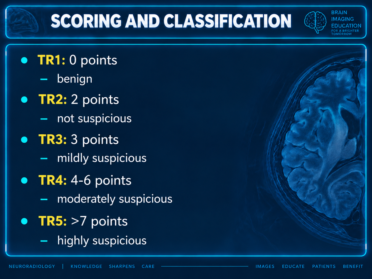
| Level | Points | Classification | Ultrasound follow-up | FNA | Published malignancy rate |
|---|---|---|---|---|---|
| TR1 | 0 | Benign | None required | None required | 0.3% |
| TR2 | 2 | Not suspicious | None required | None required | 1.5% |
| TR3 | 3 | Mildly suspicious | ≥1.5 cm: at 1, 3 and 5 years | ≥2.5 cm | 4.8% |
| TR4 | 4–6 | Moderately suspicious | ≥1.0 cm: at 1, 2, 3 and 5 years | ≥1.5 cm | 9.1% |
| TR5 | ≥7 | Highly suspicious | ≥0.5 cm: annually for up to 5 years | ≥1.0 cm | 35% |
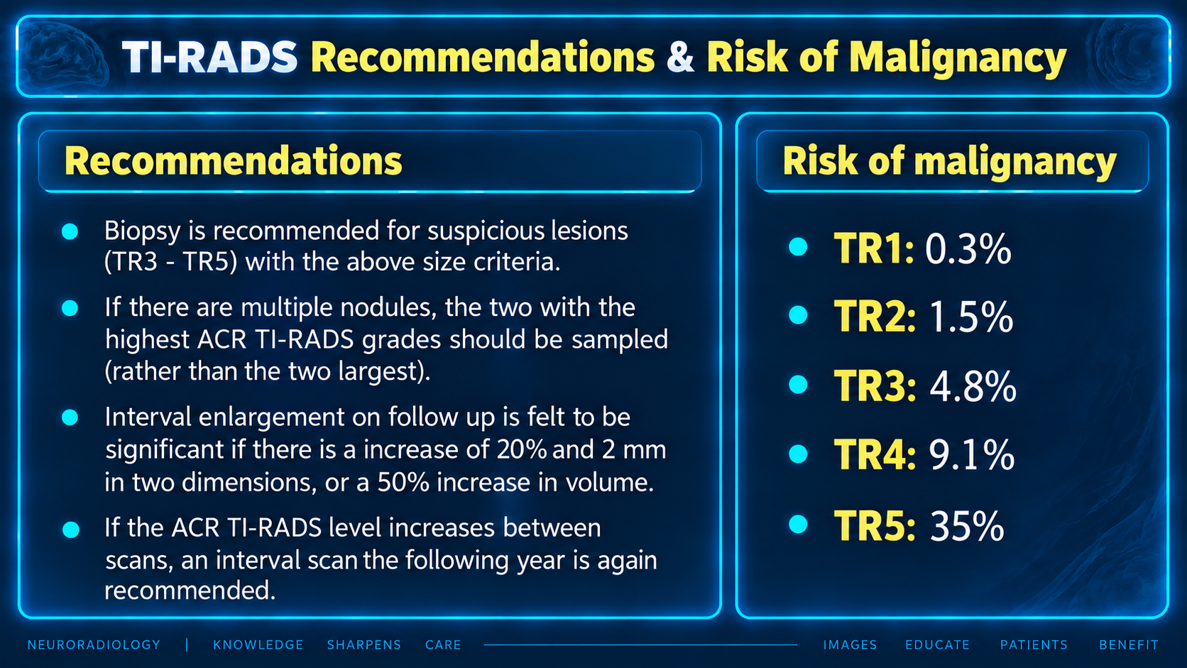
FNA biopsy is recommended for suspicious lesions (TR3–TR5) meeting these size criteria. In multiple nodules, sample the two with the highest scores rather than the two largest; largest size breaks ties within the same classification.
Interval change
The supplied prose defines significant enlargement as an increase of >20% and >2 mm in two dimensions, or a >50% increase in volume. If the ACR TI-RADS level increases between scans, an interval scan the following year is again recommended. See the growth discrepancy above.
Practical points and reporting cautions
“Punctate echogenic foci” can encompass microcalcifications and inspissated colloid, depending on technique and colloid-focus size. Unlike microcalcifications, inspissated colloid foci are not associated with malignancy in the supplied account and often look different on closer inspection. Inspissated colloid is not a high-risk feature.
ACR TI-RADS is reserved for lesion reporting; it does not grade a “normal thyroid gland.” TR1 includes benign simple and/or spongiform cysts meeting 0-point criteria. Purely anechoic/cystic lesions receive 0 points. Calling such a lesion “very hypoechoic” instead assigns 3 points and may lead to a fruitless FNA and patient/physician anxiety.
Frank invasion of surrounding structures is an unfavorable prognostic sign and receives 3 points. Use caution and experience when reporting suspected minimal extrathyroidal extension without frank invasion, particularly with otherwise benign features.
Background, malignancy risk and comparative evidence
Development of ACR TI-RADS 2017
The 2015 ACR white paper developed a descriptive lexicon. The 2017 white paper updated this to a stratified scoring system rather than a pattern-based system. The source describes support from an ACR paper on incidental thyroid nodules (citation 3) and National Cancer Institute Surveillance, Epidemiology, and End Results (SEER) data. The 2015 lexicon included six stages with subdivisions and increasing malignancy risk; these are not replicated in ACR TI-RADS.
Risk of malignancy
The original 2017 paper's projected risks came from partial analysis of 3,433 nodules with cytological results: <2% for TR1/TR2, 5% for TR3, 5–20% for TR4 and >20% for TR5. The source attributes the final category rates in the table to citation 4 and describes a stepwise increase with each point, validating each category. Full references and study details were not supplied.
Comparison with other classification systems
The source places ACR TI-RADS in 2017, preceded by K-TIRADS (2017) and followed by EU-TIRADS (2017), and describes ATA and SRU (citation 5) as also in regular use.
Reported comparative sensitivity ranges from 75–97% and specificity from 53–67%. One study describes the highest sensitivity and lowest specificity among compared systems (citation 6); two others describe the highest specificity (citations 7 and 8). In the latter studies, ACR had the greatest overall performance by area under the receiver operating characteristic curve or accuracy, with fewer unnecessary FNAs (false positives). These differing conclusions are preserved; the studies are not identified in the supplied bibliography.
Good interobserver agreement for the biopsy decision is reported (Cohen kappa 0.61; citation 9).
Revisions
The undated source states that no formal revisions had been made at the time of writing. It describes one group's artificial-intelligence refinement as maintaining high sensitivity, increasing specificity and reducing the FNA rate. No study identification or date was supplied.