At a Glance
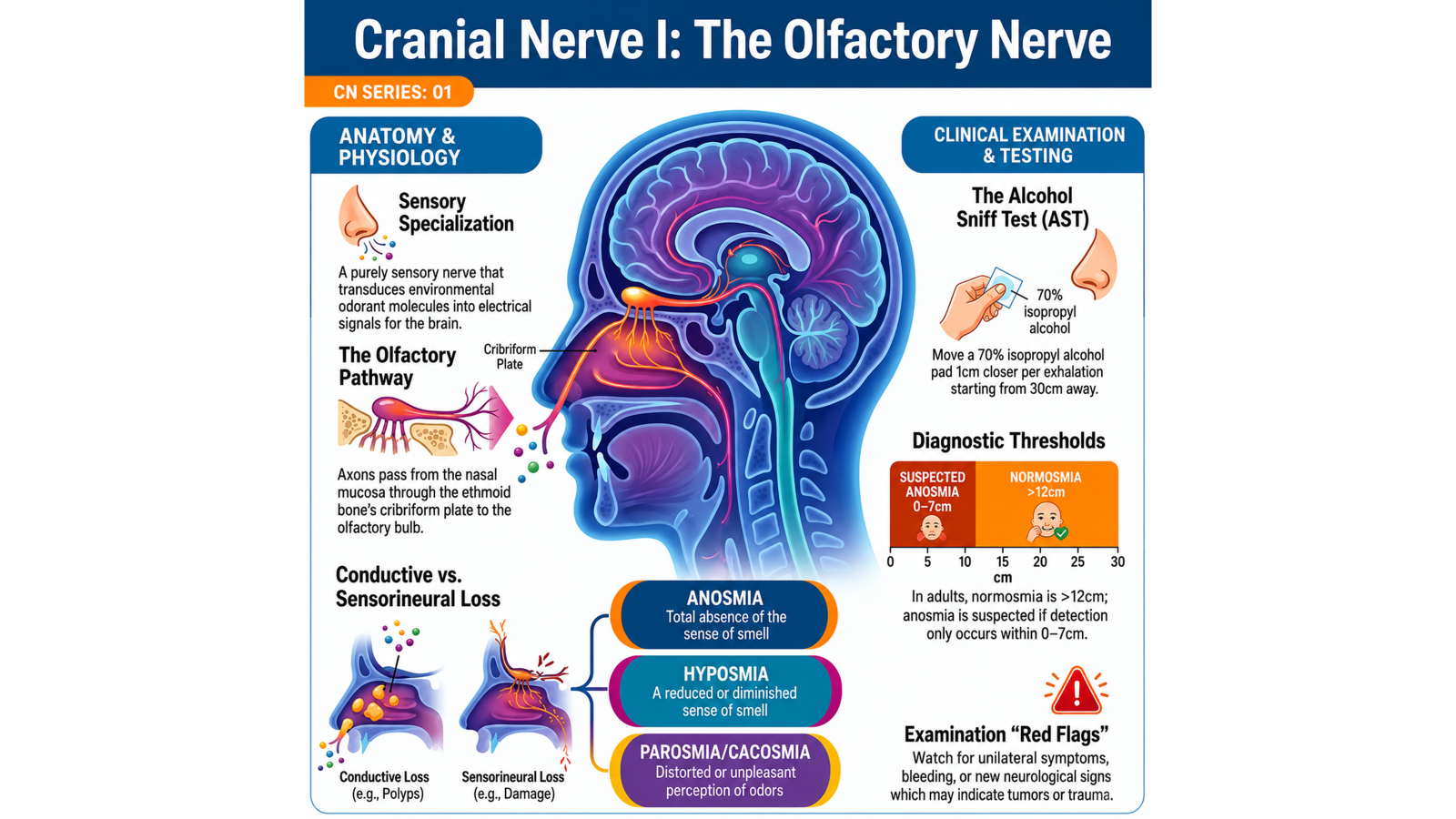
FunctionCN I conveys special sensory information for smell from the olfactory mucosa of the nasal cavity.
Location
- Olfactory cleft in the roof of the nasal cavity.
- Cribriform plate of the ethmoid bone.
- Olfactory groove of the anterior cranial fossa near the midline.
What is actually CN I?
Strictly, the olfactory nerve comprises approximately 20 bundles of first-order axons—the olfactory filaments. The bulb, tract, and striae are extensions of the central nervous system rather than peripheral nerve.
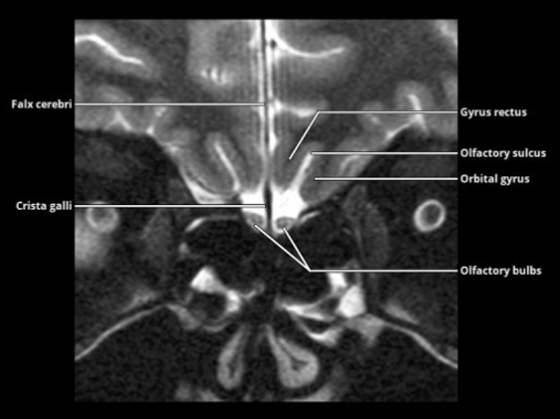
Key imaging landmarks
- Crista galli and cribriform plate.
- Paired olfactory bulbs and tracts.
- Gyrus rectus medially and orbital gyri laterally.
- Olfactory sulcus between the gyrus rectus and orbital gyri.
Course
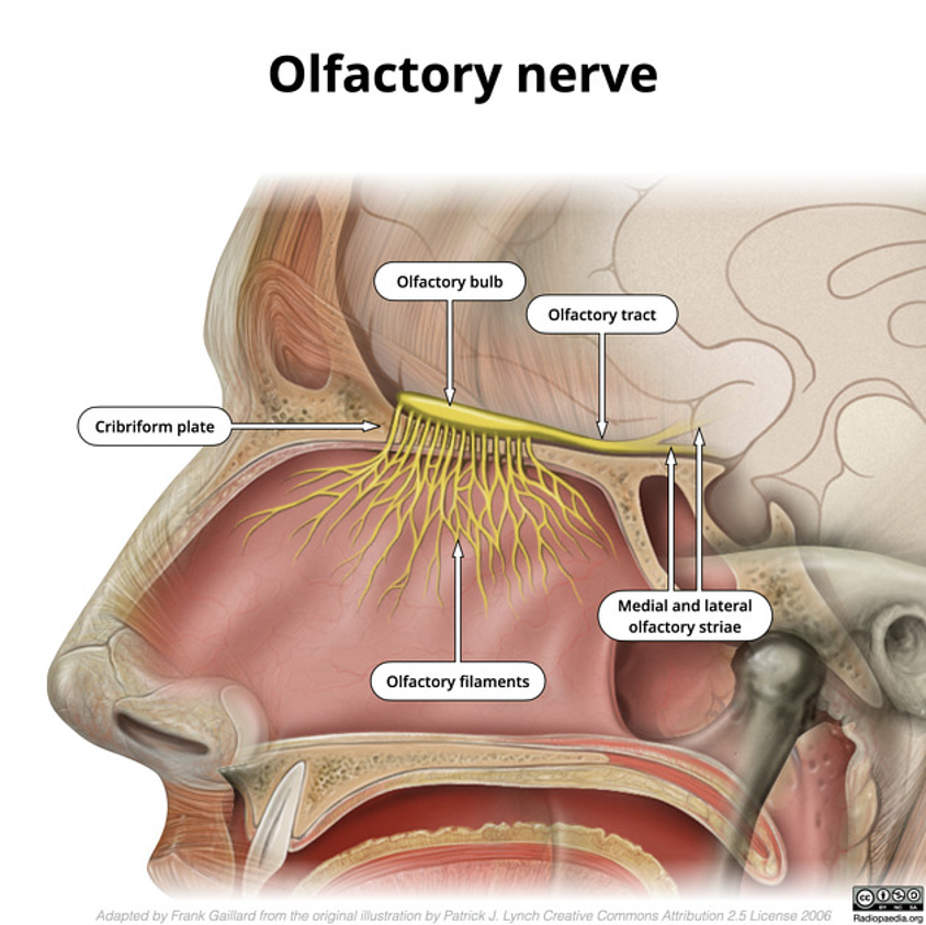
1 · Olfactory filaments
Bipolar olfactory sensory neurons lie in specialized neuroepithelium within the olfactory cleft. Their central axons form fine filaments that traverse the cribriform plate, pierce the dura, and synapse in the olfactory bulb.
2 · Olfactory bulb
The paired bulbs lie above the cribriform plate in the olfactory grooves. Mitral cells within the bulb form the principal second-order neurons.
3 · Olfactory tract
The tract courses posteriorly in the olfactory groove, inferior to the olfactory sulcus, between the gyrus rectus and orbital gyri.
4 · Olfactory striae
Near the anterior perforated substance, the tract expands at the olfactory trigone and divides into medial and lateral striae. The lateral stria projects toward the uncus and primary olfactory cortex; medial fibers connect with septal and hypothalamic pathways.
Pathway shorthandOlfactory mucosa → filaments → cribriform plate → bulb → tract → trigone → medial and lateral striae