Special sensory pathway for vision · retina to lateral geniculate body and visual cortex
At a Glance
FunctionCN II is a purely sensory pathway carrying visual information from retinal ganglion cells to the brain.
Central nervous system tract
Like CN I, the optic nerve is an extension of the central nervous system. It is myelinated by oligodendrocytes, surrounded by meninges and cerebrospinal fluid, and not invested by Schwann cells.
Key relationships
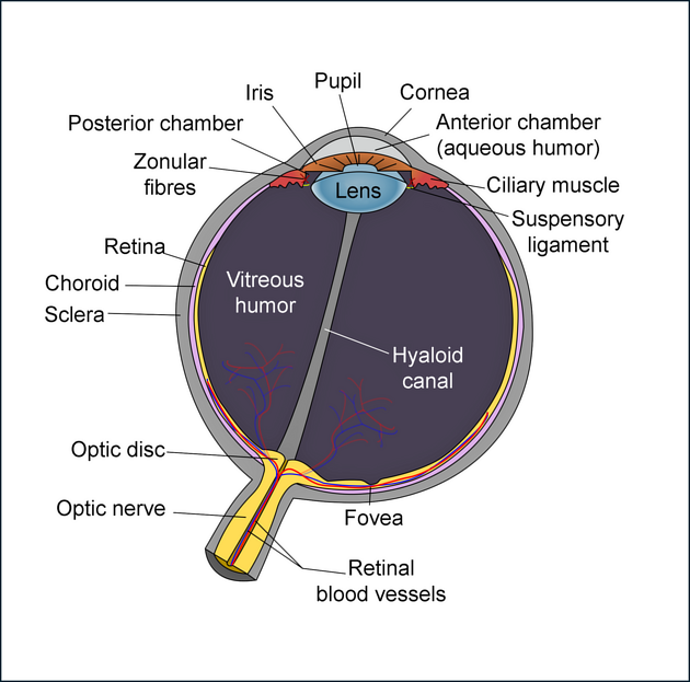
- Begins at the optic disc at the posterior globe.
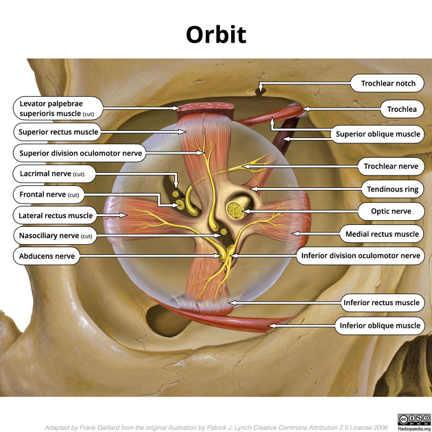
- Passes centrally through the intraconal orbit.
- Traverses the optic canal superior to the ophthalmic artery.
- Enters the suprasellar cistern and joins the opposite nerve at the optic chiasm.
Main relay
Most fibers terminate in the lateral geniculate body of the thalamus. Additional fibers reach the pretectal midbrain for pupillary reflex pathways.
Four Segments
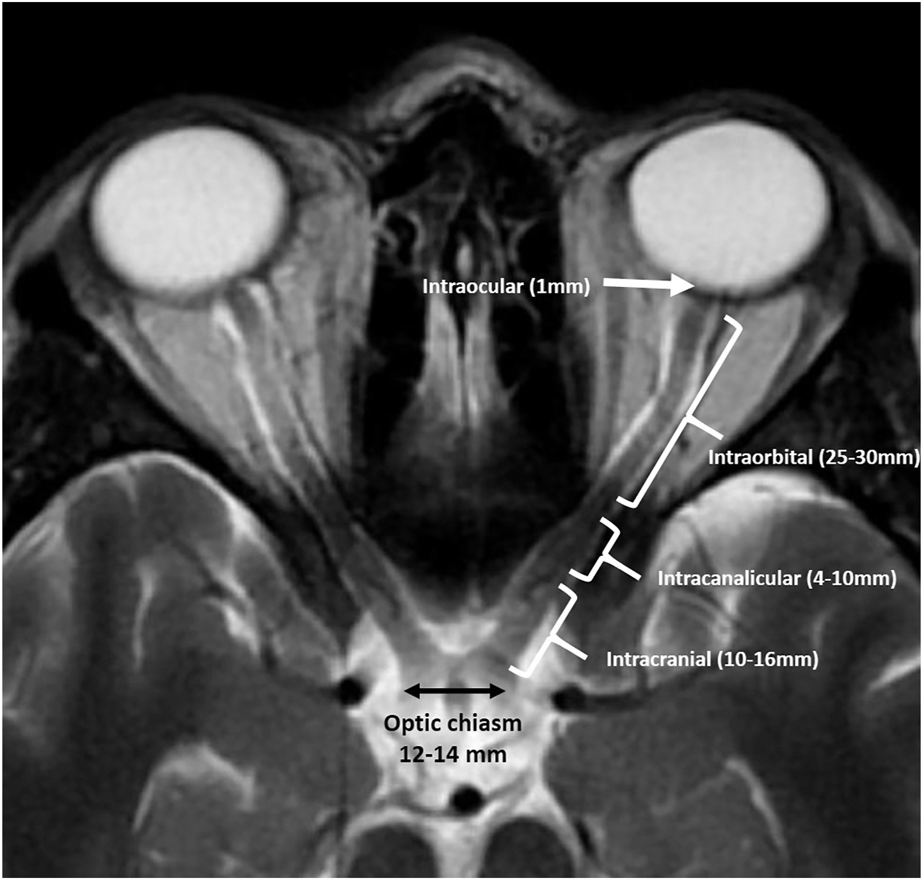
1 · Intraocular · ~1 mm
Retinal ganglion-cell axons converge at the optic disc and pass through the scleral lamina cribrosa to form the nerve.
2 · Intraorbital · ~25 mm
The longest and most mobile segment courses posteriorly within the muscle cone. Its meningeal and CSF coverings communicate directly with the intracranial subarachnoid space.
3 · Intracanalicular · ~9 mm
Passes through the common tendinous ring and optic canal, with the ophthalmic artery usually inferolateral or inferior to the nerve.
4 · Intracranial · ~16 mm
The prechiasmatic segment crosses the suprasellar cistern. The anterior cerebral artery lies superolaterally before the nerve joins the chiasm.
Clinical imaging connectionBecause the optic nerve sheath contains CSF, raised intracranial pressure can distend the sheath and produce papilledema.
Chiasm & Visual Pathway
Partial decussation
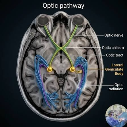
At the chiasm, fibers from each nasal hemiretina cross while temporal retinal fibers remain ipsilateral. Each optic tract therefore carries information from the contralateral visual field.
Pathway shorthandRetina → optic nerve → chiasm → optic tract → lateral geniculate body → optic radiations → visual cortex
Course beyond the chiasm
- Optic tracts wrap around the cerebral peduncles.
- Most fibers synapse in the lateral geniculate bodies.
- Optic radiations project to the calcarine cortex.
Localization pearl
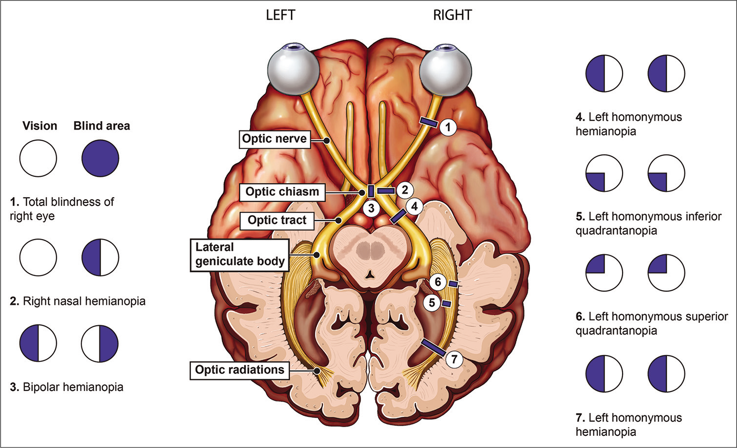
- Prechiasmatic lesion: monocular deficit.
- Midline chiasmal lesion: classically bitemporal hemianopia.
- Retrochiasmatic lesion: contralateral homonymous field deficit.
Arterial Supply
Nerve head
- Circle of Zinn–Haller, mainly from short posterior ciliary arteries.
- Additional pial and choroidal contributions.
Orbital and canalicular nerve
- Ophthalmic artery.
- Central retinal artery and pial branches.
Intracranial nerve and chiasm
- Small anterior cerebral arterial branches.
- Superior hypophyseal arteries.
Optic tract
- Anterior choroidal artery.
- Posterior communicating artery branches.
Sphenoid Sinus Variants
Why it mattersThe optic nerve may closely abut or protrude into the sphenoid sinus, increasing vulnerability during endoscopic sinus surgery.
Delano classification
- Type 1 · 76% — adjacent to the lateral or superior sinus wall without indentation.
- Type 2 · 15% — indents the lateral sphenoid sinus wall.
- Type 3 · 6% — courses through the sphenoid sinus.
- Type 4 · 3% — lies immediately lateral to the posterior ethmoid and sphenoid sinuses.
CT review
Assess optic-canal protrusion, bony dehiscence, sphenoid pneumatization, Onodi cells, and adjacent carotid anatomy before sinonasal surgery.
Imaging Checklist
MRI
- Use thin-section, fat-suppressed orbital imaging in axial and coronal planes.
- Follow both nerves continuously from globe to chiasm.
- Assess caliber, signal, enhancement, sheath distention, and symmetry.
- Inspect the chiasm, optic tracts, lateral geniculate bodies, radiations, and occipital cortex when clinically relevant.
CT
- Review optic canals and anterior clinoid processes.
- Define fractures, canal narrowing, calcification, and sphenoid-sinus relationships.
Important distinction
Optic neuritis affects the nerve; optic perineuritis and optic nerve sheath meningioma primarily involve the sheath.