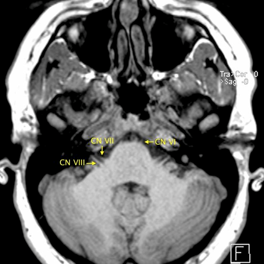
Special sensory pathways for hearing, static equilibrium, and dynamic equilibrium
At a Glance
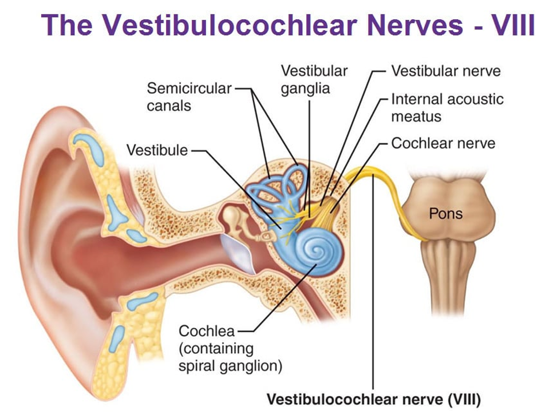
FunctionCN VIII carries special sensory afferents for hearing, static equilibrium, and rotational equilibrium.
Components
- Cochlear division — auditory information from the organ of Corti.
- Vestibular division — position and motion information from the utricle, saccule, and semicircular canals.
Course summary
Distinct cochlear and vestibular roots emerge at the lateral pontomedullary junction, unite in the cerebellopontine angle, enter the internal acoustic meatus, and divide again near its fundus.
TerminologyVestibulocochlear nerve is preferred; “acoustic nerve” is an older synonym.
Nuclei & Brainstem Exit
Cochlear nuclei
Dorsal and ventral cochlear nuclei lie in the dorsolateral upper medulla, deep to the lateral angle of the rhomboid fossa.
Vestibular nuclei
Four vestibular nuclei form paired columns medial to the cochlear nuclei, extending from upper medulla into lower pons.
Root exit
- Fibers curve around the inferior cerebellar peduncle.
- Separate vestibular and cochlear roots exit at the pontomedullary junction.
- CN VIII lies lateral to CN VII at the brainstem and within the CPA.
Cisternal Course
Cerebellopontine angle
The roots rapidly converge and pass laterally and slightly anterosuperiorly through the CPA cistern toward the porus acusticus.
Acousticofacial bundle
- Vestibulocochlear nerve — most lateral component.
- Facial nerve.
- Nervus intermedius.
- Labyrinthine artery.
Relationships
The nerve courses adjacent to the posterior petrous temporal bone, above the petro-occipital fissure and inferior petrosal sinus, before entering the internal acoustic meatus.
Pathway shorthandPontomedullary junction → CPA cistern → porus acusticus → internal acoustic meatus → cochlear and vestibular divisions
IAC Quadrants
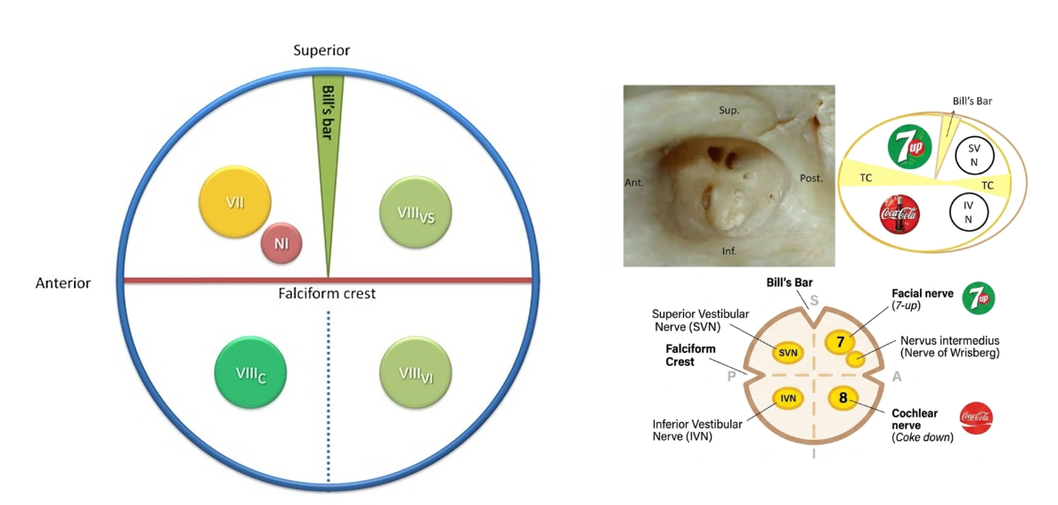
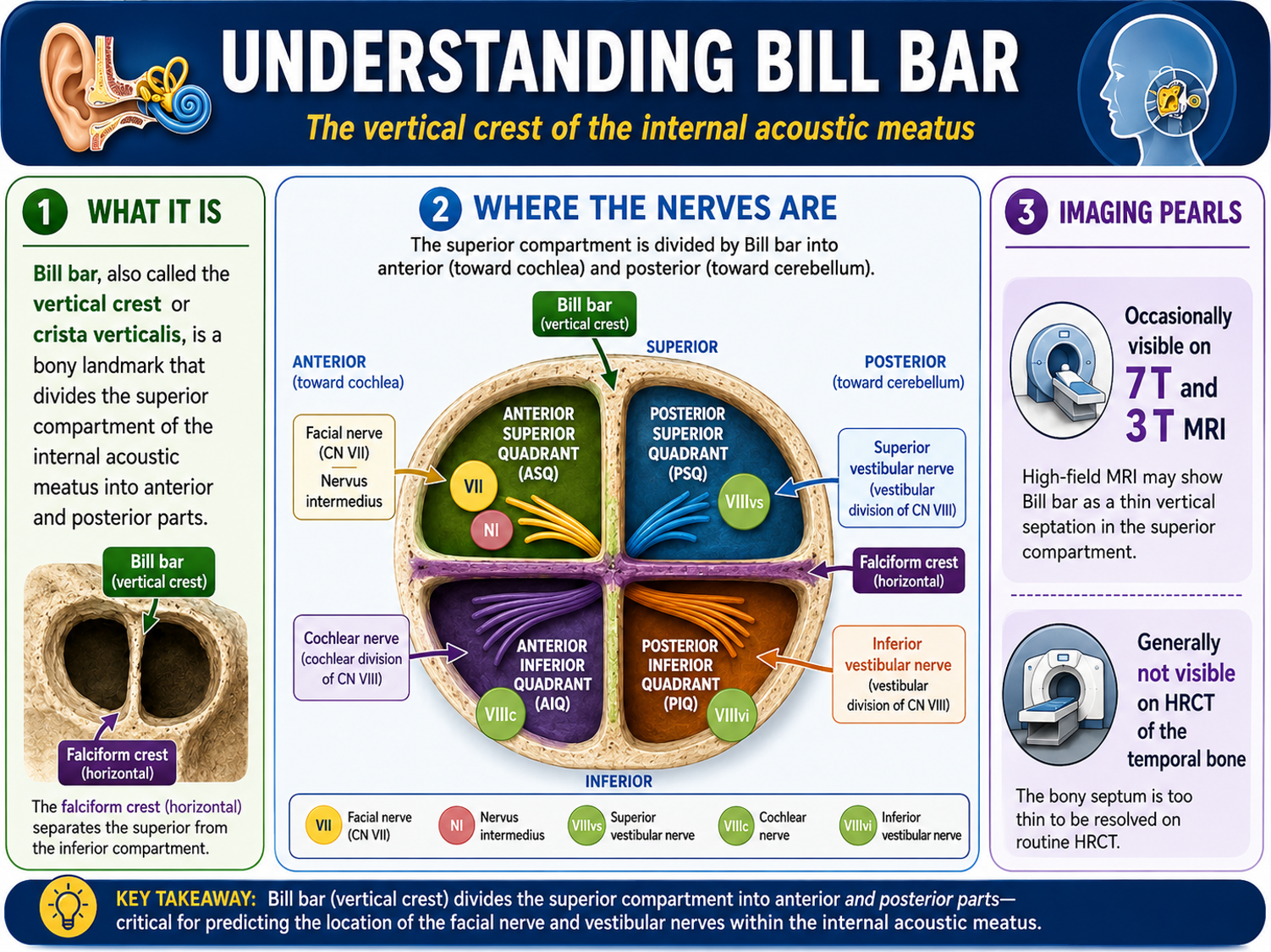
Fundus orientationThe falciform crest separates superior from inferior; Bill bar separates the two superior quadrants.
Anterior superior
Facial nerve (CN VII) with nervus intermedius.
Posterior superior
Superior vestibular nerve.
Anterior inferior
Cochlear nerve.
Posterior inferior
Inferior vestibular nerve; the singular nerve courses separately toward the foramen singulare.
Memory aid“Seven up, Coke down”: facial nerve superior, cochlear nerve inferior in the anterior half of the IAC.
Peripheral Branches
Cochlear nerve
Occupies the anteroinferior IAC quadrant and spirals into the modiolus toward the spiral ganglion and organ of Corti.
Superior vestibular division
Travels in the posterosuperior quadrant and carries afferents from the utricle and superior and lateral semicircular canals.
Inferior vestibular division
Travels in the posteroinferior quadrant and carries afferents from the saccule.
Singular nerve
The posterior ampullary nerve passes through the foramen singulare, carrying sensory fibers from the posterior semicircular canal.
Imaging Checklist
MRI
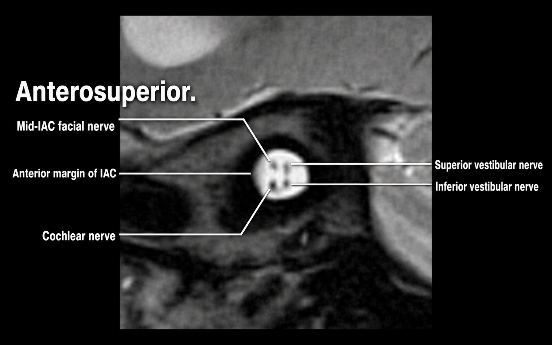
- Use thin-section heavily T2-weighted 3D imaging through the CPA and IACs.
- Confirm four discrete nerve bundles near the IAC fundus.
- Assess symmetry, caliber, continuity, displacement, and enhancement.
- Inspect the cochlea, vestibule, semicircular canals, brainstem nuclei, and CPA cistern.
CT temporal bone
- Review IAC caliber and osseous margins.
- Assess the cochlear aperture, vestibular aqueduct, labyrinth, and foramen singulare.
- Bill bar is generally too thin for routine HRCT resolution.
Radiology relevance
Vestibular schwannomas most often arise from a vestibular division and may widen the IAC or extend into the CPA cistern.