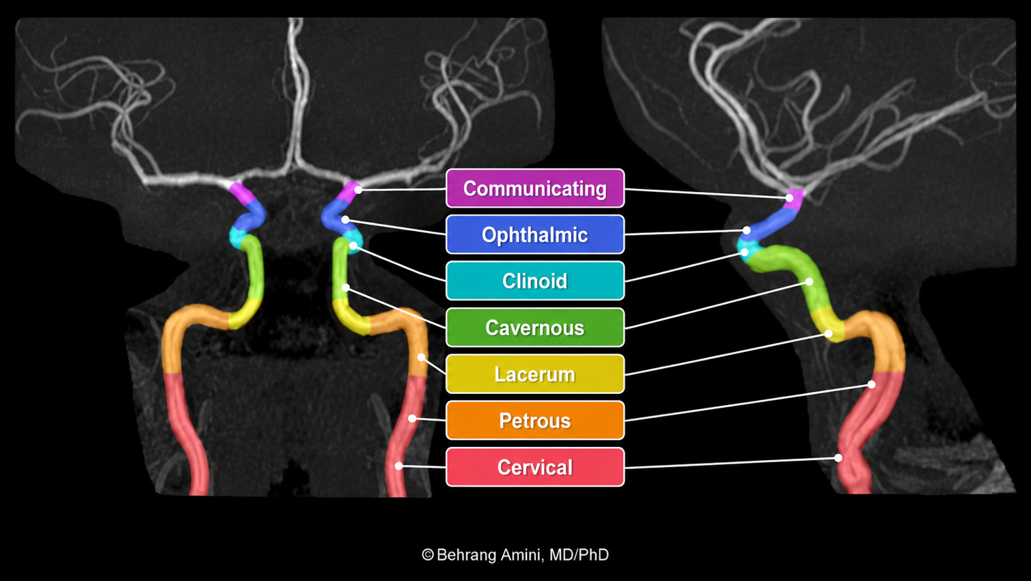
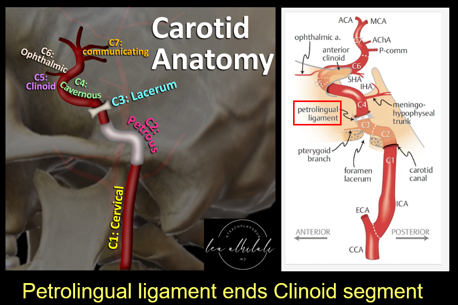
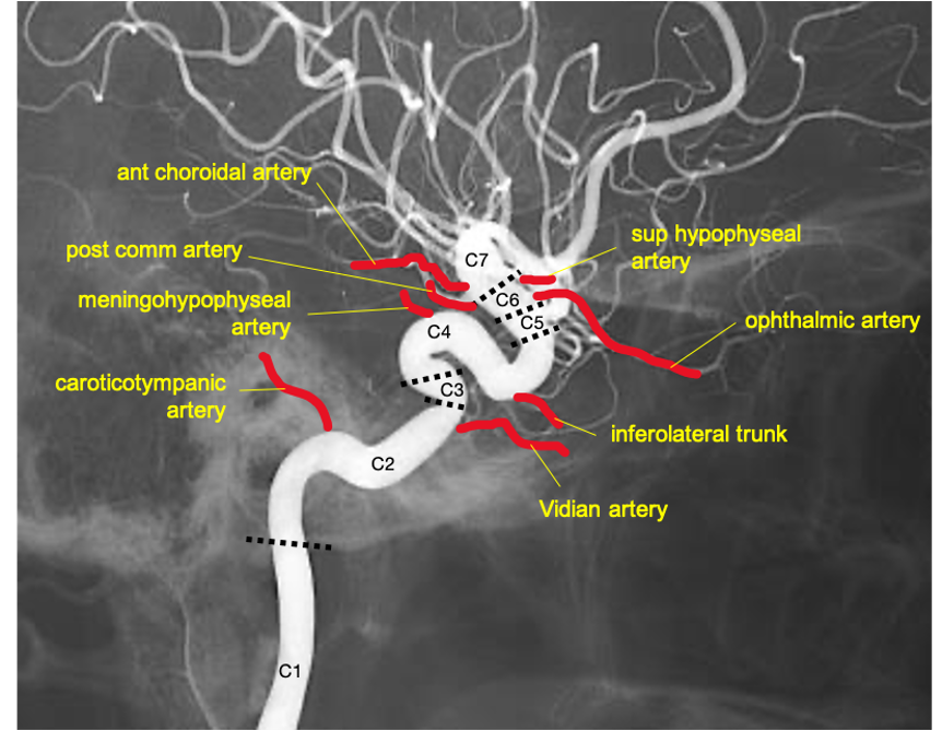
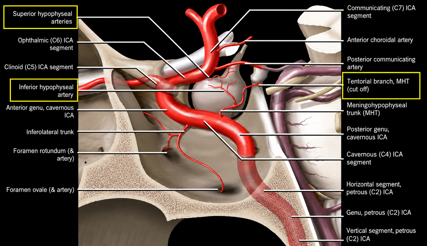
Origin · Bouthillier C1–C7 segments · branches · skull-base and angiographic landmarks
At a Glance
Origin and terminationThe ICA begins at the common carotid bifurcation, enters the skull through the carotid canal, and terminates as the ACA and MCA.
Carotid bulb
The proximal ICA expands at the carotid sinus. The carotid body is an adjacent chemoreceptor organ.
Bifurcation level
Usually near C3–C5, the hyoid, or upper thyroid cartilage; high, low, and asymmetric bifurcations occur.
Function
Major anterior-circulation supply to the brain and orbit.
C1–C3: Skull-Base Entry
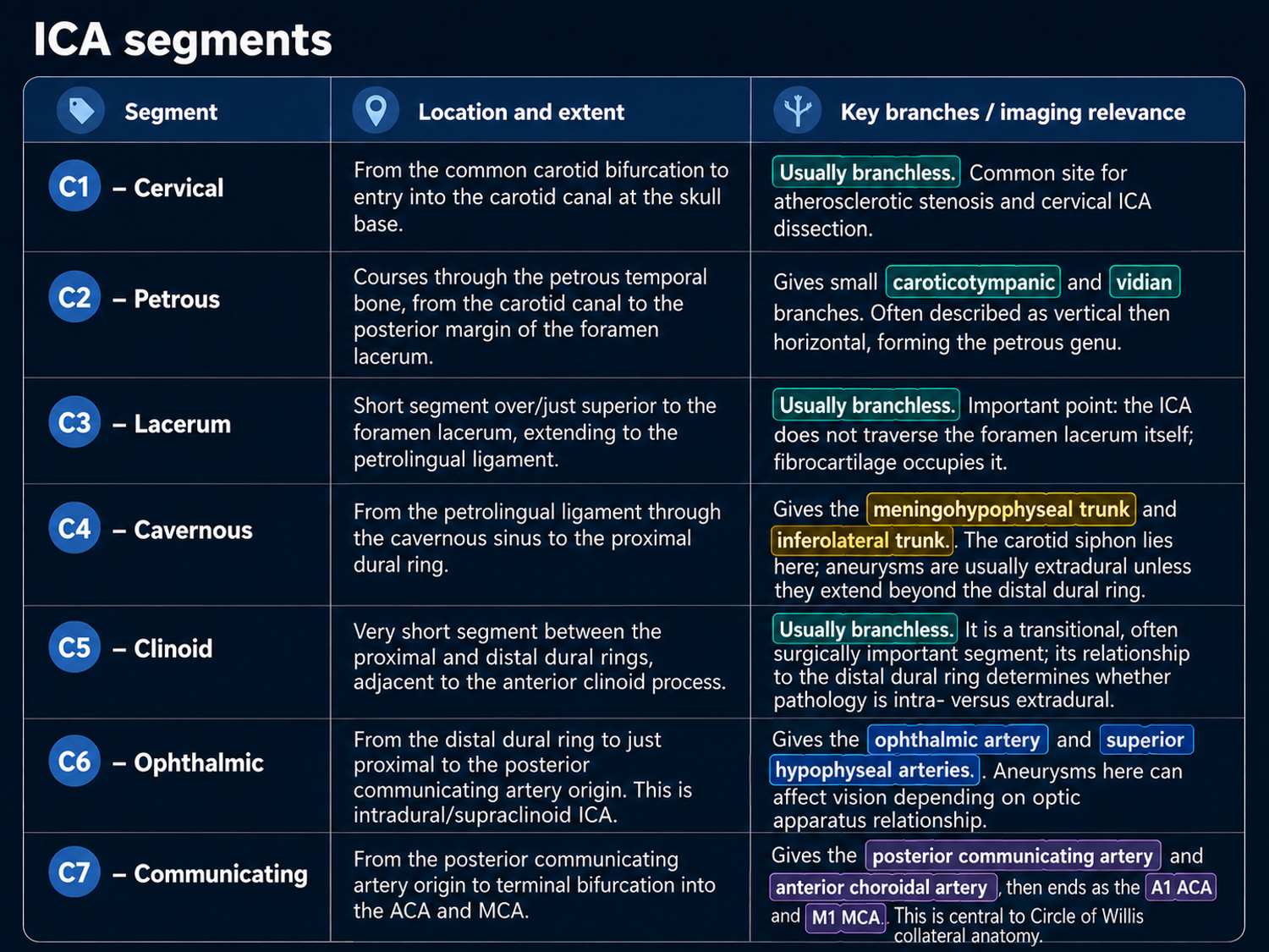
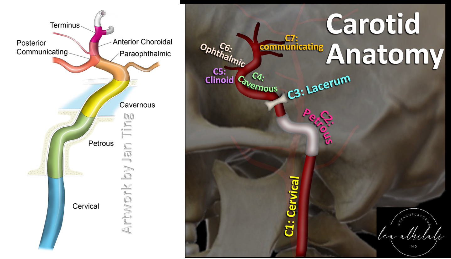
C1 Cervical
Bifurcation to carotid canal; ascends in the carotid sheath and is usually branchless.
C2 Petrous
Traverses the petrous carotid canal and turns anteromedially at the petrous genu. Branches: caroticotympanic and Vidian arteries.
C3 Lacerum
Short segment above the foramen lacerum, ending at the petrolingual ligament; usually branchless.
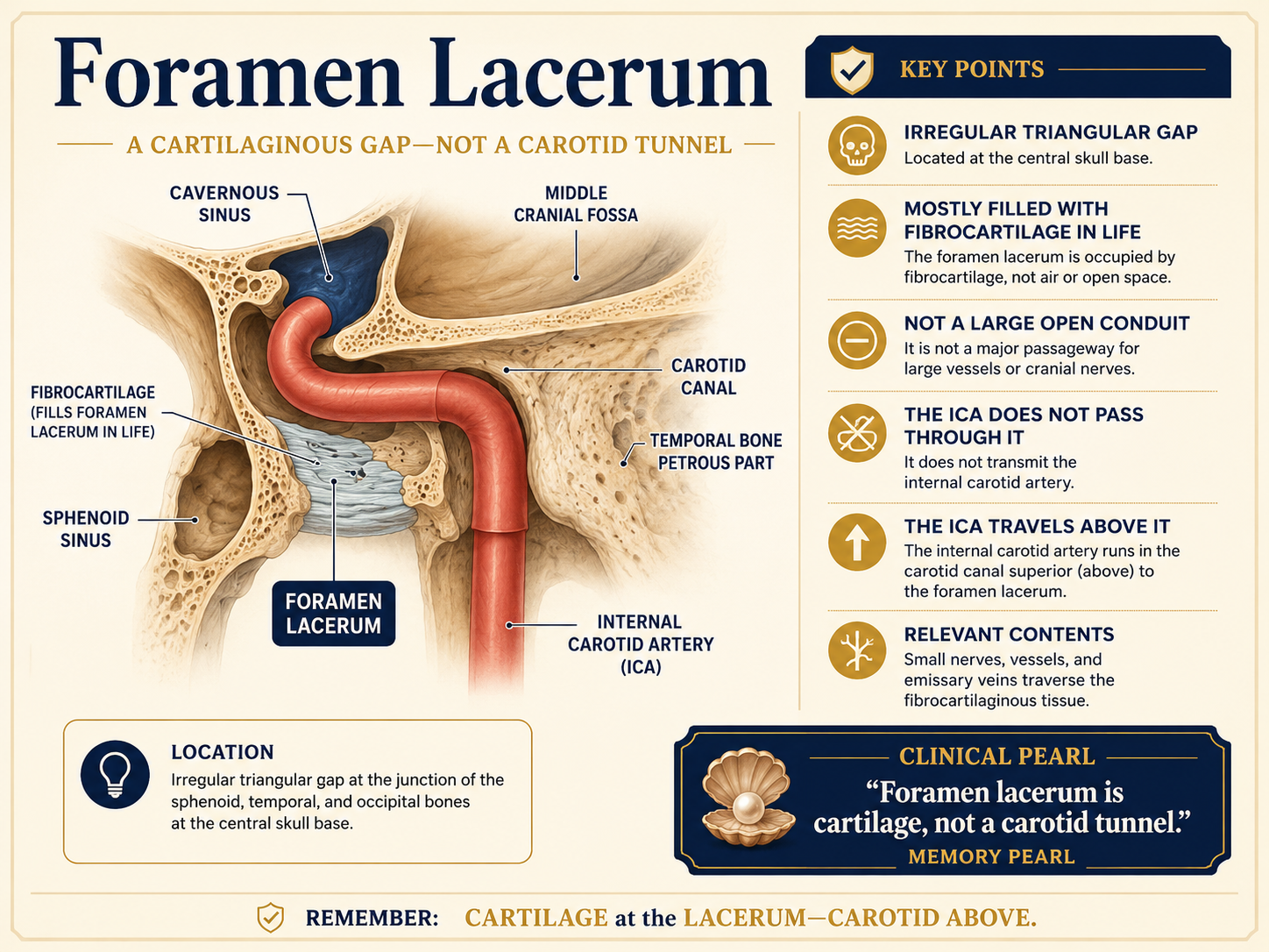
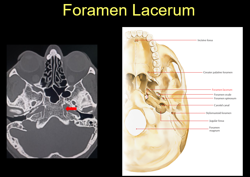
Key pearlThe ICA passes over the fibrocartilage-filled foramen lacerum—it does not traverse the foramen.
C4–C7: Intracranial Course
C4 Cavernous
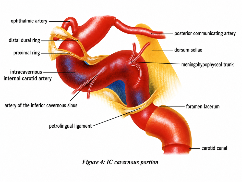
Petrolingual ligament to proximal dural ring through the cavernous sinus. CN VI lies lateral. Branches: MHT and inferolateral trunk.
C5 Clinoid
Very short, usually branchless segment between the proximal and distal dural rings.
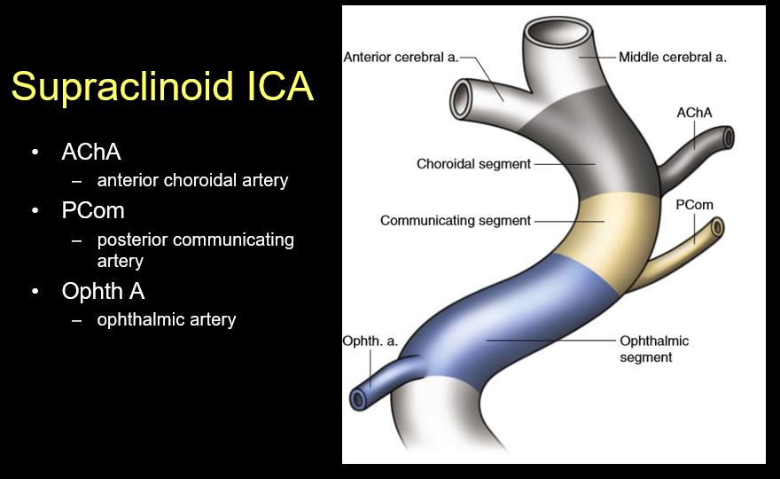
C6 Ophthalmic
Intradural supraclinoid segment giving ophthalmic and superior hypophyseal arteries.
C7 Communicating
Gives PCOM and AChA, then divides into ACA and MCA.
Branches by Segment
Even segments
- C2: caroticotympanic; Vidian.
- C4: meningohypophyseal trunk; inferolateral trunk.
- C6: ophthalmic; superior hypophyseal.
Terminal segment
- C7 side branches: PCOM and AChA.
- C7 terminal branches: ACA and MCA.
PatternOdd segments are usually branchless except C7; C2, C4, and C6 characteristically have paired branch groups.
Branch Mnemonics
A VIP'S COMMA
- Anterior choroidal
- Vidian
- Inferolateral trunk
- Posterior communicating
- Superior hypophyseal
- Caroticotympanic
- Ophthalmic
- Meningohypophyseal trunk
- Middle cerebral
- Anterior cerebral
In segment order
Calming Voices Make Intra-Operative Surgery Pleasurable And Almost Memorable
C2: C/V · C4: M/I · C6: O/S · C7: P/A/A/M.
Imaging & Reporting Pearls
Use segment names
Describe pathology by Bouthillier segment and relationship to the cavernous sinus and dural rings. “Paraclinoid” generally spans C5–C6 but is less specific.
Carotid siphon
Commonly refers to the curved lacerum, cavernous, clinoid, and adjacent supraclinoid course.
Dural rings
The distal dural ring marks entry into the subarachnoid space and helps determine whether disease is intra- or extradural.
Other schemes
Older Gibo, four-part Terminologia Anatomica, and angiographic schemes remain in use; state the classification if terminology is ambiguous.